ISSN 2234-3806 • eISSN 2234-3814
Ann Lab Med 2017;37:261-266
https://doi.org/10.3343/alm.2017.37.3.261
Novel SLC37A4 Mutations in Korean Patients With Glycogen Storage Disease Ib
Rihwa Choi, M.D.1, Hyung-Doo Park, M.D.1, Jung Min Ko, M.D.2, Jeongho Lee, M.D.3, Dong Hwan Lee, M.D.3, Suk Jin Hong, M.D.4, Chang-Seok Ki, M.D.1, Soo-Youn Lee, M.D.1, Jong-Won Kim, M.D.1, Junghan Song, M.D.5, and Yon Ho Choe, M.D.6
Department of Laboratory Medicine and Genetics1, Samsung Medical Center, Sungkyunkwan University School of Medicine, Seoul; Department of Pediatrics2, Seoul National University Children’s Hospital, Seoul National University College of Medicine, Seoul; Department of Pediatrics3, Soonchunhyang University Hospital, Seoul; Department of Pediatrics4, Catholic University of Daegu School of Medicine, Daegu; Department of Laboratory Medicine5, Seoul National University College of Medicine, Seoul National University Bundang Hospital, Seongnam; Department of Pediatrics6, Samsung Medical Center, Sungkyunkwan University School of Medicine, Seoul, Korea
Background: Molecular techniques are fundamental for establishing an accurate diagno- sis and therapeutic approach of glycogen storage diseases (GSDs). We aimed to evaluate SLC37A4 mutation spectrum in Korean GSD Ib patients.
Methods: Nine Korean patients from eight unrelated families with GSD Ib were included.
SLC37A4 mutations were detected in all patients with direct sequencing using a PCR method and/or whole-exome sequencing. A comprehensive review of previously reported SLC37A4 mutations was also conducted.
Results: Nine different pathogenic SLC37A4 mutations were identified in the nine patients with GSD Ib. Among them, four novel mutations were identified: c.148G>A (pGly50Arg), c.320G>A (p.Trp107*), c.412T>C (p.Trp138Arg), and c.818G>A (p.Gly273Asp). The most common mutation type was missense mutations (66.7%, 6/9), followed by nonsense mu- tations (22.2%, 2/9) and small deletion mutations (11.1%, 1/9). The most common muta- tion identified in the Korean population was c.443C>T (p.Ala148Val), which comprised 39.9% (7/18) of all tested alleles. This mutation has not been reported in GSD Ib patients in other ethnic populations.
Conclusions: This study expands knowledge of the SLC37A4 mutation spectrum in Ko- rean patients with GSD Ib.
Key Words: Glycogen storage disease, GSD Ib, Korean population, mutation, SLC37A4
Received: July 13, 2016
Revision received: October 12, 2016 Accepted: January 2, 2017
Corresponding author: Hyung-Doo Park Department of Laboratory Medicine and Genetics, Samsung Medical Center, Sungkyunkwan University School of Medicine, 81 Irwon-ro, Gangnam-gu, Seoul 06351, Korea
Tel: +82-2-3410-0290 Fax: +82-2-3410-2719 E-mail: [email protected]
Co-corresponding author: Junghan Song Department of Laboratory Medicine, Seoul National University Bundang Hospital, 82 Gumi-ro 173-beon-gil, Bundang-gu, Seongnam 13620, Korea Tel: +82-31-787-7691
Fax: +82-2-3410-4015 E-mail: [email protected]
© Korean Society for Laboratory Medicine This is an Open Access article distributed under the terms of the Creative Commons Attribution Non-Commercial License (http://creativecom- mons.org/licenses/by-nc/4.0) which permits unrestricted non-commercial use, distribution, and reproduction in any medium, provided the original work is properly cited.
INTRODUCTION
Glycogen storage disease type I (GSD I) is a group of rare auto- somal recessive disorders caused by deficiencies in the activi- ties of glucose-6-phosphatase-α (G6Pase-α)/glucose-6-phos- phate transporter (G6PT) complexes. The disease has an overall incidence of approximately 1 in 100,000 individuals [1, 2]. In
this complex, G6Pase-α and G6PT are functionally coupled;
G6PT transports G6P from the cytoplasm into the lumen of the endoplasmic reticulum, where it is hydrolyzed to glucose and inorganic phosphate by G6Pase-α [3]. A functional G6Pase-α/
G6PT complex maintains interprandial glucose homeostasis.
Specifically, the complex serves as a catalyst in the hydrolysis of intracellular G6P to glucose in the terminal step of gluconeogen-
esis and glycogenolysis in the liver, kidney, and intestine [2, 3].
Mutations in the G6PC gene, which encodes G6Pase-α, are re- sponsible for approximately 80% of all GSD I cases, classified as GSD Ia. Mutations in the SLC37A4 gene, which encodes G6PT, are responsible for the remaining ~20% of GSD I cases, and are classified as GSD Ib [4].
GSD is a clinically and genetically heterogeneous group of dis- eases that differs according to the site of abnormal glycogen me- tabolism (i.e., the liver, muscle, heart, or brain) [5]. Different types of GSDs can be clinically indistinguishable. For example, patients with GSD I, GSD III, GSD 0, and GSD XI present with hepatomegaly and/or hypoglycemia, and manifest as hepatic GSDs [5]. Patients with GSD Ib have symptoms similar to those of patients with GSD Ia; however, those with GSD Ib also have neutropenia and inflammatory bowel disease, which require dif- ferent therapeutic options for management [4, 6]. The molecu- lar diagnosis of GSD avoids the need for invasive liver biopsies [7]. Furthermore, the molecular diagnosis of GSD is important for establishing appropriate therapeutic and monitoring plans [8]. A recent clinical practice guideline recommended that the diagnosis of GSD I should be confirmed by using full-gene se- quencing of the G6PC (GSD Ia) and SLC37A4 (GSD Ib) genes [4]. The guideline also mentioned that although full-gene sequen- cing of both genes is available for clinical testing, targeted muta- tion analysis would be helpful for some ethnic groups. Testing for specific, common mutations can identify up to 100% of af- fected individuals, depending on the ethnic group [4]. In this con-
text, identifying the mutation spectrum in specific ethnic popu- lations is important for patient care [4, 8].
Since the GSD enzyme activity tests were first introduced in Korea, there have only been a few case reports of SLC37A4 mu- tations in Korean patients with GSD Ib [9-12]. Therefore, the aim of this study was to evaluate the mutation spectrum in Ko- rean patients with GSD Ib for the first time, and to further com- pare the spectrum to previously reported mutation spectra re- ported for other ethnic populations.
METHODS
1. Study populationBetween April 2003 and September 2015, nine Korean children from eight unrelated families who have been identified as hav- ing SLC37A4 variants were included in this study. Two of the patients were previously reported (cases 1 and 2 in Table 1) [9, 10]. All of these patients were identified as having SLC37A4 mu- tations at Samsung Medical Center, Seoul, Korea. Written inform ed consent was obtained from all subjects and/or their parents. This study was conducted according to the guidelines of the Decla- ration of Helsinki. All procedures involving human subjects were approved by the Institutional Review Board of Samsung Medical Center.
2. SLC37A4 mutation analysis
Human genomic DNA was prepared from peripheral blood sam-
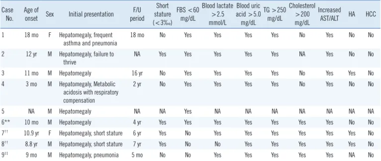
Table 1. Clinical manifestations of Korean patients with GSD Ib with identified SLC37A4 mutations Case
No.
Age of
onset Sex Initial presentation F/U period
Short stature (<3‰)
FBS <60 mg/dL
Blood lactate
>2.5 mmol/L
Blood uric acid >5.0
mg/dL
TG >250 mg/dL
Cholesterol
>200 mg/dL
Increased
AST/ALT HA HCC 1 18 mo F Hepatomegaly, frequent
asthma and pneumonia
18 mo No Yes Yes Yes Yes No Yes No No
2 12 yr M Hepatomegaly, failure to
thrive NA Yes Yes Yes Yes Yes NA Yes No No
3 11 mo M Hepatomegaly 16 yr No Yes Yes Yes Yes No Yes Yes No
4 3 mo M Hepatomegaly, Metabolic acidosis with respiratory compensation
2 yr No Yes Yes Yes Yes No Yes No No
5 NA M Hepatomegaly NA NA Yes NA NA NA NA NA NA NA
6** 10 mo M Hepatomegaly 4 yr Yes Yes Yes Yes Yes Yes Yes No No
7†† 10.9 yr F Hepatomegaly, short stature 6 yr Yes No Yes Yes Yes Yes Yes Yes No
8†† 8.8 yr M Hepatomegaly, short stature 7 yr Yes No No Yes Yes Yes Yes Yes No
9‡‡ 9 mo M Hepatomegaly, pneumonia 5 mo No No Yes Yes Yes Yes Yes NA No
(continued to the next page)
ples by using a Wizard genomic DNA purification kit (Promega, Madison, WI, USA) according to the manu- facturer’s recommendations. All nine exons and flank- ing regions of the SLC37A4 gene were amplified with PCR using primers designed by the authors (sequences available upon request). PCR was performed by using a thermal cycler (Model 970; Applied Biosystems, Fos- ter City, CA, USA). Direct sequencing of the DNA was performed using the ABI Prism 3100 Genetic Analyzer (Applied Biosystems) with the BigDye Terminator Cycle Sequencing-Ready Reaction Kit (Applied Biosystems).
The nucleotides were numbered from the first adenine of the ATG translation initiation codon in the SLC37A4 cDNA Reference Sequence NM_001164277.1.
Whole-exome sequencing (WES) was applied for one patient (case 6 in Table 1). Exonic sequences were enriched in the DNA sample using the SureSelect Target Enrichment kit (Agilent Technologies, Santa Clara, CA, USA). Sequences were determined on a HiSeq2000 system (Illumina, San Diego, CA, USA). A total of 150–
200 bp were obtained as paired-end reads. The vari- ants of the patients that passed the quality filtering step were compared against those in public databases [Na- tional Heart, Lung, and Blood Institute Exome Sequenc- ing Project; 1000 Genomes Project; dbSNP; Exome Aggregation Consortium (ExAC, exac.broadinstitute.
org/)] with a cut-off of a global minor allele frequency
<1.0%. Protein-altering variants were then selected.
The variants derived from the variant filtering strategy were then prioritized on the basis of their likelihood to affect protein function, and/or to completely or partially match the patient’s phenotype. The variants’ effects on protein function were predicted by using public algo- rithms such as Sorting Intolerant from Tolerant (SIFT;
http://sift.jcvi.org/).
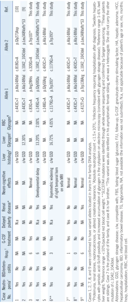
In addition, a comprehensive review of the literature on previously reported SLC37A4 mutations was con- ducted. The Human Gene Mutation Database (HGMD, http://www.hgmd.org/) was checked for previously re- ported sequence variants [13]. The pathogenicity of missense variants was evaluated by in silico analyses with SIFT and Polymorphism Phenotyping v.2 (Poly- Phen-2) (http://genetics.bwh.harvard.edu/pph2/). The variants that were previously unreported with unknown pathogenicity were classified as the standards accord- ing to the American College of Medical Genetics and Case No.Neutro penia†IBD/entero colitisHosp.‡GCSF treatmentDelayed pubertyRenal disease*Neurocognitive effectsLiver histology§Liver Glycogen||RBC Glycogen¶Allele 1Allele 2Ref. 1YesNoNANAN.aNANAc/w GSDNANAc.443C>Tp.Ala148Valc.818G>Ap.Gly273Asp[10] 2YesYesNANANANANAc/w GSD12.30%NAc.443C>Tp.Ala148Valc.1042_1043delp.Leu348Valfs*53[9] 3YesNo6YesNoNoNormalc/w GSDNANAc.83G>Ap.Arg28Hisc.320G>Ap.Trp107*This study 4YesYes6NoN.aYesDevelopmental delayc/w GSD13.20%2.00%c.149G>Ap.Gly50Gluc.1042_1043delp.Leu348Valfs*53This study 5NANANANANANANAc/w GSDNANAc.148G>Ap.Gly50Argc.443C>Tp.Ala148ValThis study 6**YesNo3YesN.aYesAsymmetric widening of right temporal horn on sella MRI
c/w GSD16.72%4.05%c.1179G>Ap.Trp393*c.1179G>Ap.Trp393*This study 7††YesNo1YesYesNoNormalc/w GSDNANAc.443C>Tp.Ala148Valc.443C>Tp.Ala148ValThis study 8††YesNo0NoNoNoNormalc/w GSDNANAc.443C>Tp.Ala148Valc.443C>Tp.Ala148ValThis study 9‡‡NANA6NoNAYesNormalc/w GSDNANAc.412T>Cip.Trp138Argc.1042_1043delp.Leu348Valfs*53This study Cases 1, 2, 7, 8, and 9 were confirmed to have variant alleles located in trans in the family DNA analysis. *Proteinuria, renal stones, nephrocalcinosis, or altered creatinine clearance; †Absolute neutrophil count <1.5×10⁹/L; ‡Infection frequency requiring hospitalization after diagnosis; §Swollen hepato- cytes with periodic acid–Schiff-positivity and increased accumulation of glycogen in the cytoplasm on electron microscopy (consistent with glycogen storage disease); ||Reference range 1–6% /wet liver weight; ¶Reference range <10%/packed red blood cell weight; **SLC37A4 mutations were identified through whole-exome sequencing and confirmed by Sanger sequencing; ††These patients are siblings. Case 7 is the proband of the family, and case 8 is her brother; ‡‡This variant was also identified in his asymptomatic female sibling, who was a heterozygote. She did not carry the other mutation of c.1042_1043del. Abbreviations: GSD, glycogen storage disease; c/w, compatible with; FBS, fasting blood sugar (glucose); F/U, follow-up; G-CSF, granulocyte-colony stimulating factor; HA, hepatic adenoma; HCC, hepatocellular carcinoma; IBD, inflammatory bowel disease; TG, triglycerides; NA, not available (the information was not submitted); N.a, not applicable because of patient’s age or sex; mo, months; hosp, hospitalization; RBC, red blood cell.
Table 1. Continued
Genomics (ACMG) guidelines for the interpretation of sequence variants [14].
RESULTS
Nine patients from eight unrelated families, including two previ- ously reported Korean patients, were identified to have SLC37A4 mutations. Among the nine patients, 77.8% were male. The clinical information and specific SLC37A4 mutations identified in the nine Korean GSD Ib patients are shown in Table 1. The median age of onset was 14.5 months (range 3 months to 10.9 yr). The median age at molecular diagnostic work-up was 10.4 yr (range 6 months to 19 yr). All patients presented with hepa- tomegaly and elevated AST and ALT levels as the first clinical signs. All of the patients also had neutropenia (absolute neutro- phil count <1.5×10⁹/L). Because of limited clinical information, the genotype–phenotype correlation could not be assessed.
Among 18 mutant alleles, nine different SLC37A4 mutations were identified (Table 2). These mutations were distributed among all of the coding exons of SLC37A4, except for exon 11. Among the nine mutations, four were novel variants: c.148G>A (p.Gly- 50Arg), c.320G>A (p.Trp107*), c.412T>C (p.Trp138Arg), and c.818G>A (p.Gly273Asp). The remaining five mutations were previously reported: c.83G>A (p.Arg28His), c.149G>A (p.Gly- 50Glu), c.443C>T (p.Ala148Val), c.1042_1043del (p.Leu348 - Valfs*53), and c.1179G>A (p.Trp393*) [1, 15-21]. Two of the
novel variants were classified as “pathogenic”, including c.148G>A (p.Gly50Arg) and c.320G>A (p.Trp107*). The other two novel variants, c.412T>C (p.Trp138Arg) and c.818G>A (p.Gly273Asp), were classified as “likely pathogenic” according to the ACMG sequence variants interpretation guidelines [14]. Among the nine mutations, the most common were missense mutations (66.7%, 6/9) followed by nonsense mutations (22.2%, 2/9) and small deletion mutations (11.1%, 1/9).
Among the 18 tested alleles, the variants c.443C>T (p.Ala- 148Val), c.1042_1043del (p.Leu348Valfs*53), and c.1179G>A (p.Trp393*) were repeatedly identified in different individuals (7 times, 3 times, and 2 times, respectively). Notably, the most common mutation identified in Korean patients was c.443C>T (p.Ala148Val), accounting for 55.6% (5/9 patients) of all GSD Ib patients and 38.9% of the tested alleles (7/18 alleles). WES fol- lowed by Sanger sequencing was used to confirm that one pa- tient (case 6) carried the known homozygous pathogenic muta- tion c.1179G>A (p.Trp393*). Cases 7 and 8 are siblings from the same family; case 7 is the proband of the family and case 8 is her brother.
DISCUSSION
To our knowledge, this is the first study to summarize the clini- cal characteristics of Korean patients with GSD Ib. The SLC37A4 mutation spectrum is known to be distributed widely across the Table 2. SLC37A4 mutation spectrum in nine Korean GSD type Ib patients
Exon No. Nucleotide
change Amino acid
change Mutation
type Location PolyPhen2 score SIFT
score Allele
count Previously
reported References Variants category*
3 c.83G>A p.Arg28His Missense Luminal loop 1 1.000 0.00 1 Yes [1, 15, 16, 18]
3 c.148G>A p.Gly50Arg Missense Luminal loop 1 1.000 0.00 1 No [15, 16, 20]† Pathogenic
4 c.149G>A p.Gly50Glu Missense Luminal loop 1 1.000 0.00 1 Yes [17]
4 c.320G>A p.Trp107* Nonsense Helix2 N/A N/A 1 No Pathogenic
5 c.412T>C p.Trp138Arg Missense Helix3 0.741 0.00 1 No Likely pathogenic
5 c.443C>T p.Ala148Val Missense Helix3 0.921 0.00 7 Yes [9]
7 c.818G>A p.Gly273Asp Missense Helix6 1.000 0.00 1 No [21]‡ Likely pathogenic
9 c.1042_1043del p.Leu348Valfs*53 Frameshift (small deletion)
Helix8 N/A N/A 3 Yes [19, 25]
10 c.1179G>A p.Trp393* Nonsense Luminal loop 5 N/A N/A 2 Yes [26]
*Previously unreported variants were classified by the American College of Medical Genetics and Genomics (ACMG) standards and guidelines for the inter- pretation of sequence variants [14]. †c.148G>A has not been reported previously; however, c.148G>C has been reported as a pathogenic mutation that re- sults in the same amino acid change of p.Gly50Arg. This mutation could be categorized as a pathogenic variant by the ACMG standards and guidelines for the interpretation of sequence variants [14]. ‡c.818G>A has not been reported previously; however, c.817G>A (p.Gly273Ser) has been reported in a GSD patient [21].
Abbreviations: GSD, glycogen storage disease; N/A, not applicable.
SLC37A4 gene. In this study, the most common mutations were missense mutations, which is consistent with the data in the HGMD database.
We identified four novel pathogenic [c.148G>A (p.Gly50Arg) and c.320G>A (p.Trp107*)] or likely pathogenic [c.412T>C (p.
Trp138Arg) and c.818G>A (p.Gly273Asp)] SLC37A4 mutations.
Among them, c.148G>A has not been reported previously. In contrast, c.148G>C has been previously reported as a pathoge- nic mutation. The c.148G>C mutation results in the same amino acid change (p.Gly50Arg), abolishes the microsomal G6P uptake activity, and compromises G6PT stability [15]. The c.148G>A mutation could be categorized as a pathogenic variant accord- ing to the ACMG standards and guidelines for the interpretation of sequence variants [14].
Notably, the most common mutation identified in the Korean population was c.443C>T (p.Ala148Val), which was found in 55.6% of the GSD Ib patients and in 38.9% of the tested alleles.
This mutation has not been reported in other ethnic patients with GSD Ib. It has only been reported in two alleles in East Asia, and was identified as heterozygous among 43,554 individuals (87,108 tested alleles) with an allele frequency of 2.296e-5 in the ExAC database. However, this site is covered in <80% of the individu- als in the ExAC database, which may indicate a low-quality site.
These findings suggest that this variant might be an important marker for the diagnosis of GSD Ib in the East Asian population specifically. Considering that no Japanese or Chinese patients have been reported to have c.443C>T, it is possible that this represents a recurrent mutation specific to Koreans. Although this is a very rare single nucleotide variant (SNV) in the public database, this variation was detected in most cases simultane- ously with another SNV that seems to have a greater impact on protein function. Therefore, further studies are needed to con- firm the pathogenicity of this recurrent SNV.
The pathogenic variant of c.1042_1043del (p.Leu348Valfs*53) was the second most frequent mutation (33.3%, 3/9 of GSD Ib patients; 16.7%, 3/18 tested alleles). This mutation has been frequently reported in mixed Caucasian (27–31%) and German (32%) populations [4]. Other mutations that have been frequently reported in other ethnic populations have not been identified in the Korean population [4]. These include c.352T>C (p.Trp118Arg) identified in 37–50% of Japanese people, and c.1015G>T (p.Gly - 339Cys) identified in 19–21% of mixed Caucasians and in 29%
of Germans. These results suggest that Korean SLC37A4 muta- tions differ from those of other ethnic populations owing to the genetic divergence of Homo sapiens. Furthermore, the c.443C>T mutation may be relatively new, as compared with c.1042_1043del
[22], given that identification of a large proportion of rare alleles can be a signature of recent expansion, as mutations that have occurred since the expansion will not have had sufficient time to spread throughout the population [23].
In this study, cases 7 and 8, both of whom have homozygous c.443C>T mutant alleles, are siblings from the same family. These patients also showed similar clinical manifestations, including short stature and hepatic adenoma. Granulocyte-colony stimu- lating factor treatment was only used in case 7. However, both cases 7 and 8 tolerated dietary management (including corn starch) and only presented with minor infections (such as chronic otitis media or mucosal infection) that did not require hospital- ization.
In this study, all patients with SLC37A4 mutations had neutro- penia, except for two cases whose clinical information was not available. In the literature, no correlation could be established between the presence of “leaky” mutations and the absence of neutropenia, in both, homozygous and compound heterozygous patients [24]. Further studies are needed to take into account clinical and biochemical data, which are integral for assessing genotype-phenotype correlations in the Korean population. How- ever, recent molecular diagnostic approaches based on muta- tion analysis for the disease-causing genes of each type of GSD provide the advantages of avoiding invasive liver biopsies or en- zymatic studies. These latter methods are historical diagnostic methods that can potentially introduce ambiguity when differen- tiating between several types of GSD with similar clinical find- ings [4]. Despite its limitations, this study is valuable to improve understanding of the SLC37A4 mutation spectrum in the Korean population.
In conclusion, we identified four novel pathogenic and likely pathogenic SLC37A4 variations. The c.443C>T (p.Ala148Val) variant was novel and the most common mutation identified. The SLC37A4 mutation spectrum in Korean GSD Ib patients tends to differ from that of other ethnic populations. Direct sequence analysis of full-gene sequences is needed to provide accurate molecular diagnoses, and WES could be an effective approach in the diagnosis of GSD Ib.
Authors’ Disclosures of Potential Conflicts of Interest
No potential conflicts of interest relevant to this article were re- ported.
Acknowledgments
This study was supported by a grant from the Korea Health Tech- nology R&D Project, Ministry of Health & Welfare, Republic of Korea (A120030).
REFERENCES
1. Chou JY, Matern D, Mansfield BC, Chen YT. Type I glycogen storage diseases: disorders of the glucose-6-phosphatase complex. Curr Mol Med 2002;2:121-43.
2. Chou JY, Jun HS, Mansfield BC. Type I glycogen storage diseases: dis- orders of the glucose-6-phosphatase/glucose-6-phosphate transporter complexes. J Inherit Metab Dis 2015;38:511-9.
3. Chou JY, Jun HS, Mansfield BC. Glycogen storage disease type I and G6Pase-β deficiency: etiology and therapy. Nat Rev Endocrinol 2010;6:
676-88.
4. Kishnani PS, Austin SL, Abdenur JE, Arn P, Bali DS, Boney A, et al. Di- agnosis and management of glycogen storage disease type I: a practice guideline of the American College of Medical Genetics and Genomics.
Genet Med 2014;16:e1.
5. Saudubray JM, Van den Berghe G, et al, eds. Inborn metabolic diseas- es. Berlin: Springer, 2012.
6. Visser G, Rake JP, Labrune P, Leonard JV, Moses S, Ullrich K, et al. Con- sensus guidelines for management of glycogen storage disease type 1b - European Study on Glycogen Storage Disease Type 1. Eur J Pediatr 2002;161(S1):S120-3.
7. Davit-Spraul A, Piraud M, Dobbelaere D, Valayannopoulos V, Labrune P, Habes D, et al. Liver glycogen storage diseases due to phosphorylase system deficiencies: diagnosis thanks to non invasive blood enzymatic and molecular studies. Mol Genet Metab 2011;104:137-43.
8. Bali DS, Chen YT, Goldstein JL. Glycogen Storage Disease Type I. In:
Pagon RA, Adam MP, et al., eds. GeneReviews. Seattle, WA: University of Washington, 1993.
9. Han SH, Ki CS, Lee JE, Hong YJ, Son BK, Lee KH, et al. A novel muta- tion (A148V) in the glucose 6-phosphate translocase (SLC37A4) gene in a Korean patient with glycogen storage disease type 1b. J Korean Med Sci 2005;20:499-501.
10. Kim MS, Park JB, Ki CS, Kim JK. A case of glycogen storage disease type Ib. Korean J Pediatr 2009;52:1383-7.
11. Jeong YJ, Kang B, Choi SY, Ki CS, Lee SY, Park HD, et al. Does type I truly dominate hepatic glycogen storage diseases in Korea?: a single cen- ter study. Pediatr Gastroenterol Hepatol Nutr 2014;17:239-47.
12. Kim JW, Kim JQ, Cho HI, Kim SI. Experience on the confirmatory en- zyme tests for the diagnosis of glycogen storage disease. Korean J Clin Pathol 1990;10:257-64.
13. Stenson PD, Mort M, Ball EV, Shaw K, Phillips A, Cooper DN. The Hu-
man Gene Mutation Database: building a comprehensive mutation re- pository for clinical and molecular genetics, diagnostic testing and per- sonalized genomic medicine. Hum Genet 2014;133:1-9.
14. Richards S, Aziz N, Bale S, Bick D, Das S, Gastier-Foster J, et al. Stan- dards and guidelines for the interpretation of sequence variants: a joint consensus recommendation of the American College of Medical Genet- ics and Genomics and the Association for Molecular Pathology. Genet Med 2015;17:405-24.
15. Chen LY, Pan CJ, Shieh JJ, Chou JY. Structure-function analysis of the glucose-6-phosphate transporter deficient in glycogen storage disease type Ib. Hum Mol Genet 2002;11:3199-207.
16. Chen SY, Pan CJ, Lee S, Peng W, Chou JY. Functional analysis of muta- tions in the glucose-6-phosphate transporter that cause glycogen stor- age disease type Ib. Mol Genet Metab 2008;95:220-3.
17. Dissanayake VH, Jayasinghe JD, Thilakaratne V, Jayasekara RW. A nov- el mutation in SLC37A4 gene in a Sri Lankan boy with glycogen storage disease type Ib associated with very early onset neutropenia. J Mol Gen- et Med 2011;5:262-3.
18. Hiraiwa H, Pan CJ, Lin B, Moses SW, Chou JY. Inactivation of the glu- cose 6-phosphate transporter causes glycogen storage disease type 1b.
J Biol Chem 1999;274:5532-6.
19. Melis D, Balivo F, Della Casa R, Romano A, Taurisano R, Capaldo B, et al. Myasthenia gravis in a patient affected by glycogen storage disease type Ib: a further manifestation of an increased risk for autoimmune dis- orders? J Inherit Metab Dis 2008;31(S2):S227-31.
20. Veiga-da-Cunha M, Gerin I, Chen YT, Lee PJ, Leonard JV, Maire I, et al.
The putative glucose 6-phosphate translocase gene is mutated in es- sentially all cases of glycogen storage disease type I non-a. Eur J Hum Genet 1999;7:717-23.
21. Wang J, Cui H, Lee NC, Hwu WL, Chien YH, Craigen WJ, et al. Clinical application of massively parallel sequencing in the molecular diagnosis of glycogen storage diseases of genetically heterogeneous origin. Genet Med 2013;15:106-14.
22. Flanagan JM, McMahon G, Brendan Chia SH, Fitzpatrick P, Tighe O, O’Neill C, et al. The role of human demographic history in determining the distribution and frequency of transferase-deficient galactosaemia mutations. Heredity (Edinb) 2010;104:148-54.
23. Keinan A, Mullikin JC, Patterson N, Reich D. Measurement of the hu- man allele frequency spectrum demonstrates greater genetic drift in East Asians than in Europeans. Nat Genet 2007;39:1251-5.
24. Melis D, Fulceri R, Parenti G, Marcolongo P, Gatti R, Parini R, et al. Gen- otype/phenotype correlation in glycogen storage disease type 1b: a mul- ticentre study and review of the literature. Eur J Pediatr 2005;164:501-8.
25. Marcolongo P, Barone V, Priori G, Pirola B, Giglio S, Biasucci G, et al.
Structure and mutation analysis of the glycogen storage disease type 1b gene. FEBS Lett 1998;436:247-50.
26. Santer R, Rischewski J, Block G, Kinner M, Wendel U, Schaub J, et al.
Molecular analysis in glycogen storage disease 1 non-A: DHPLC detec- tion of the highly prevalent exon 8 mutations of the G6PT1 gene in Ger- man patients. Hum Mutat 2000;16:177.