ISSN 2234-3806 • eISSN 2234-3814
http://dx.doi.org/10.3343/alm.2015.35.1.141
Non-Homologous End Joining Repair Mechanism- Mediated Deletion of CHD7 Gene in a Patient with Typical CHARGE Syndrome
Seung Jun Lee, M.D.1, Jong Hee Chae, M.D.2, Jung Ae Lee, M.T.1, Sung Im Cho, M.T.1, Soo Hyun Seo, M.D.1, Hyunwoong Park, M.D.1, Moon-Woo Seong, M.D.1, and Sung Sup Park, M.D.1
Department of Laboratory Medicine1, Seoul National University Hospital; Department of Pediatrics2, Seoul National University Children’s Hospital, Seoul, Korea
CHARGE syndrome MIM #214800 is an autosomal dominant syndrome involving multiple congenital malformations. Clinical symptoms include coloboma, heart defects, choanal atresia, retardation of growth or development, genital hypoplasia, and ear anomalies or deafness. Mutations in the chromodomain helicase DNA binding protein 7 (CHD7) gene have been found in 65-70% of CHARGE syndrome patients. Here, we describe a 16-month- old boy with typical CHARGE syndrome, who was referred for CHD7 gene analysis. Se- quence analysis and multiplex ligation-dependent probe amplification were performed. A heterozygous 38,304-bp deletion encompassing exon 3 with a 4-bp insertion was identi- fied. There were no Alu sequences adjacent to the breakpoints, and no sequence micro- homology was observed at the junction. Therefore, this large deletion may have been medi- ated by non-homologous end joining. The mechanism of the deletion in the current case differs from the previously suggested mechanisms underlying large deletions or complex genomic rearrangements in the CHD7 gene, and this is the first report of CHD7 deletion by this mechanism worldwide.
Key Words: CHARGE syndrome, CHD7, Large deletion, Non-homologous end joining
Received: August 14, 2014 Revision received: August 28, 2014 Accepted: October 28, 2014 Corresponding author: Sung Sup Park Department of Laboratory Medicine, Seoul National University Hospital, 101 Daehak- ro, Jongno-gu, Seoul 110-744, Korea Tel: +82-2-2072-3206
Fax: +82-2-747-0359 E-mail: [email protected]
© The Korean Society for Laboratory Medicine This is an Open Access article distributed under the terms of the Creative Commons Attribution Non-Commercial License (http://creativecom- mons.org/licenses/by-nc/3.0) which permits unrestricted non-commercial use, distribution, and reproduction in any medium, provided the original work is properly cited.
INTRODUCTION
CHARGE syndrome MIM #214800 is an autosomal dominant genetic disorder with multiple congenital anomalies. The syn- drome derives its name from the first letters of its main clinical manifestations: Coloboma, Heart defects, Atresia of choanae, Retardation, Genitourinary malformation, and Ear abnormalities.
The clinical criteria for CHARGE syndrome were first described by Blake et al. [1], and then modified by Verloes [2]. According to Verloes’ criteria, CHARGE syndrome can be classified as typi- cal, partial, or atypical.
The chromodomain helicase DNA binding protein 7 (CHD7) gene located on chromosome 8q12.1 is 188 kb in length and
consists of 38 exons. The CHD7 protein functions as a regulator of DNA transcription [3]. CHD7 gene mutations have been identified in 65-70% of patients with CHARGE syndrome [4-6].
As most of cases of CHARGE syndrome are caused by a de novo mutation [7], familial cases are rarely reported.
CHD7 gene mutations have been identified throughout the coding exons, and most of them are point mutations. Large de- letions and duplications account for only 2% of the observed mutations, while translocations account for <1%. As they are rare, large CHD7 gene deletions and duplications have not been reported previously in the Korean population. Here, we report a typical CHARGE syndrome patient with a large deletion in the CHD7 gene and a presumptive relevant mechanism.
CASE REPORT
A 16-month-old boy was referred for genetic workup because of typical manifestations of the CHARGE syndrome. Facial asymme- try was observed at birth, and auditory and visual dysfunctions were also noted. Heart defects, including aortic stenosis, persis- tent ductus arteriosus, atrial septal defect, and pulmonary ste- nosis, were identified on cardiological examination. Multiple ab- normal findings, including incomplete cochlear turn and dyspla- sia of the vestibule and semicircular canal, were found on a computerized tomography scan. Two major signs (coloboma and hypoplastic semicircular canals) and four minor signs (rhomb- encephalic dysfunction, abnormal middle or external ear, mal- formation of mediastinal organs, and mental retardation) were identified according to Verloes’ criteria. The patient was diag- nosed as having typical CHARGE syndrome. A CHD7 gene analysis was requested for a confirmative diagnosis.
Informed consent was obtained from his legal representative, and genomic DNA was extracted from whole blood. PCR was performed by using primers specific for the 37 coding exons of the CHD7 gene. The sequencing reaction was performed with an ABI 3730 analyzer (Applied Biosystems, Foster City, CA, USA) by using a BigDye Terminator v3.1 Cycle sequencing kit (Ap- plied Biosystems). Sequencher 5.0 software (Gene Codes Cor- poration, Ann Arbor, MI, USA) was used for the sequencing data analysis. No mutations were identified from this CHD7 gene sequence analysis.
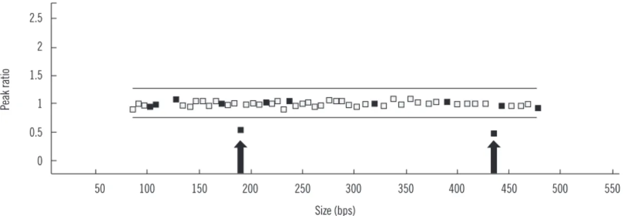
Gene dosage analysis was performed by using a multiplex li- gation-dependent probe amplification (MLPA) kit (SALSA MLPA P201-C1 CHARGE probemix; MRC-Holland, Amsterdam, Neth- erlands). MLPA analysis of the CHD7 gene revealed a heterozy- gous exon 3 deletion (Fig. 1). Genetic analyses of the patient’s parents were not available. Further experiments were then per-
formed in order to identify the precise breakpoints of this large deletion. A total of nine Alu sequences were located in intron 2 and three were located in intron 3 (Fig. 2C). Long range PCR was performed by using five forward primers adjacent to AluSx3, AluSg, AluSx1 (the more distal of the two), AluY, and AluSx, and one common reverse primer adjacent to AluSq2. The proximal breakpoint was localized between AluSg and AluSx1 (the more distal of the two). Another long range PCR was performed by using one common forward primer adjacent to AluSg, and two reverse primers adjacent to AluJr and AluSq2. The distal break- point was localized between AluJr and AluSq2. An additional long range PCR was performed by using forward primers lo- cated 1 kb, 2 kb, and 3 kb distal to AluSg and reverse primers located 1 kb, 2 kb, and 3 kb proximal to AluSq2. The proximal breakpoint was located between 3 kb distal to AluSg and AluSx1 (more distal of the two), while the distal breakpoint was located between 3 kb and 2 kb proximal to AluSq2. Finally, a 1kb-sized PCR fragment was obtained by using the following primers:
GGTGGGCTGTGAAGTGTTCTGGC (forward primer; located in intron 2) and ACCCACAGTGCACTCCTCCCC (reverse primer; lo- cated in intron 3) (Fig. 2A). Sequence analysis revealed the ex- act breakpoints (Fig. 2B). The deleted region totaled 38,304 bp from c.1665+10039 in intron 2 to c.2097-3547 in intron 3, and it was accompanied by a TAAC insertion (Fig. 2D).
DISCUSSION
The majority of CHD7 gene mutations are point mutations—
nonsense mutations, 44%; frameshift deletions or insertions, 34%; splice site mutations, 11%; and missense mutations, 8%
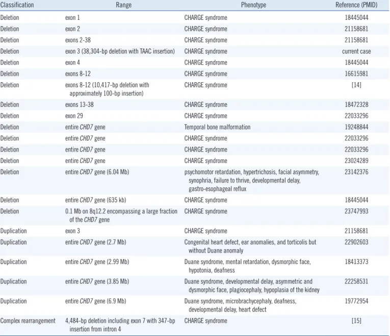
[8]. Large deletions or duplications account for only 2% of total cases. To date, 15 cases of large deletions and 5 cases of large duplications in the CHD7 gene region have been reported (Table
Fig. 1. Multiplex ligation-dependent probe amplification analysis of the CHD7 gene. Arrows indicate the reduced ratio of exon 3.
2.5 2 1.5 1 0.5 0
50 100 150 200 250 300 350 400 450 500 550 Size (bps)
Peak ratio
1). Of these 20 cases, 10 cases involved whole gene deletions and duplications. To date, 16 index cases of CHD7 gene muta- tions have been reported in Korea [9-12]. However, large dele- tions and duplications have not yet been reported in the Korean population.
Copy number variations including both additions and dele- tions are mediated by homologous recombination and non-ho- mologous repair mechanisms [13]. Homologous recombination is composed of non-allelic homologous recombination and sin- gle-strand annealing. Non-homologous repair mechanisms are classified into non-replicative non-homologous repair (non-ho- mologous end joining [NHEJ] and microhomology-mediated end joining), and replicative non-homologous repair (replication slippage or template switching, fork stalling and template switching [FosTes], and microhomology-mediated break-in- duced replication [MMBIR]).
Two different mechanisms underlying CHD7 gene deletions have been previously reported. In the first case, a large deletion spanning exons 8-12 was detected in a Japanese girl [14]. The deletion encompassed 10,417 bases from intron 7 to intron 12.
A polyadenine tract of approximately 100 bases was inserted into the junction. Therefore, this deletion was attributed to an
Alu retrotransposition-mediated mechanism. In the second case, a complex genomic rearrangement was detected in a Caucasian girl [15]. A deletion of approximately 4,484 bases, including exon 7, was accompanied by an insertion of 347 bases from intron 4 between the breakpoints. This deletion was attributed to a FosTes or MMBIR mechanism.
The CHD7 gene mutations in the current study were mediated by a distinctly different mechanism from the aforementioned cases. Alu sequences were not adjacent to the breakpoints of the deleted region in the current case. The nearest Alu se- quences from the proximal and distal breakpoints were located 469 bp and 2,400 bp away, respectively. There was a 4-bp, non- template insertion at the junction site, and there was no se- quence microhomology between the upstream and downstream sequences near the junction. Therefore, NHEJ is the most prob- able mechanism responsible for the current case. NHEJ is a part of the double-strand break repair pathway and is the predomi- nant repair mechanism used in mammals [16]. Furthermore, it is not uncommonly related to large deletions [17]. One or more nucleotide bases can be inserted into the junctions during the repair procedure in NHEJ [18, 19]. In summary, we report the first case of a CHD7 gene deletion mediated by NHEJ worldwide.
A B
C
D Fig. 2. Results of the sequence analysis and genomic structure encompassing exons 2-4 of the CHD7 gene. (A) Electrophoresis of a PCR product using the forward and reverse primers designated in (C) showed an apparent band in the patient. (B) The proximal and distal parts had no homologous sequences near the breakpoints. (C) Schematic diagram showing the range of the 38,304-bp deletion from c.1665+10039 to c.2097-3547. Alu sequences (blue triangles) were not adjacent to the breakpoints. Orange, green, and violet arrows indi- cate stepwise long-range PCR primers. (D) Sequence analysis revealed a 4-bp insertion (TAAC) between the breakpoints.
Abbreviations: Pt., patient; NC, normal control.
Table 1. Previously reported cases of gross deletion or duplication of the CHD7 gene
Classification Range Phenotype Reference (PMID)
Deletion exon 1 CHARGE syndrome 18445044
Deletion exon 2 CHARGE syndrome 21158681
Deletion exons 2-38 CHARGE syndrome 21158681
Deletion exon 3 (38,304-bp deletion with TAAC insertion) CHARGE syndrome current case
Deletion exon 4 CHARGE syndrome 18445044
Deletion exons 8-12 CHARGE syndrome 16615981
Deletion exons 8-12 (10,417-bp deletion with
approximately 100-bp insertion) CHARGE syndrome [14]
Deletion exons 13-38 CHARGE syndrome 18472328
Deletion exon 29 CHARGE syndrome 22033296
Deletion entire CHD7 gene Temporal bone malformation 19248844
Deletion entire CHD7 gene CHARGE syndrome 22033296
Deletion entire CHD7 gene CHARGE syndrome 22033296
Deletion entire CHD7 gene CHARGE syndrome 23024289
Deletion entire CHD7 gene (6.04 Mb) psychomotor retardation, hypertrichosis, facial asymmetry, synophria, failure to thrive, developmental delay, gastro-esophageal reflux
23142376
Deletion entire CHD7 gene (635 kb) CHARGE syndrome 18445044
Deletion 0.1 Mb on 8q12.2 encompassing a large fraction of the CHD7 gene
CHARGE syndrome 23747993
Duplication exon 3 CHARGE syndrome 21158681
Duplication entire CHD7 gene (2.7 Mb) Congenital heart defect, ear anomalies, and torticolis but
without Duane anomaly 22902603
Duplication entire CHD7 gene (2.99 Mb) Duane syndrome, mental retardation, dysmorphic face, hypotonia, deafness
18413373 Duplication entire CHD7 gene (3.85 Mb) Duane syndrome, developmental delay, asymmetric and
dysmorphic face, plagiocephaly, hypoplasia of the kidney 22258531 Duplication entire CHD7 gene (6.9 Mb) Duane syndrome, microbrachycephaly, deafness,
developmental delay, heart defect
19772954 Complex rearrangement 4,484-bp deletion including exon 7 with 347-bp
insertion from intron 4 CHARGE syndrome [15]
Abbreviations: PMID, PubMed identifier; Mb, megabase.
Authors’ Disclosures of Potential Conflicts of Interest
No potential conflicts of interest relevant to this article were re- ported.
REFERENCES
1. Blake KD, Davenport SL, Hall BD, Hefner MA, Pagon RA, Williams MS, et al. CHARGE association: an update and review for the primary pedia- trician. Clin Pediatr (Phila) 1998;37:159-73.
2. Verloes A. Updated diagnostic criteria for CHARGE syndrome: a pro-
posal. Am J Med Genet A 2005;133A:306-8.
3. Woodage T, Basrai MA, Baxevanis AD, Hieter P, Collins FS. Character- ization of the CHD family of proteins. Proc Natl Acad Sci USA 1997;94:
11472-7.
4. Aramaki M, Udaka T, Kosaki R, Makita Y, Okamoto N, Yoshihashi H, et al. Phenotypic spectrum of CHARGE syndrome with CHD7 mutations. J Pediatr 2006;148:410-4.
5. Zentner GE, Layman WS, Martin DM, Scacheri PC. Molecular and phe- notypic aspects of CHD7 mutation in CHARGE syndrome. Am J Med Genet A 2010;152A:674-86.
6. Jongmans MC, Admiraal RJ, van der Donk KP, Vissers LE, Baas AF, Ka- pusta L, et al. CHARGE syndrome: the phenotypic spectrum of muta- tions in the CHD7 gene. J Med Genet 2006;43:306-14.
7. Sanlaville D and Verloes A. CHARGE syndrome: an update. Eur J Hum
Genet 2007;15:389-99.
8. Janssen N, Bergman JE, Swertz MA, Tranebjaerg L, Lodahl M, Schoots J, et al. Mutation update on the CHD7 gene involved in CHARGE syn- drome. Hum Mutat 2012;33:1149-60.
9. Lee YW, Kim SC, Shin YL, Kim JW, Hong HS, Lee YK, et al. Clinical and genetic analysis of the CHD7 gene in Korean patients with CHARGE syndrome. Clin Genet 2009;75:290-3.
10. Song MH, Cho HJ, Lee HK, Kwon TJ, Lee WS, Oh S, et al. CHD7 muta- tional analysis and clinical considerations for auditory rehabilitation in deaf patients with CHARGE syndrome. PLoS One 2011;6:e24511.
11. Cho HJ, Song MH, Choi SY, Kim J, Lee J, Kim UK, et al. Genetic analy- sis of the CHD7 gene in Korean patients with CHARGE syndrome. Gene 2013;517:164-8.
12. Kim Y, Lee HS, Yu JS, Ahn K, Ki CS, Kim J. Identification of a novel mu- tation in the CHD7 gene in a patient with CHARGE syndrome. Korean J Pediatr 2014;57:46-9.
13. Hastings PJ, Lupski JR, Rosenberg SM, Ira G. Mechanisms of change in gene copy number. Nat Rev Genet 2009;10:551-64.
14. Udaka T, Okamoto N, Aramaki M, Torii C, Kosaki R, Hosokai N, et al.
An Alu retrotransposition-mediated deletion of CHD7 in a patient with CHARGE syndrome. Am J Med Genet A 2007;143A:721-6.
15. Vatta M, Niu Z, Lupski JR, Putnam P, Spoonamore KG, Fang P, et al.
Evidence for replicative mechanism in a CHD7 rearrangement in a pa- tient with CHARGE syndrome. Am J Med Genet A 2013;161A:3182-6.
16. Guirouilh-Barbat J, Huck S, Bertrand P, Pirzio L, Desmaze C, Sabatier L, et al. Impact of the KU80 pathway on NHEJ-induced genome rear- rangements in mammalian cells. Mol Cell 2004;14:611-23.
17. Roth DB, Porter TN, Wilson JH. Mechanisms of nonhomologous re- combination in mammalian cells. Mol Cell Biol 1985;5:2599-607.
18. Roth DB, Proctor GN, Stewart LK, Wilson JH. Oligonucleotide capture during end joining in mammalian cells. Nucleic Acids Res 1991;19:
7201-5.
19. Little KC and Chartrand P. Genomic DNA is captured and amplified during double-strand break (DSB) repair in human cells. Oncogene 2004;23:4166-72.