ABSTRACT
The musculoskeletal system can be involved as an extra-intestinal manifestation of inflammatory bowel disease. Among these, myositis in ulcerative colitis (UC) is very rare.
A 14-year-old girl was admitted due to severe shoulder tenderness. She had complained of left jaw pain and swelling for the past 10 days. Inflammatory markers were elevated with no evidence of infectious etiology. Myositis was suspected by shoulder magnetic resonance imaging. Three days after admission, she developed hematochezia. Muscle biopsy and colonoscopy was performed due to worsening left mandibular area pain and persistent hematochezia. Colonoscopy showed consistent findings with UC. She was finally diagnosed with UC with myositis as an extra-intestinal manifestation. She showed a dramatic response to UC treatment. Gastrointestinal symptoms were well-controlled. After 14 months, UC symptoms and muscle pain were aggravated, which were relieved after steroid and cyclosporin treatment. We report a unique case of UC initially presented with myositis, preceding gastrointestinal symptoms.
Keywords: Pediatric ulcerative colitis; Myositis
INTRODUCTION
Ulcerative colitis (UC) is a chronic inflammatory bowel disease (IBD), characterized by inflammation in the mucosa or submucosa of the colon, with accompanying diarrhea, hematochezia, and abdominal pain as the main symptoms. The occurrence of extra-intestinal symptoms in IBD is common, and the musculoskeletal system is often involved in 9–53% of patients with IBD [1]. However, arthritis accounts for almost all musculoskeletal symptoms as extra-intestinal symptoms and there have been few reports on the diagnosis of myositis in IBD patients [2]. Also, there have been no reports on the symptom onset of myositis before hematochezia or diarrhea. We report a case of UC initially presented with myositis, preceding gastrointestinal symptoms.
Case Report
Received: Aug 2, 2019 1st Revised: Oct 18, 2019 2nd Revised: Feb 26, 2020 Accepted: Mar 6, 2020 Correspondence to Yae-Jean Kim
Division of Pediatric Infectious Diseases and Immunodeficiency, Department of Pediatrics, Samsung Medical Center, Sungkyunkwan University School of Medicine, 81 Irwon-ro, Gangnam-gu, Seoul 06351, Korea.
E-mail: yaejeankim@skku.edu
Copyright © 2020 by The Korean Society of Pediatric Gastroenterology, Hepatology and Nutrition
This is an open-access article distributed under the terms of the Creative Commons Attribution Non-Commercial License (https://
creativecommons.org/licenses/by-nc/4.0/) which permits unrestricted non-commercial use, distribution, and reproduction in any medium, provided the original work is properly cited.
ORCID iDs Doo Ri Kim
https://orcid.org/0000-0001-5233-4043 DongSub Kim
https://orcid.org/0000-0002-9836-6769 SangJoon Choi
https://orcid.org/0000-0003-2108-0575 Yeon-Lim Suh
https://orcid.org/0000-0002-1171-0951 So-Young Yoo
https://orcid.org/0000-0002-8203-3441
Doo Ri Kim ,1 DongSub Kim ,2 SangJoon Choi ,3 Yeon-Lim Suh ,3 So-Young Yoo ,4 Mi Jin Kim ,1 Yon Ho Choe ,1 and Yae-Jean Kim 1
1 Department of Pediatrics, Samsung Medical Center, Sungkyunkwan University School of Medicine, Seoul, Korea
2Department of Pediatrics, School of Medicine, Kyungpook National University, Daegu, Korea
3 Department of Pathology, Samsung Medical Center, Sungkyunkwan University School of Medicine, Seoul, Korea
4 Department of Radiology and Center for Imaging Science, Sungkyunkwan University School of Medicine, Korea
Myositis as an Initial Presentation of Ulcerative Colitis before
Gastrointestinal Symptoms
Mi Jin Kim
https://orcid.org/0000-0002-4505-4083 Yon Ho Choe
https://orcid.org/0000-0003-1525-7688 Yae-Jean Kim
https://orcid.org/0000-0002-8367-3424 Conflict of Interest
The authors have no financial conflicts of interest.
CASE REPORT
A previously healthy 14-year-old female started to take oral non-steroidal anti-inflammatory drugs for left mandible area pain with swelling. Six days later, oral antibiotics were prescribed for persistent pain. At that time, vomiting and diarrhea occurred. Four days later, she visited the emergency department for newly developed left shoulder pain. There had been no fever since the initial symptom onset. At the time of the emergency department visit, her shoulder pain was 6–7 on the numeric rating scale and previously complained of pain on the left mandible area had improved, with mild swelling and redness remained. A physical examination showed a range of motion limitation of the left shoulder with swelling and tenderness. There were no abnormalities with motor, sensory, and perfusion in the distal area from the elbow. Up to that point, intermittent abdominal pain, vomiting, and diarrhea (three–four times/day) persisted, but these symptoms were considered as one of the side effects of antibiotics use.
Blood test results showed that the hemoglobin level was 14.1 g/dL; white blood cell count was 10,770/μL with 80% neutrophils, 13% band neutrophils, and 6% lymphocytes; platelet count was 137,000/μL; erythrocyte sedimentation rate was 26 mm/hr; and C-reactive protein level was 7.92 mg/dL (reference range, 0–0.5 mg/dL). Creatinine kinase (CK) level was 111 IU/L.
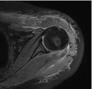
Left shoulder magnetic resonance imaging (MRI) revealed myositis in the deltoid muscle, teres minor muscle, and inflammatory changes in the adjacent subcutaneous areas (Fig. 1).
No abnormalities were shown in the left shoulder joint. Intravenous antibiotic treatment was started because the possibility of myositis, cellulitis, and bacterial infection could not be eliminated. On the second day of hospitalization, newly developed hematochezia occurred and persisted. Oral metronidazole was added in consideration of pseudomembranous colitis after antibiotic use; but on hospital day four, the stool exam revealed that Clostridium difficile toxin was negative. However, hematochezia with abdominal pain persisted, so we discontinued intravenous antibiotic (cefazolin, clindamycin) treatment and maintained oral metronidazole treatment for infectious colitis, such as pseudomembranous colitis.
Fig. 1. Left shoulder magnetic resonance imaging shows increased signal intensity with swelling in the anterior deltoid, teres minor muscle, and subcutaneous fat layer without remarkable findings in the joint space on fat saturated proton density axial image.
On hospital day five, abdominal computed tomography (CT) was performed because of persistent hematochezia, but no bleeding point was found.
On hospital day six, the left shoulder pain was somewhat improved, but the left mandible area pain developed again and worsened. Cervical CT revealed myositis on the left masseter and pterygoid muscle.
On hospital day eight, an additional blood test was performed for autoantibodies and were all negative except for the perinuclear anti-neutrophil cytoplasmic antibody. The CK level, which was in the normal range upon admission, increased to 1,720 IU/L.
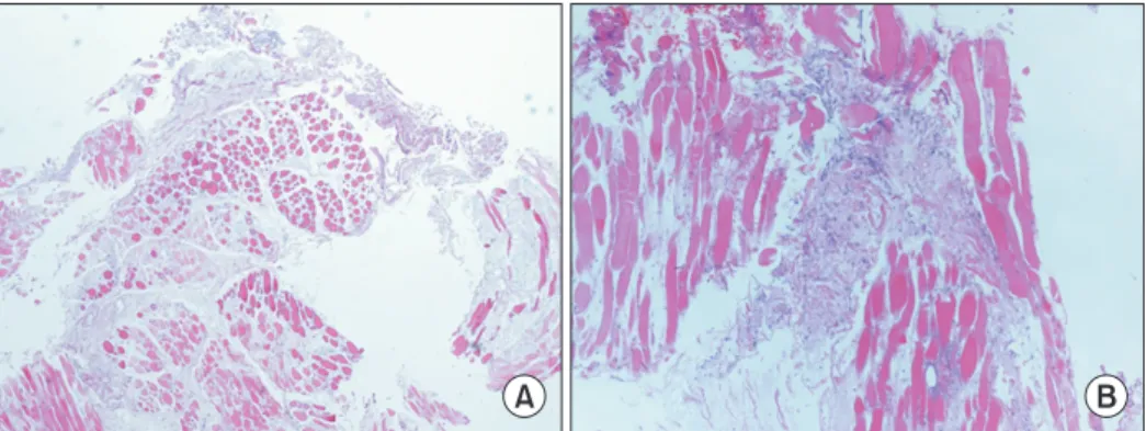
On hospital day 11, esophagogastroduodenoscopy and colonoscopy were performed for persistent hematochezia. Muscle biopsy on the left masseter muscle was performed to assess the left mandible area pain. Colonoscopy showed complete loss of the vascular pattern, some free liquid blood in the lumen, and larger (>5 mm) defects in the mucosa, compatible with UC. Muscle biopsy on the left master muscle showed significant size variation, degeneration, atrophy, and necrosis of the muscle fibers. Interstitial edema was observed, but no inflammatory cell infiltration was shown (Fig. 2). Although there was no histologically significant inflammation, evidence such as edema and degeneration and necrosis of muscle fibers suggested myositis. All things considered, we diagnosed the condition as UC with polymyositis (left masseter, deltoid, teres minor muscle), and initiated steroid treatment with mesalazine and azathioprine (AZA).
Without responding to treatment, hematochezia persisted, fever developed, and the C-reactive protein level increased to 22.08 mg/dL. Pyoderma gangrenosum, another extra- intestinal symptom of UC, was observed in the right shoulder and left buttock area.
On hospital day 13, cyclosporine and three-day intravenous immunoglobulin (400 mg/kg) were added in consideration of steroid-resistant UC and pyoderma gangrenosum, another extra-intestinal symptom of UC. After that, hematochezia as well as the extra-intestinal symptoms dramatically improved.
On hospital day 19, the CK level decreased to within the normal range, and on hospital day 30, the C-reactive protein level decreased to 0.97 mg/dL.
A B
Fig. 2. Microscopic findings of masseter muscle biopsy. (A) Significant size variation, degeneration, and atrophy of muscle fibers are identified. Marker interstitial edema is found; however, inflammatory cell infiltration or fibrosis are not remarkable. There is no evidence of vasculitis (H&E, ×10). (B) Multifocal ischemic necrosis of the muscle fibers with nuclear dusts are shown (H&E, ×100).
On hospital day 64, gastrointestinal, muscle and skin symptoms improved, and the patient was discharged only with UC medication (mesalazine, AZA), while cyclosporine was tapered off.
The Pediatric Ulcerative Colitis Activity Index [3] decreased to 5, from 45, the maximum point on hospital day 12. After discharge, regular outpatient department follow-ups were performed, and no aggravation of gastrointestinal and myositis symptoms was observed.
Occasional nausea and vomiting occurred, but there was no evidence of disease aggravation, and it improved with temporal supportive care.
After 14 months since the initial diagnosis of UC, the patient was admitted because of aggravated nausea and vomiting. Considering it as the symptoms of UC progression, we started additional steroid treatment, and symptoms improved. But three days later, the patient complained of severe pain in the left shoulder area, with tapering of the steroid dose.
Left shoulder MRI was performed again, and it revealed myositis which was considered as a relapse of an extra-intestinal symptom with UC aggravation, so we restarted cyclosporine.
Symptoms improved after two weeks of treatment, and the patient was discharged planning to discontinue cyclosporin and steroid, with follow-up in the outpatient department.
DISCUSSION
In this case, the patient initially complained of muscle pain followed by gastrointestinal symptoms, such as diarrhea and hematochezia, which occurred later. UC with myositis, as an uncommon extra-intestinal symptom, was finally diagnosed. Her muscle symptoms as well as hematochezia improved with treatment for UC. This case is distinguished from other reports in that the extra-intestinal symptoms developed earlier than the intestinal symptoms of UC.
Extra-intestinal symptoms are often associated with IBD [4]. However, few cases of myositis as an extra-intestinal symptom have been reported. There were only 11 cases found in the literature [2,4-13]. Compared to the other 11 cases, this patient demonstrated several distinctive features in characteristics of myositis (Table 1).
First, in this case, the diagnosis point of myositis was different from others. According to the current literature, almost all myositis in UC patients developed during UC treatment or after remission. It took an average of 7.9 years for myositis onset after initial UC diagnosis (minimum 0 yr; maximum, 15 years). But in this case, sudden muscle pain with swelling, which was suspected myositis, developed 13 days earlier from the onset of hematochezia. Six days after the onset of muscle symptoms, vomiting and diarrhea developed, but it coincided with antibiotic administration. Considering that the patient was a previously healthy female, it was reasonable that her gastrointestinal symptoms may have been associated with symptoms of antibiotic use. In this case, myositis, an extra-intestinal symptom, preceded the gastrointestinal symptoms of UC and is a distinguishing point from other cases.
Second, myositis in UC involved the large skeletal and deltoid muscles which have often been involved in previous cases. The patient in this case initially complained of muscle pain in the left masseter muscle and then the right deltoid muscle. In reference to the previous 11 reports of myositis in UC, all cases except one involved skeletal muscle. The other one case involved the ocular muscle, and the patient complained of diplopia [13]. This is a unique case in point
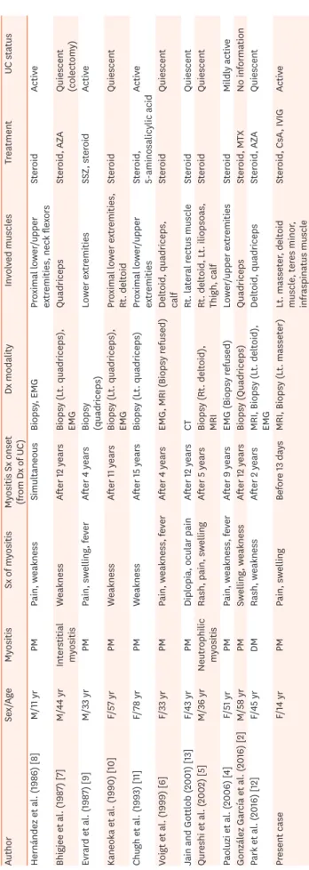
Table 1. Summary of case reports of myositis in patients with UC AuthorSex/AgeMyositisSx of myositisMyositis Sx onset (from Dx of UC)Dx modalityInvolved musclesTreatmentUC status Hernández et al. (1986) [8]M/11 yrPMPain, weaknessSimultaneousBiopsy, EMGProximal lower/upper extremities, neck flexorsSteroidActive Bhigjee et al. (1987) [7]M/44 yrInterstitial myositisWeaknessAfter 12 yearsBiopsy (Lt. quadriceps), EMGQuadricepsSteroid, AZA Quiescent (col
ectomy) Evrard et al. (1987) [9]M/33 yrPMPain, swelling, feverAfter 4 yearsBiopsy (quadriceps)Lower extremitiesSSZ, steroidActive Kaneoka et al. (1990) [10]F/57 yrPMWeaknessAfter 11 yearsBiopsy (Lt. quadriceps), EMGProximal lower extremities, Rt. deltoidSteroidQuiescent Chugh et al. (1993) [11]F/78 yrPMWeaknessAfter 15 yearsBiopsy (Lt. quadriceps)Proximal lower/upper extremitiesSteroid, 5-aminosalicylic acidActive Voigt et al. (1999) [6]F/33 yrPMPain, weakness, feverAfter 4 yearsEMG, MRI (Biopsy refused)Deltoid, quadriceps, calfSteroidQuiescent Jain and Gottlob (2001) [13]F/43 yrPMDiplopia, ocular painAfter 12 yearsCTRt. lateral rectus muscleSteroidQuiescent Qureshi et al. (2002) [5]M/36 yrNeutrophilic myositisRash, pain, swellingAfter 5 yearsBiopsy (Rt. deltoid), MRIRt. deltoid, Lt. iliopsoas, Thigh, calfSteroidQuiescent Paoluzi et al. (2006) [4]F/51 yrPMPain, weakness, feverAfter 9 yearsEMG (Biopsy refused)Lower/upper extremitiesSteroidMildly active González García et al. (2016) [2]M/58 yrPMSwelling, weaknessAfter 12 yearsBiopsy (Quadriceps)QuadricepsSteroid, MTXNo information Park et al. (2016) [12]F/45 yrDMRash, weaknessAfter 2 yearsMRI, Biopsy (Lt. deltoid), EMGDeltoid, quadricepsSteroid, AZAQuiescent Present caseF/14 yrPMPain, swellingBefore 13 daysMRI, Biopsy (Lt. masseter)Lt. masseter, deltoid muscle, teres minor, infraspinatus muscle
Steroid, CsA, IVIGActive UC: ulcerative colitis, Sx: symptom, Dx: diagnosis, M: male, F: female, PM: polymyositis, DM: dermatomyositis, EMG: electromyography, Lt: left, MRI: magnetic resonance imaging, CT: computed tomography, Rt: right, AZA: azathioprine, SSZ: sulfasalazine, MTX: methotrexate, CsA: cyclosporin A.
because the masseter muscle, which has not been reported before, was involved in myositis as an extra-intestinal manifestation of UC.
Third, comparing all 12 myositis cases in UC, including this case, only two cases were pediatric patients. As mentioned previously, myositis in UC is rare. Among them, myositis in pediatric UC patients is even rarer. More interestingly, two pediatric cases including this case are distinguished from the other 10 adult cases in the onset point of myositis. In adult UC cases (10 cases), myositis developed in a minimum of two years to maximum of fifteen years (average, 8.7 years) after initial UC diagnosis. However, in this case myositis preceded gastrointestinal symptoms, and in the other pediatric case, although not described in detail regarding the onset time, myositis and gastrointestinal symptoms developed at a similar time [8]. Through the analysis of similar cases, we observed that myositis in pediatric UC is rarer and occurred almost simultaneously, or even preceded gastrointestinal symptoms.
In cases when the symptoms such as muscle pain or weakness develop in patients who have already been diagnosed with UC, it may have been considered an extra-intestinal symptom.
However, as in this case, if the muscle symptoms developed alone as an extra-intestinal symptom of UC, it may be difficult or take a lengthy time for precise diagnosis. Thus, it would be prudent to consider the possibility of myositis associated with IBD, especially UC, if a patient with muscle symptoms complains of newly developed gastrointestinal symptoms.
REFERENCES
1. Levine JS, Burakoff R. Extraintestinal manifestations of inflammatory bowel disease. Gastroenterol Hepatol (N Y) 2011;7:235-41.
PUBMED
2. González García A, Sifuentes-Giraldo WA, Diz Fariña S, Pian H. Polymyositis in a patient with ulcerative colitis. Reumatol Clin 2016;12:360-2.
PUBMED | CROSSREF
3. Turner D, Otley AR, Mack D, Hyams J, de Bruijne J, Uusoue K, et al. Development, validation, and evaluation of a pediatric ulcerative colitis activity index: a prospective multicenter study. Gastroenterology 2007;133:423-32.
PUBMED | CROSSREF
4. Paoluzi OA, Crispino P, Rivera M, Iacopini F, Palladini D, Consolazio A, et al. Skeletal muscle disorders associated with inflammatory bowel diseases: occurrence of myositis in a patient with ulcerative colitis and Hashimoto's thyroiditis--case report and review of the literature. Int J Colorectal Dis 2006;21:473-7.
PUBMED | CROSSREF
5. Qureshi JA, Staugaitis SM, Calabrese LH. Neutrophilic myositis: an extra-intestinal manifestation of ulcerative colitis. J Clin Rheumatol 2002;8:85-8.
PUBMED | CROSSREF
6. Voigt E, Griga T, Tromm A, Henschel MG, Vorgerd M, May B. Polymyositis of the skeletal muscles as an extraintestinal complication in quiescent ulcerative colitis. Int J Colorectal Dis 1999;14:304-7.
PUBMED | CROSSREF
7. Bhigjee AI, Bill PL, Cosnett JE. Ulcerative colitis and interstitial myositis. Clin Neurol Neurosurg 1987;89:261-3.
PUBMED | CROSSREF
8. Hernández MA, Díez Tejedor E, Barbado J, Morales C. [Polymyositis and ulcerative colitis. A new form of association?]. Neurologia 1986;1:134-5. Spanish.
PUBMED
9. Evrard P, Lefebvre C, Brucher JM, Coche E. [Myositis associated with ulcero-hemorrhagic rectocolitis.
Apropos of a case]. Acta Gastroenterol Belg 1987;50:675-9. French.
PUBMED
10. Kaneoka H, Iyadomi I, Hiida M, Yamamoto K, Kisu T, Tokunaga O, et al. An overlapping case of ulcerative colitis and polymyositis. J Rheumatol 1990;17:274-6.
PUBMED
11. Chugh S, Dilawari JB, Sawhney IM, Dang N, Radotra BD, Chawla YK. Polymyositis associated with ulcerative colitis. Gut 1993;34:567-9.
PUBMED | CROSSREF
12. Park CH, Myong NH, Joo HD, Kang MI. Dermatomyositis: a rare extra-intestinal manifestation of ulcerative colitis. J Rheum Dis 2016;23:183-6.
CROSSREF
13. Jain S, Gottlob I. Orbital myositis associated with ulcerative colitis. Am J Gastroenterol 2001;96:3442-4.
PUBMED | CROSSREF