PGHN
Review Article
Abnormality on Liver Function Test
Ki-Soo Kang
Department of Pediatrics, Jeju National University School of Medicine, Jeju, Korea
Children with abnormal liver function can often be seen in outpatient clinics or inpatients wards. Most of them have respiratory disease, or gastroenteritis by virus infection, accompanying fever. Occasionally, hepatitis by the viruses causing systemic infection may occur, and screening tests are required. In patients with jaundice, the tests for differ- ential diagnosis and appropriate treatment are important. In the case of a child with hepatitis B virus infection vertically from a hepatitis B surface antigen positive mother, the importance of the recognition of immune clearance can’t be overstressed, for the decision of time to begin treatment. Early diagnosis changes the fate of a child with Wilson disease. So, screening test for the disease should not be omitted. Non-alcoholic fatty liver disease, which is mainly discovered in obese children, is a new strong candidate triggering abnormal liver function. Muscular dystrophy is a representative disease mimicking liver dysfunction. Although muscular dystrophy is a progressive disorder, and early diagnosis can’t change the fate of patients, it will be better to avoid parent’s blame for delayed diagnosis.
Key Words: Liver function tests, Child
Received:November 29, 2013, Accepted:December 10, 2013
Corresponding author: Ki-Soo Kang, Division of Pediatric Gastroenterology, Hepatology and Nutrition, Department of Pediatrics, Jeju National University School of Medicine, 15 Aran 13-gil, Jeju 690-767, Korea. Tel: +82-64-754-8146, Fax: +82-64-754-3114, E-mail: [email protected] Copyright ⓒ 2013 by The Korean Society of Pediatric Gastroenterology, Hepatology and Nutrition
This is an openaccess article distributed under the terms of the Creative Commons Attribution NonCommercial License (http://creativecommons.org/licenses/by-nc/3.0/) which permits unrestricted noncommercial use, distribution, and reproduction in any medium, provided the original work is properly cited.
INTRODUCTION
We often see children with abnormal liver function in pediatric outpatient clinics [1]. The children have variable complaints, such as incidental finding of ab- normal liver function test on routine health check-up in their school, and jaundice or hepatomegaly sus- pected dysfunction of the hepatobiliary system.
Occasionally, we have to identify the possibility of liv- er dysfunction in some children, to whom long-term medication had been undertaken, for treatment of some intractable disease. During the growth of chil- dren who have chronic infection of hepatitis B virus
(HBV) caused by vertical infection, liver injury may occur without hepatobiliary symptoms. Recently, as childhood obesity has increased, liver dysfunction may accompany obesity.
More frequently, hospitalized children develop an abnormal liver function. We have to find which liver functions are abnormal in children, who are admit- ted to be diagnosed or treated for their major hep- atobiliary symptoms. Variable febrile illness caused by viral or bacterial infection in children is often ac- companied by some liver dysfunction.
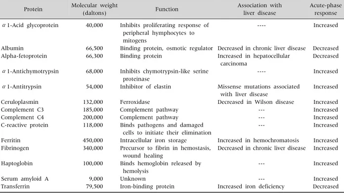
Table 1. Some Serum Proteins Produced by the Liver Protein Molecular weight
(daltons) Function Association with
liver disease
Acute-phase response α1-Acid glycoprotein
Albumin Alpha-fetoprotein
α1-Antichymotrypsin
α1-Antitrypsin
Ceruloplasmin Complement C3 Complement C4 C-reactive protein
Ferritin Fibrinogen
Haptoglobin
Serum amyloid A Transferrin
40,000
66,500 66,300
68,000
54,000
132,000 185,000 200,000 118,000
450,000 340,000
100,000
9,000 79,500
Inhibits proliferating response of peripheral hymphocytes to mitogens
Binding protein, osmotic regulator Binding protein
Inhibits chymotrypsin-like serine proteinase
Inhibitor of elastin
Ferroxidase
Complement pathway Complement pathway
Binds pathogens and damaged cells to initiate their elimination Intracellular iron storage Precursor to fibrin in hemostasis, wound healing
Binds hemoglobin released by hemolysis
Unknown
Iron-binding protein
----
Decreased in chronic liver disease Increased in hepatocellular carcinoma
----
Missense mutations associated with liver disease
Decreased in Wilson disease ---
--- ---
Increased in hemochromatosis Decreased in chronic liver disease
---
--- Increased iron deficiency
Increased
Decreased Decreased
Increased
Increased
Increased Increased Increased Increased
Increased Increased
Increased
Increased Decreased Adapted from Roy-Chowdhury and Roy-Chowdhury. Liver physiology and energy metabolism. Table 72-1. In: Feldman M, Friedman LS, Brandt LJ, eds. Sleisenger and Fordtran's gastrointestinal and liver disease. 9th ed. Philadelphia: Elsevier Saunders, 2010:1215.
Permission from Elsevier Limited was given to the author [5].
BASIC UNDERSTANDING ABOUT THE LIVER
In a brief review of the anatomy of the liver, it was traditionally divided into four lobes, of right, left, caudate and quadrate lobe. According to Couinaud nomenclature, the division of the liver into 8 seg- ments is frequently used. The liver is divided into right and left lobe by the line between the gall- bladder and inferior vena cava. Each lobe is parti- tioned into 2 sub-lobes, and each sub-lobe into 2 segments. This divides the liver into 8 segments clockwise from the caudate lobe [2]. Blood and oxy- gen supply to the liver is attributed to the portal vein from the superior vena cava, and hepatic artery from the heart. While the portal vein supplies 70% blood and 40% oxygen, the hepatic artery is responsible for 30% blood and 40% oxygen [2]. The pathway of bile excretion from the liver to the duodenum is the com- mon hepatic duct. The portal vein, hepatic artery and
common hepatic duct are triple structures of the por- ta hepatis. In the internal structure of the liver, the porta hepatis is connected to the portal tract, one of three components of the hepatic lobule [2,3].
The core structure of liver histology is a hepatic lo- bule with hexagonal shape [2,3]. The central vein and portal tract are located at the center, and three angular points of the hepatic lobule, respectively.
Liver cells compose three groups [3,4]. The first is the parenchymal cells, consisting of hepatocytes and bile duct epithelia. The second is sinusoidal cells, includ- ing the hepatic sinusoidal endothelial cells and Kupper cells (hepatic macrophages). The third is perisinusoidal cells, consisting of hepatic stellate cells and pit cells.
Major functions of the liver are protein synthesis, bilirubin metabolism associated with bile pro- duction, carbohydrates metabolism, and fat metabo- lism [5]. Important proteins excreted after synthesis in the liver are as in Table 1 [5].
MAJOR TESTS FOR LIVER FUNCTION
To evaluate the degree of liver injury or liver dis- ease, the most common ‘liver function tests’ are as- partate aminotransferase (AST), and alanine amino- transferase (ALT). But, they represent ‘liver bio- chemical tests’, rather than tests for the known func- tions of the liver.
The most useful biochemical test to discover liver disease is the standard battery test. The test consists of total bilirubin, albumin, prothrombin time, and se- rum enzymes. Serum enzymes, which include AST, ALT and alkaline phosphatase (ALP), are usually measured. Gamma glutamyl transpeptidase (GGTP) and 5’-Nucleotidase (5’NT) are occasionally meas- ured [6].
Bilirubin
Total bilirubin ranges 1.0 to 1.5 mg/dL normally, and decreases to the level 0.2 to 0.9 mg/dL in 95% of the population. The normal value of indirect bilir- ubin is 0.8 to 1.2 mg/dL. The normal upper limit of di- rect bilirubin is 0.3 mg/dL. Even a small increase of direct bilirubin means the possibility of liver injury.
In patients with jaundice, the ratio of direct bilirubin to total bilirubin does not differentiate obstructive jaundice from liver parenchymal jaundice. A ratio over 20% traditionally means cholestasis in children.
The degree and duration of hyperbilirubinemia is not a prognostic factor for liver disease. But, the higher the serum bilirubin is, the deeper the severity of liver injury is.
Aminotransferase
Serum aminotransferases was called transaminases in the past. It is the most sensitive marker for acute liv- er injury. AST and ALT catalyze the α-amino group of L-aspartic acid and alanine, respectively, to move to the α-keto group. AST, which was previously called serum glutamic oxaloacetic transaminase, are in the cytosol and mitochondria of cells. It most commonly distributes to cardiac muscle, followed by the skeletal muscle, kidney, brain, pancreas, lung, leukocyte and erythrocytes. ALT, which was previously called serum
glutamic pyruvic transaminase, is cytoplasmic en- zyme, and exists most commonly in hepatocyte. So, it is a more specific marker for the evaluation of liver in- jury, than AST. The normal value of ALT is generally less than 30 U/L in men, and less than 19 U/L in women.
But, the value is dependent on the laboratories.
Alkaline phosphatase
Most serum ALP is made in liver and bones. The normal value of ALP depends on the age.
Adolescents have two times higher level than adults.
The difference between adolescents and adults seems to be due to bone growth. A high level of ALP, when the increase of GGTP and 5’NT is identified, must originate from liver, rather than the bone.
Gamma glutamyl transpeptidase
GGTP is on the cell membrane of the liver (hepatocyte and bile duct cell), kidney, pancreas, spleen, heart and brain, etc. A high concentration of serum GGTP has a limitation for clinical use; because although the sensitivity is high, the specificity is low for hepatobiliary diseases. The increase of GGTP can be detected in patients taking phenytoin and barbiturates.
5'-Nucleotidase
5’-NT is associated with canalicular among hep- atocytes, and sinusoidal plasma membrane neigh- boring hepatocytes. The function of 5’NT is not well known. 5’NT exists in the small bowel, brain, heart, blood vessel and pancreas. The normal value of se- rum 5’NT increases with aging. 5’NT, as well as GGTP, is used for differential diagnosis of high se- rum ALP level alone.
Albumin
Albumin is the most important plasma protein, in terms of quantity. It is responsible for 75% of plasma colloid osmotic pressure, and is synthesized only in hepatocytes. When albumin loss occurs rapidly, the liver can make 2 times the usual production. The half life of albumin is 14 to 20 days. The final site of break down is not known. Albumin synthesis is regulated
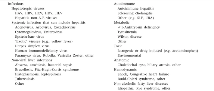
Table 2.Causes and Differential Diagnosis of Hepatitis in Children Infectious
Hepatotropic viruses
HAV, HBV, HCV, HDV, HEV Hepatitis non-A-E viruses
Systemic infection that can include hepatitis Adenovirus, Arbovirus, Coxackievirus Cytomegalovirus, Enterovirus Epstein-barr virus
"Exotic" viruses (e.g., yellow fever) Herpes simplex virus
Human immunodeficiency virus
Paramyxo virus, Rubella, Varicella Zoster, other Non-viral liver infections
Abscess, amebiasis, bacterial sepsis Brucellosis, Fitz-Hugh-Curtis syndrome Histoplasmosis, leptospirosis
Tuberculosis Other
Autoimmune
Autoimmune hepatitis Sclerosing cholangitis Other (e.g. SLE, JRA) Metabolic
α1-Antitrypsin deficiency Tyrosinemia
Wilson disease Other Toxic
Iatrogenic or drug induced (e.g. acetaminophen) Environmental
Anatomic
Choledochal cyst, biliary atresia, other Hemodynamic
Shock, Congestive heart failure Budd-Chiari syndrome, other Non-alcoholic fatty liver diseases Idiopathic, Rye syndrome, other
HAV: hepatitis A virus, HBV: hepatitis B virus, HCV: hepatitis C virus, HDV: hepatitis D virus, HEV: hepatitis E virus, SLE: systemic lupus erythematosus, JRA: juvenile rheumatoid arthritis. Modified from Yazigi and Balistreri. Viral hepatitis. Table 350-2. In:
Kliegman RM, Stanton BF, St. Geme lll JW, Schor NF, Behrman RE, eds. Nelson textbook of pediatrics. 19th ed. Philadelphia:
Elsevier Saunders, 2011:1394. Permission from Elsevier Limited was given to the author [1].
by nutritional status, osmotic pressure, systemic in- flammation, and hormone concentration in the blood. Therefore, when hypoalbuminemia is de- tected, differential diagnosis should include live cell dysfunction, protein-losing enteropathy, nephrotic syndrome, chronic systemic inflammation, and im- balance of hormone.
A long half-life of albumin is the cause of low us- ability for liver synthetic function, when acute liver injury has developed. In chronic liver disease or liver cirrhosis; however, albumin is an excellent marker for the synthetic function of the liver.
Prothrombin time
All coagulation factors, except factor VIII, are syn- thesized in the liver. Prothrombin time measures the extrinsic pathway of hemosatasis. Factors II, V, VII and X are clotting factors involved in prothrombin production. Prolongation of prothrombin time may occur from other liver diseases, beside of liver syn- thetic dysfunction. Vitamin K deficiency and dis- seminated intravascular coagulation are representa- tive causes of prolonged prothrombin time. The
measuring of prothrombin time is most useful in pa- tients with acute liver disease. In contrast to serum albumin, prothrombin time can evaluate the actual liver synthetic function. Prothrombin time is also a valuable prognostic factor of liver failure.
DISEASES CAUSING LIVER DYSFUNTION
When liver dysfunction occurs, the most common laboratory finding is an increase of AST and ALT, rep- resentative of serum enzyme associated with liver in- jury [1,7]. Abnormal AST and ALT levels are occasion- ally accompanied by cholestatic jaundice in variable liver diseases. Without the increase of AST and ALT;
however, jaundice alone may appear [8]. Diseases of abnormal bilirubin metabolism are classified to two types such as the conjugated hyperbilirubinemia and unconjugated hyperbilirubinemia without hepatitis.
The former include Gilbert syndrome, and Crigller Najjar types I and II. The latter contains Rotor syn- drome and Dubin-Johnson syndrome.
The review will focus on diseases with increased AST and ALT (Table 2) [1]. The representative disease
is hepatitis, caused by viral infection, such as hepato- trophic viruses and other viruses inducing systemic fe- brile infection [1]. Besides of hepatitis caused by virus infection, there is hepatitis caused by bacterial sepsis or parasitic infection. Other diseases are autoimmune hepatitis, metabolic liver disease, such as Wilson dis- ease, toxic hepatitis caused by drugs, cholestatic hep- atitis from anatomic problems of the hepatobiliary sys- tem, and idiopathic hepatitis caused by non-alcoholic fatty liver disease (NAFLD) of obese children.
Hepatitis A virus
In the domestic area, the prevalence of hepatitis A virus (HAV) infection has shown several outbreaks since the second half of the 1990s, and tremendous increase since year 2000 [9-11]. The possibility of meeting the patients is rising in outpatient clinics.
The earlier child may be a little bit sick, without jaundice. In contrast, the older child and adults prominently complain of hepatobiliary symptoms [9,11]. Symptoms include fever, anorexia, nausea, vomiting, fatigue and jaundice. The typical symp- toms may continue for 1 to 2 weeks. AST, ALT, bilir- ubin, ALP, 5’NT and GTTP become over the normal limits. The disease can be easily confirmed by pos- itive anti-HAV antibody (immunogrobulin M [IgM]). ALT rapidly increases to the top, before symptom development. From this point, symptoms such as jaundice begin. After this, ALT gradually de- creases, and normalizes, when jaundice disappears.
Hepatitis B virus
The hepatitis B surface antigen (HBsAg) positive rate of school ages, born before the induction of HBV vaccine, was 3.2 % in 1988. But, the rate decreased to 0.9% in a survey (Seoul area, 1995) of infant and tod- dlers born after the vaccine induction [12,13]. At pres- ent, domestic children of preschool ages have 70% to 80% of positive hepatitis B surface antibody (HBsAb).
The positive rates of HBsAg are 0.4% in 20s, and 0.2 % in adolescents [14]. But, we can occasionally meet children with HBV infection in outpatient clinics.
The most common pathway of HBV transmission in childhood is vertical infection from an HBsAg pos-
itive mother [15]. Over 90% of vertically infected children develop chronic HBV infection. In their nat- ural history, the change from the immune tolerance phase to the immune clearance phase occurs in 15%
of patients before 20 years of age [16]. The immune tolerance phase is a period of normal AST, ALT, pos- itive HBeAg and high concentration of HBV DNA in the serum. The immune clearance phase means the period of elevated AST, ALT, positive HBeAg, and de- creasing concentration of HBV DNA. In the immune clearance phase, the mean of AST and ALT can in- crease 3 to 4 times over that of the immune tolerance phase [15,17].
Hepatitis caused by other viral systemic infection
We can often see the disease in children with viral respiratory infection and viral gastroenteritis [1].
The disease is usually accompanied by fever. Most patients don’t have other liver dysfunction, except for elevated AST and ALT. Rarely, AST and ALT of 10 to 20 times higher than normal value can be seen. In that case, it may take 6 to 12 months, until the en- zymes normalize. Viruses can be identified, using polymerase chain reaction for respiratory infection or gastroenteritis. Hepatitis caused by cytomegalovi- rus or Epstein bar virus etc. may occur, and the tests to identify these viruses are necessary [1].
Wilson disease
Wilson disease is an autosomal recessive genetic disorder, which is caused by difficulty of copper ex- cretion to bile duct from the liver cell, and is accom- panied by liver and neurologic disease [18]. The do- mestic prevalence of children is approximately 1 per 37,000 persons. In East Asia, including Korea, the most common mutation is R778L (Arg778Leu) of the ATP7B gene [19]. The disease usually does not show abnormal liver function until 5 years old. So, most patients visit outpatient clinics with abnormal liver function in the health check-up of elementary or middle school. In particular, when siblings with ab- normal liver function visit the clinic together, we can easily suspect Wilson disease. In most patients, other
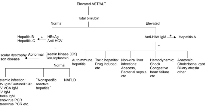
Fig. 1. Suggesting diagnostic algorithm for the children with elevated AST and ALT. The first step is the measure of serum total bilirubin. When it is normal, serum HBsAg and anti-HCV can be checked. In case of the viral marker negative, the measurement of serum creatin kinase and ceruloplasmin may be performed. In addition, other viral markers can be tested. The markers are CMV IgM/Culture/PCR, EBV VCA IgM, and adenovirus PCR etc. When serum total bilirubin is elevated, serum anti-HAV IgM can be measured. In this way, we can differentiate variable diseases with hepatitis. AST: aspartate aminotransferase, ALT: alanine aminotransferase, HBsAg: hepatitis B surface antigen, HCV: hepatitis C virus, CMV IgM: cytomegalovirus immunoglobulin M, PCR:
polymerase chain reaction, EBV VCA: Epstein bar virus viral capsid antigen, HAV: hepatitis A virus, NAFLD: non-alcoholic fatty liver disease, HSV: herpes simplex virus.
liver dysfunctions, other than elevated AST and ALT, are not detected. The screening test is the measure of serum ceruloplasmin. If the value is under 20 mg/dL, the confirmative test should be done. The patients should take drugs for Wilson disease throughout life.
Fortunately, if the disease is diagnosed before it is ac- companied by neurologic complication, most pa- tients can maintain health through life. Earlier diag- nosis for Wilson disease can prevent severe neuro- logic complication. Therefore, suspicion and diag- nosis is crucial for Wilson disease.
Non-alcoholic fatty liver disease
Most NAFLDs are discovered in obese children.
Yang et al. [20] reported that 33 of 111 children with NAFLD had elevated hepatic enzymes and non- alcoholic steatohepatitis. In this way, obese children visiting clinics tend to have the possibility of abnor- mal liver function. Other abnormal liver functions
are rare, except for elevated hepatic enzymes. If hab- its of diet, exercise, life and mind are appropriately managed, the obesity will be improved, and followed by normalization of the liver function.
Diseases with abnormal bilirubin metabolism Diseases that have only jaundice, without elevated hepatic enzymes, are divided into two groups [8].
One is the disease with increased indirect bilirubin.
These include Gilbert syndrome, and Crigller Najjar type I and II. Another is the disease with increased direct bilirubin. That includes Rotor syndrome and Dubin-Johnson syndrome.
DISEASE MIMICKING LIVER DYSFUNCTION
Muscular dystrophy
In the earlier child with Duchenne muscular dys-
trophy, elevated levels of AST and ALT can always be seen [7]. Muscular dystrophy can be easily dis- covered in the child with marked delay of motor de- velopment, and musculoskeletal symptoms. With only elevated AST and ALT levels, and without per- ception of sign of motor dysfunction; however, some children can be referred to the gastroenterologist.
The elevation of AST and ALT level in these children originates in excessive excretion from the muscu- loskeletal muscle. Serum creatine kinase (CK) ex- creted from muscle usually ranges 15,000 to 35,000 IU/L (normal<160 IU/L) [21]. For this reason, serum CK should be included in the screening test for the earlier child with abnormal AST and ALT levels.
CONCLUSION
The screening tests for children with abnormal liv- er function usually consist of anti-HAV IgM, HBsAg/Ab, anti-hepatitis C virus, cytomegalovirus IgM/culture, Epstein bar virus viral capsid antigen IgM, Rubella IgM, herpes simplex virus IgM, CK/lac- tate dehydronase, and ceruloplasmin and liver so- nography, etc (Fig. 1).
The etiologies of abnormal liver function are variable. We can first think hepatitis is caused by vi- ral infection, followed by non-viral infection, auto- immune, metabolic, toxic and anatomic liver diseases. Finally, NAFLDs can be considered. Once abnormal liver function is detected, screening tests should immediately be done for differential diagnosis.
REFERENCES
1. Yazigi N, Balistreri WF. Viral hepatitis. In: Kliegman RM, Stanton BF, St. Geme lll JW, Schor NF, Behrman RE, eds. Nelson textbook of pediatrics. 19th ed.
Philadelphia: Elsevier Saunders, 2011:1393-4.
2. Misdraji J. Embriology, anatomy, histology, and devel- opmental anomalies of the liver. In: Feldman M, Friedman LS, Brandt LJ, eds. Sleisenger and Fordtran's gastrointestinal and liver disease. 9th ed.
Philadelphia: Elsevier Saunders, 2010:1201-6.
3. McLin VA, Yagzi N. Developmental anatomy and phys-
iology of the liver and bile ducts. In: Wyllie R, Hyams JS, eds. Pediatric gastrointestinal disease. 4th ed.
Philadelphia: Elsevier Saunders, 2011:718-27.
4. Davenport M. Anatomy and embriology. In: Kleinman RE, Sanderson IR, Goulet O, Sherman PM, Mieli- Vergani G, Shneider BL, et al, eds. Walker's pediatric gastrointestinal disease: physiology, diagnosis, management. 5th ed. Hamilton: BC Decker, 2008:
749-66.
5. Roy-Chowdhury N, Roy-Chowdhury J. Liver physiol- ogy and energy metabolism. In: Feldman M, Friedman LS, Brandt LJ, eds. Sleisenger and Fordtran's gastro- intestinal and liver disease. 9th ed. Philadelphia:
Elsevier Saunders, 2010:1207-25.
6. Pratt DS. Liver chemistry and function tests. In:
Feldman M, Friedman LS, Brandt LJ, eds. Sleisenger and Fordtran's gastrointestinal and liver disease. 9th ed. Philadelphia: Elsevier Saunders, 2010:1227-37.
7. Kim KM. The interpretation of abnormal liver function test in children. In: 2010 Spring Symposium, Seoul: The Society of Korean Pediatric Gastroenteroloy and Nutrition, 2010:70-6.
8. Bergeron M, Gourley GR. Bilirubin metabolism. In:
Kleinman RE, Sanderson IR, Goulet O, Sherman PM, Mieli-Vergani G, Shneider BL, et al, eds. Walker's pe- diatric gastrointestinal disease: physiology, diagnosis, management. 5th ed. Hamilton: BC Decker, 2008:
749-66.
9. Cho KY. Hepatitis A. Korean J Pediatr Gastroenterol Nutr 2010;13(Suppl 1):70-7.
10. Kim JH. Recent epidemiological status and vaccination of hepatitis A in Korea. J Korean Med Assoc 2008;51:
110-8.
11. Youn HS. Current status of hepatitis A virus infections in Korea. Korean J Pediatr 2008;51:690-5.
12. Choe BH. Hepatitis B vaccine: prevention of perinatal infection and management of nonresponder. Korean J Pediatr Gastroenterol Nutr 2007;10(Suppl 1):91-100.
13. Choe YH, Seo JK, Yun JH, Lee HS. Recent changesin prevalence of hepatitis B viral markers in preschool children in Seoul, 1995. Korean J Pediatr 1996;39:
1254-9.
14. Choe BH. Hepatitis B. In: An HS, ed. Hong Change Yee Pediatrics. 10th ed. Seoul: MiraeN Inc., 2012:551-5.
15. Kang HS, Kang KS, Song BC. Precore and core pro- moter mutations of the hepatitis B virus gene in chronic genotype C-infected children. J Korean Med Sci 2011;26:546-50.
16. Park HJ, Chu MA, Hong SJ, Choe B. The rate of con- version to immune-reactive phase form immune-toler- ant phase in children with chronic hepatitis B. 4th
WCPGHAN 2012 [abstract] book p. 68(OP-1-4-3).
17. Robert P. Hepatitis B and D. In: Feldman M, Friedman LS, Brandt LJ, eds. Sleisenger and Fordtran's gastro- intestinal and liver disease. 9th ed. Philadelphia:
Elsevier Saunders, 2010:1287-312.
18. Seo JK. Diagnosis of Wilson disease in young children:
molecular genetic testing and a paradigm shift from the laboratory diagnosis. Pediatr Gastroenterol Hepatol Nutr 2012;15:197-209.
19. Seo JK, Kim JW. Mutation analysis of Wilson disease gene: Arg778Leu mutation in Korean children. Korean
J Pediatr Gastroenterol Nutr 1999;2:164-8.
20. Yang HR, Ko JS, Seo JK. Role of tumor necrosis factor- α promoter polymorphism and insulin resistance in the development of non-alcoholic fatty liver disease in obese hildren. Pediatr Gastroenterol Hepatol Nutr 2012;15:44-51.
21. Sarnat HB. Muscular dystrophies. In: Kliegman RM, Stanton BF, St. Geme lll JW, Schor NF, Behrman RE, eds. Nelson textbook of pediatrics. 19th ed.
Philadelphia: Elsevier Saunders, 2011:2119-22.