ISSN 0378-6471 (Print)⋅ISSN 2092-9374 (Online)
http://dx.doi.org/10.3341/jkos.2016.57.4.595
Original Article
정상안에서 파장가변 빛간섭 단층촬영을 이용한 유리체 피질과 후유리체박리의 관찰
Evaluation of the Cortical Vitreous Using Swept-Source Optical Coherence Tomography in Normal Eyes
박승훈⋅이정욱⋅이민지⋅이주향⋅송인석⋅이병로
Seung Hun Park, MD, Jung Wook Lee, MD, Min Ji Lee, MD, Ju Hyang Lee, MD, PhD, In Seok Song, MD, PhD, Byung Ro Lee, MD, PhD
한양대학교 의과대학 한양대학교병원 안과학교실
Department of Ophthalmology, Hanyang University Hospital, Hanyang University College of Medicine, Seoul, Korea
Purpose: To observe the shape of posterior vitreous spaces using swept-source optical coherence tomography (SS-OCT) in normal eyes.
Methods: The posterior vitreous of 80 eyes of 80 volunteers without ocular disease was imaged. The DRI OCT-1 Atlantis (Topcon, Oakland, NJ, USA) was used to acquire scans of the posterior vitreous over an 18 x 18-mm2 area using the 12-mm hori- zontal line scan protocol. The size of the premacularis bursa was measured by the aliper function of the OCT.
Results: A boat-shape bursa was found in most cases. The prevalence of detected bursa fell with further increases in the extent of posterior vitreous detachment. The mean width of the bursa premacularis was 7,679.1 µm and the mean depth was 471.4 µm.
Conclusions: Deep range imaging optical coherence tomography will provide improvement for in vivo anatomic characterization of the cortical vitreous, and allow better visualization of the dimensions of the bursa premacularis.
J Korean Ophthalmol Soc 2016;57(4):595-600
Keywords: Posterior vitreous, Swept-source optical coherence tomography
■Received: 2015. 9. 24. ■ Revised: 2015. 12. 29.
■Accepted: 2016. 2. 9.
■Address reprint requests to Byung Ro Lee, MD, PhD Department of Ophthalmology, Hanyang University Hospital,
#222-1 Wangsimni-ro, Seongdong-gu, Seoul 04763, Korea Tel: 82-2-2290-8570, Fax: 82-2-2291-8517
E-mail: [email protected]
* This study was presented as a poster at the 111th Annual Meeting of the Korean Ophthalmological Society 2014.
ⓒ2016 The Korean Ophthalmological Society
This is an Open Access article distributed under the terms of the Creative Commons Attribution Non-Commercial License (http://creativecommons.org/licenses/by-nc/3.0/) which permits unrestricted non-commercial use, distribution, and reproduction in any medium, provided the original work is properly cited.
망막질환에서 유리체는 그 중요성에 비해 해부학적 구조 에 대해 자세히 알려져 있지 않다. 유리체황반경계면은 망 막앞막, 유리체황반견인증후군 등 망막질환의 병태생리에 큰 영향을 주며, 이 부분에 대한 연구는 다양하게 이루어져
왔다.1-5 그러나 유리체와 망막의 경계면에 비해 유리체 중 심부의 구조는 상대적으로 잘 알려져 있지 않다. 유리체는 성인이 될 때까지 변하게 되는데, Cloquet`s canal은 이차유 리체인 후유리체 중 일차유리체가 남아 있는 부분으로 시 신경 유두 앞에 위치한다. 1970년대 Worst6는 Indian ink를 vitreous 내 주입하여 특정 공간으로 흐름을 확인하였는데, 황반부 앞쪽의 오목한 공간을 “bursa premacularis”라 칭하 였고, 이후 연구에서 posterior precortical vitreous pocket (PPVP)가 발견되었으며, 많은 연구에서 PPVP를 bursa pre- macularis로 간주하였다.4,7,8
세극등현미경 검사는 유리체를 확인하기에 해상도가 떨 어지며, 유리체의 투명도 때문에 정확히 관찰하기 어렵다.
또한 유리체를 확인할 수 있는 안구 초음파 검사는 0.2 mm
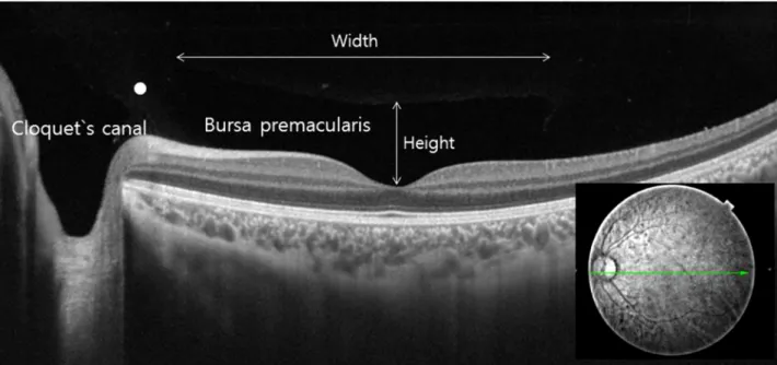
Figure 1. Evaluation of cortical vitreous using SS-OCT. An OCT 12-mm horizontal scan through the fovea in the left eye of a
57-year-old man. To improve visualization, the brightness and contrast were increased. The height of the bursa premacularis is de- fined as the distance between the fovea and the anterior border, and the width is the maximum diameter in the 12-mm horizontal scan through the fovea and the disc. The white dot indicates the septum between the bursa premacularis and Cloquet's canal. SS-OCT = swept-source optical coherence tomography.파장의 높은 주파수에도 불구하고 유리체 구조를 확인함에 있어 한계가 있다.9,10 빛간섭 단층촬영 검사(optical coher- ence tomography, OCT)는 해상도와 민감도가 뛰어나 안구 내부 구조를 정량적으로 평가할 수 있으며, spectral-domain OCT (SD-OCT)는 850 nm의 파장을 이용하여, 5 µm의 해 상도와 초당 20,000 이상의 A-scan 속도로 망막내부 구조를 정밀하게 촬영한다. 이를 이용하여 후유리체 구조를 확인한 연구가 있다.1-3 최근 등장한 Swept source OCT (SS-OCT) 는 1,050 nm의 더 긴 파장을 이용하기 때문에 depth of field 가 뛰어나고, 맥락막, 공막, 안와 지방뿐만 아니라 후극부의 유리체 구조를 더 명확히 평가할 수 있다.11,12 따라서 이를 이용한 유리체의 해부학적 구조에 대한 연구가 이루어지고
있으며,5,13,14 SS-OCT를 통한 우리나라 정상안의 후유리체
의 해부학 구조를 확인해 보고자 하였다.
대상과 방법
대상 환자
본 연구는 후향적 횡단적 단면연구로서, 본원에 2014년 11월부터 2015년 9월까지 내원한 80명 80안의 환자(남자 52명, 여자 28명)를 대상으로 연구하였다. 연령분포는 8세 부터 78세까지이며 평균 44.0 ± 13.8세였다. 유리체망막 수 술을 받은 병력, 망막앞막, 유리체망막견인, 황반원공, 고도 근시인 경우 연구에서 제외하였으며, 양안 모두 대상이 될
경우 영상의 질이 높은 쪽을 연구에 포함하였다.
검사
SS-OCT는 Deep range imaging optical coherence tomog- raphy (DRI OCT-1 Atlantis; Topcon, Oakland, NJ, USA)를 사용하였으며, 이는 초당 10만 회 A-scan을 조사하고, 축 해상도는 8 μm, imaging depth는 2.3 mm이다. 12-mm- hori- zontal scan으로 시행하였으며, 후유리체 구조를 선명하게 보기 위해 유리체에 초점을 맞추어 촬영하였다. Scan line 중 중심오목과 시신경을 지나는 영상을 선별하여, 유리체 가 잘 보이도록 대비감도와 색상을 조정하였다.
지표
Bursa premacularis, Cloquet`s canal의 존재 유무, 후유리 체 경계면을 확인하였으며, SS-OCT에 내장된 caliper 기능 을 이용하여 bursa premacularis의 너비과 깊이를 측정하였 다(Fig. 1).15 후유리체박리의 정도는 Uchino et al16에 근거 하여, ‘후유리체박리가 전혀 없는 경우(No posterior vitre- ous detachment [PVD])’, ‘중심와와 시신경유두에는 후유 리체막이 붙어있으나, 중심와 코쪽 또는 귀쪽은 떨어져 있 는 경우(PVD stage 1)’, ‘중심와와 시신경유두에는 후유리 체막이 붙어있으나, 중심와 코쪽과 귀쪽은 떨어져 있는 경 우(PVD stage 2)’, ‘시신경유두에만 후유리체막이 붙어있는 경우(PVD stage 3)’, ‘완전 후유리체박리(PVD stage 4)’로
Figure 2. Swept-source optical coherence tomography of the cortical vitreous. A horizontal scan through the fovea and the optic disc
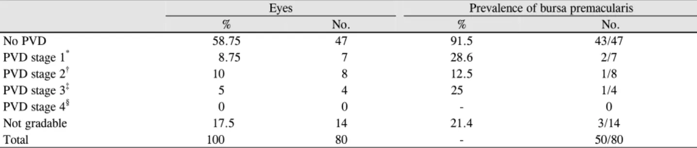
showed a boat-shaped bursa premacularis (*). In front of the optic disc, the Cloquet's canal (**) was connected to the bursa premacularis.Table 1. Number of eyes and prevalence of the bursa premacularis in the cortical vitreous according to PVD stage
Eyes Prevalence of bursa premacularis
% No. % No.
No PVD 58.75 47 91.5 43/47
PVD stage 1* 8.75 7 28.6 2/7
PVD stage 2† 10 8 12.5 1/8
PVD stage 3‡ 5 4 25 1/4
PVD stage 4§ 0 0 - 0
Not gradable 17.5 14 21.4 3/14
Total 100 80 - 50/80
PVD = posterior vitreous detachment.
*Focal perifoveal PVD, limited to either the temporal or nasal side of the fovea, with persistent attachment to the fovea and optic nerve head;
†Focal perifoveal PVD, involving both the temporal and nasal side of the fovea, with persistent attachment to the fovea and optic nerve head;
‡PVD over the fovea with persistent attachment to the optic nerve head; §Complete PVD over the macula and optic nerve head.
분류하였다.
통계
통계학적 분석은 SPSS (version 18.0, SPSS Inc., Chicago, IL, USA)를 사용하였다. 연령에 따른 bursa premacularis 크기의 상관관계를 확인하기 위해 선형회귀분석을 하였다.
결 과
Bursa premacularis는 80안 중 50안(62.5%)에서 관찰되 었고, 대부분 보트 모양이었다. Cloquet’s canal은 80안 중 48안(60%)에서 확인되었으며, 96% (50안 중 48안)에서 bursa premacularis와 Cloquet`s canal이 동시에 관찰되었다 (Fig. 2). 후유리체박리의 정도에 따른 bursa premacularis의
관계는 Table 1에 정리되어 있다. 전체 80안 중 47안(58.8%) 에서 후유리체박리는 관찰되지 않았으며, 19안(23.6%)에서 후유리체박리가 관찰되었다(Fig. 3). bursa premacularis는 후유리체박리가 없는 경우(47안 중 43안, 91.5%)가 있는 경우(19안 중 4안, 21.0%)보다 더 많이 관찰되었다(Table 1).
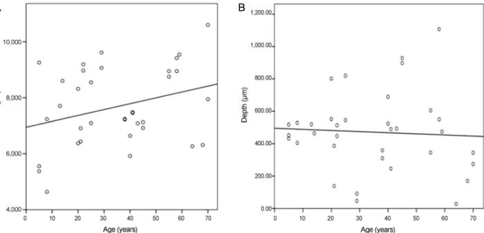
Bursa premacularis가 관찰된 50안 중 35안에서 너비와 깊 이의 측정이 가능하였으며, 너비는 7,679.1 ± 1,402.2 µm (4,640-10,604 µm), 깊이는 471.4 ± 243.9 µm (28-1,108 µm)였다. Bursa premacularis의 크기는 연령과는 연관성이 없었다(R2 width = 0.09, R2 depth = 0.03, Fig. 4).
고 찰
본 연구에서는 SS-OCT를 이용하여 정상안의 후유리체
** *
**
Figure 3. Classification of posterior vitreous detachment (PVD). ‘◇’ indicates the border of posterior vitreous. (A) No PVD. (B) PVD
stage 1. (C) PVD stage 2. (D) PVD stage 3.구조를 확인하였다. Mojana et al17의 연구에서 SD-OCT와 scanning laser ophthalmoscopy를 이용하여 bursa premacularis 를 42%에서 확인한데 비해 본 연구에서는 62.5%에서 발견하 였고, 이는 SS-OCT를 이용해 bursa premacularis를 확인한 Stanga et al14의 57.1%와 비슷한 정도이다. 우리나라에서는 2013년 You et al3이 유리체황반경계면을 Time domain OCT (TD-OCT)와 SD-OCT를 이용하여 확인하였고, TD-OCT 로 약 6%, SD-OCT로 2/3에서 후유리체면을 발견하였다.
이에 비해, 본 연구에서는 82.5%에서 후유리체막을 확인할 수 있어 SS-OCT가 SD-OCT보다 후유리체 구조를 잘 파악
할 수 있음을 알 수 있다.
OCT가 개발되기 전에는 사체부검으로 형광염색하여 유 리체 구조를 확인하였고,8 이후 in vivo 상태에서 유리체 확 인은 안저검사와 초음파검사로 하였다.9,10 안저검사는 매체 혼탁에 영향을 받고 주관적이며, 초음파검사는 유리체 세 부 구조를 확인하기에는 해상도가 낮다는 단점이 있다. SD-OCT는 망막 및 후유리체 평가에 큰 도움이 되었으나 유리체에 대한 낮은 민감도와 파장 길이의 한계로 bursa premacularis를 확인하는 데 어려움이 있다. 최근 개발된 SS-OCT는 긴 파장(1,050 nm)으로 유리체 섬유에 의한 빛
A
B
C
D
Figure 4. Correlation of variation in width and depth of the bursa premacularis with patient age. There was no correlation between var-
iation in width and patient age (R2 = 0.09; A), and between variation in depth and patient age (R2 = 0.03; B).산란을 줄이고, 매우 빠른 조사 속도(초당 10만 회 A-scan)로 유리체 섬유의 움직임을 잡아낼 수 있다. 또한 12-mm-long scan으로 황반부와 시신경을 동시에 확인할 수 있다는 장 점이 있다.
Bursa premacularis의 너비와 깊이는 각각 7,679.1 ± 1,402.2 µm, 471.4 ± 243.9 µm로 측정되었다. SD-OCT를 이용한 연구18의 너비 6,000 µm, 깊이 500 µm, SS-OCT를 이용한 Itakura et al13의 너비 6,420.6 µm, 깊이 708.1 µm, Stanga et al14의 너비 7,001 µm, 깊이 416 µm와 비교하면, 길이 측정 시 caliper 위치가 검사자마다 차이가 있음을 고 려할 때 bursa premacularis의 너비와 크기가 이전 연구들과 비슷하게 측정된 것으로 사료된다. Bursa premacularis의 너비와 깊이 모두 나이와 상관 관계를 보이지 않아, bursa premacularis의 크기는 나이와 관련하여 큰 변화가 없었다.
그러나 Li et al19이 3-11세 소아를 대상으로 SS-OCT를 이 용하여 bursa premacularis 크기를 측정하였을 때, 너비 4,834.4 ± 228.1 µm, 깊이 426.4 ± 38.2 µm의 결과가 나왔 으며, 이 결과는 환자군 대부분이 성인인 본 연구와는 차이 가 있었다. 결과가 다른 이유는 성장하면서 길어지는 안축 장 길이 변화와 연관이 있을 것으로 사료되나, 추가 연구가 필요할 것이다.
Mojana et al17은 후유리체박리가 없는 사람의 91%, 불완 전 후유리체박리의 60%에서 bursa premacularis를 확인했 고, 완전 후유리체박리가 있을 시 bursa premacularis를 확 인하지 못한 것으로 보고했다. 또한 Stanga et al14은 후유리 체박리가 없을 시 84.3%, 불완전 후유리체박리일 때 52%,
완전 후유리체박리일 때 25%에서 bursa premacularis를 발 견하였다. 본 연구에서는 후유체박리가 없을 때는 91.5%, 부분 박리 시에는 21%, 완전 박리 시 0%에서 발견되었다.
후유리체박리가 없을수록 bursa premacularis가 더 많이 발 견되고, 이는 후유리체박리로 인한 후유리체의 앞쪽 이동 으로 OCT에서 신호를 못 잡아내기 때문으로 생각된다.
본 연구는 양안 검사를 하여 비교하지 못한 점, 세극등현 미경 검사 소견과 SS-OCT 소견을 비교하지 못한 것이 제 한점이며, 정상안에 국한된 분석을 했기 때문에 유리체망 막질환이 있을 경우 적용이 어려운 한계가 있다. 이번 연구 를 통해 bursa premacularis는 대부분 배 모양이며, 후유리 체박리가 없거나 일부만 있을 경우 완전 유리체 박리가 일 어난 경우보다 bursa premacularis가 더 잘 관찰됨을 알 수 있었다. 최근 SS-OCT를 이용하여 유리체 피질을 확인한 연구가 많으나, 국내에서는 아직 보고된 바 없으며 파장가 변 빛간섭 단층촬영은 후유체박리와 유리체피질을 in vivo 상태에서 관찰 가능하게 하며 향후 망막질환에 따른 유리 체피질 변화를 연구하는 데 도움을 줄 것으로 생각된다.
REFERENCES
1) Barteselli G, Bartsch DU, El-Emam S, et al. Combined depth imag- ing technique on spectral-domain optical coherence tomography.
Am J Ophthalmol 2013;155:727-32, 732.e1.
2) Hoehn F, Mirshahi A, Hattenbach LO. Optical coherence tomog- raphy for diagnosis of posterior vitreous detachment at the macular region. Eur J Ophthalmol 2009;19:442-7.
3) You JY, Kim HC, Moon JW. Comparison of the efficacy between
A B
= 국문초록 =
정상안에서 파장가변 빛간섭 단층촬영을 이용한 유리체 피질과 후유리체박리의 관찰
목적: 정상안에서 파장가변 빛간섭 단층촬영을 이용하여 유리체 피질 및 후유리체박리를 관찰하고 분석해 보았다.
대상과 방법: 정상안 80안을 파장가변 빛간섭 단층촬영의 12-mm horizontal line scan 프로토콜을 이용해 검사를 시행하였다. 내장된 caliper를 이용하여 bursa premacularis의 크기를 측정하고, 후유리체박리 정도를 평가하였다.
결과: Bursa premacularis의 모양은 대체적으로 보트 모양을 보였으며 약 63%의 환자에서 관찰되었다. 후유리체박리가 없거나 일부 인 경우에서 완전 유리체 박리가 일어난 경우보다 더 bursa premacularis가 잘 관찰되었다. Bursa premacularis의 크기는 평균 폭 7,679.1 μm 깊이는 471.4 μm였다.
결론: 파장가변 빛간섭 단층촬영은 후유체박리와 유리체 피질을 in vivo 상태에서 관찰 가능하게 하며 향후 망막질환에 따른 유리체 피질 변화를 연구하는 데 도움을 줄 것으로 생각된다.
<대한안과학회지 2016;57(4):595-600>
time and spectral domain optical coherence tomography for the identification of vitreomacular interface. J Korean Ophthalmol Soc 2013;54:97-103.
4) Spaide RF. Measurement of the posterior precortical vitreous pock- et in fellow eyes with posterior vitreous detachment and macular holes. Retina 2003;23:481-5.
5) Park HY, Shin HY, Park CK. Imaging the posterior segment of the eye using swept-source optical coherence tomography in myopic glaucoma eyes: comparison with enhanced-depth imaging. Am J Ophthalmol 2014;157:550-7.
6) Worst JG. Cisternal systems of the fully developed vitreous body in the young adult. Trans Ophthalmol Soc U K 1977;97:550-4.
7) Yokoi T, Toriyama N, Yamane T, et al. Development of a pre- macular vitreous pocket. JAMA Ophthalmol 2013;131:1095-6.
8) Kishi S, Shimizu K. Posterior precortical vitreous pocket. Arch Ophthalmol 1990;108:979-82.
9) Fisher YL, Slakter JS, Friedman RA, Yannuzzi LA. Kinetic ultrasound evaluation of the posterior vitreoretinal interface. Ophthalmology 1991;98:1135-8.
10) Perichon JY, Brasseur G, Uzzan J. Ultrasonographic study of pos- terior vitreous detachment in emmetropic eyes. J Fr Ophtalmol 1993;16:538-44.
11) Shin YU, Lee BR, Lim HW. A comparison of image quality be- tween swept source optical coherence tomography and spectral do- main optical coherence tomography according to ocular media
opacity. Invest Ophthalmol Vis Sci 2014;55:3359.
12) Park SM, Cho SW, Lee TG. Swept source optical coherence to- mography findings in gas-filled eyes after macular hole surgery. J Korean Ophthalmol Soc 2015;56:1386-91.
13) Itakura H, Kishi S, Li D, Akiyama H. Observation of posterior pre- cortical vitreous pocket using swept-source optical coherence tomography. Invest Ophthalmol Vis Sci 2013;54:3102-7.
14) Stanga PE, Sala-Puigdollers A, Caputo S, et al. In vivo imaging of cortical vitreous using 1050-nm swept-source deep range imaging optical coherence tomography. Am J Ophthalmol 2014;157:397- 404.e2.
15) Itakura H, Kishi S. Alterations of posterior precortical vitreous pockets with positional changes. Retina 2013;33:1417-20.
16) Uchino E, Uemura A, Ohba N. Initial stages of posterior vitreous detachment in healthy eyes of older persons evaluated by optical coherence tomography. Arch Ophthalmol 2001;119:1475-9.
17) Mojana F, Kozak I, Oster SF, et al. Observations by spectral-do- main optical coherence tomography combined with simultaneous scanning laser ophthalmoscopy: imaging of the vitreous. Am J Ophthalmol 2010;149:641-50.
18) Itakura H, Kishi S. Aging changes of vitreomacular interface.
Retina 2011;31:1400-4.
19) Li D, Kishi S, Itakura H, et al. Posterior precortical vitreous pock- ets and connecting channels in children on swept-source optical co- herence tomography. Invest Ophthalmol Vis Sci 2014;55:2412-6.