ISSN 0378-6471 (Print)⋅ISSN 2092-9374 (Online)
https://doi.org/10.3341/jkos.2019.60.10.946
Original Article
펨토초레이저 백내장수술에서 활모양각막절개술 시행의 장기간 임상결과
Long-term Results of Arcuate Keratotomy in Femtosecond Laser-assisted Cataract Surgery
방찬우⋅최재원⋅한상엽
Chan Woo Bang, MD, Jae Won Choi, MD, Sang Youp Han, MD
성모안과병원
Sungmo Eye Hospital, Busan, Korea
Purpose: To investigate the long-term follow-up results of arcuate keratotomy in femtosecond laser-assisted cataract surgery.
Methods: The medical records of 78 patients (99 eyes) with corneal astigmatism >0.5 diopters (D) who underwent arcuate kera- totomy concurrent with femtosecond laser-assisted cataract surgery were retrospectively reviewed. Uncorrected and corrected visual acuities and keratometric and higher order aberration outcomes were compared preoperatively and 2 years postoperatively.
Results: The mean uncorrected visual acuities and corrected visual acuities changed from 0.55 ± 0.31 D and 0.33 ± 0.26 D, pre- operatively to 0.13 ± 0.19 D and 0.07 ± 0.09 D, 2 years postoperatively. The mean preoperative corneal astigmatism was -1.15
± 0.66 D. This was reduced to -0.79 ± 0.38 D at 1-2 days after surgery (p < 0.001), followed by no significant change for 2 years.
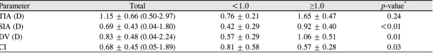
The mean target-induced astigmatism was 1.15 ± 0.66 D preoperatively, and the mean surgically-induced astigmatism and dif- ference vector were 0.69 ± 0.43 D and 0.83 ± 0.48 D, 2 years postoperatively. The mean correction index was 0.68 ± 0.45, 2 years postoperatively. There was no significant difference in higher order aberrations except 4 mm and 6 mm total higher order aberrations and 6 mm trefoil between preoperatively and 2 years postoperatively.
Conclusions: Arcuate keratotomy concurrent with femtosecond laser-assisted cataract surgery is a safe and effective way to re- duce corneal astigmatism which remained stable over 2 years of follow-up with below moderate astigmatism.
J Korean Ophthalmol Soc 2019;60(10):946-952
Keywords: Arcuate keratotomy, Astigmatism, Corneal cylinder, Femtosecond laser, Higher order abberation
■Received: 2019. 2. 7. ■ Revised: 2019. 4. 2.
■Accepted: 2019. 9. 24.
■Address reprint requests to Sang Youp Han, MD
Sungmo Eye Hospital, #409 Haeun-daero, Haeundae-gu, Busan 48064, Korea
Tel: 82-51-743-0775, Fax: 82-51-743-0776 E-mail: [email protected]
*Conflicts of Interest: The authors have no conflicts to disclose.
ⓒ2019 The Korean Ophthalmological Society
This is an Open Access article distributed under the terms of the Creative Commons Attribution Non-Commercial License (http://creativecommons.org/licenses/by-nc/3.0/) which permits unrestricted non-commercial use, distribution, and reproduction in any medium, provided the original work is properly cited.
1970년대 레이저가 백내장수술 적용 목적으로 소개된 이래 진화를 거듭하였고, 난시교정을 원하는 환자들의 요 구를 충족시키기 위해 펨토초레이저를 이용한 난시교정
수술들이 개발되었다.1,2 백내장수술 후 15-29% 환자에서 1-3 diopter (D)의 난시 발생이 보고된 바 있으며, 난시교정 을 위해 최근에는 펨토초레이저를 이용하여 백내장수술 중 윤부이완절개, 활모양각막절개 등을 동시 시행하거나, 난시 교정인공수정체를 삽입함으로써 근시 혹은 원시 교정과 더 불어 난시교정까지 시도하고 있다.3,4
펨토초레이저를 이용한 수술은 고식적인 난시교정수술 이나 백내장수술과 비교해 정확한 깊이, 길이 및 위치에 절 개가 가능하고 빠른 창상치유 효과가 있으며, 각막내피세 포 손상 및 염증 반응을 줄여 수술의 정확성과 안정성을 높 여 주었다.5-8 국내 및 국외 여러 연구에서 펨토초레이저 백
Characteristic Value
Number of patients (eyes) 78 (99)
Age (years) 60.47 ± 11.89 (26-85)
Sex (male:female) 39:60
Preoperative UCVA (logMAR) 0.55 ± 0.31 (0.22-1.40) Preoperative BCVA (logMAR) 0.33 ± 0.26 (0.00-1.00) Preoperative corneal astigmatism (D) 1.15 ± 0.66 (0.50-2.97) Values are presented as mean ± standard (range) or number.
UCVA = uncorrected visual acuity; logMAR = logarithm of mini- mal angle of resolution; BCVA = best corrected visual acuity; D = diopter.
Table 1. Demographics of patients
내장수술과 활모양각막절개의 동시 시행 후 난시교정 효과에 대해 보고하고 있고, 자연 발생 난시뿐만 아니라 각막성 형술 후 유발된 난시에서도 효과가 있다는 연구 결과들도 나오고 있다.9-13
하지만 현재까지 국내에 발표된 연구들은 1년 이내의 단 기간 임상결과에 대한 내용들이었고 장기간의 난시교정 효 과에 대해 보고된 바가 없다. 이에 본 연구에서는 수술 전 0.5D 이상의 각막난시를 가진 99안에 대해 수술 후 난시교 정 효과 및 고위수차 변화를 확인하고, 2년간의 장기간 임 상결과를 통해 효과의 지속성에 대해 분석해 보고자 한다.
대상과 방법
2015년 2월부터 2016년 3월까지 한 명의 술자(S.Y.H)에 의해 펨토초레이저 백내장수술과 활모양각막절개를 동시 에 시행받은 78명 99안을 대상으로 후향적 연구를 시행하 였다. 본 연구는 성모안과병원 기준윤리심의위원회의 승인 후 진행되었다(승인 번호: 201809-BM-04-1). 0.5D 이상 각 막난시가 있었던 환자를 대상으로 하였고 각막이나 망막질 환 및 녹내장환자, 그리고 이전에 각막난시나 고위수차에 영향을 줄 수 있는 수술을 받았던 환자는 제외하였다. 수술 전 검사는 병력청취, 안압검사, 세극등검사, 안저검사, 안구 광학단층촬영 및 각막내피세포검사를 시행하였고, top- ography (KR-8100PA®, Topcon, Tokyo, Japan)로 수술 전, 수술 후 1-2일, 2개월, 6개월, 1년 그리고 2년의 각막난시를 측정하였다. 자동굴절검사기(ARK-1a®, Nidek Co., Gamagori, Japan)와 수차분석기(KR1W, Topcon)로 수술 전, 수술 후 2개 월 그리고 2년의 굴절검사 및 고위수차를 측정하였으며, 수 술 전 인공수정체 선택을 위해 Optical biometer OA2000® (Tomey co., Nagoya, Japan)와 A-scan (Quantel medical, rue du bois joli, France)의 결과값을 비교 분석하여 결정하였다.
LenSx® (Alcon LenSx, Fort Worth, TX, USA)를 이용하 여 수술하였으며 수술 전 측정된 각막난시 축을 기준으로 마주보는 한 쌍의 활모양각막절개를 시행하였으나, 투명각 막절개 부분에 난시축이 위치해 있는 경우에는 반대쪽 하 나의 절개만 시행하였다. 수술 전 측정한 각막난시값을 Donnenfeld nomogram (www.Iricalculator.com)에 적용하여 절개 위치와 길이를 결정하였고, energy 3.00 μJ, sopt sepa- ration 4 μm , layer separation 4 μm, diameter 9 mm, depth 85% 설정값으로 활모양각막절개를 시행하였다. 모든 환자 는 0.5% proparacaine hydrocholoride 점안액으로 점안마취 한 후 장비에 부착된 Soft fitTM 인터페이스를 각막에 흡착 시켰다. 펨토초레이저를 이용해 수정체전낭절개, 수정체 분 할 및 활모양각막절개까지 시행하고 인터페이스를 제거한
후 수술실의 무균 환경으로 이동하여 투명각막절개, 수정 체유화술, 인공수정체삽입술을 마무리하였으며, 활모양각 막절개창은 추가적으로 열지 않았다. 수술 후 2개월간 항생 제, 스테로이드제제, 비스테로이드성 항염증제제를 점안하 였다.
수술 후 1일, 2개월, 6개월, 1년, 2년의 굴절검사 및 고위 수차를 조사하였고 각막의 4 mm, 6 mm 영역에서 각각 총 고위수차, 트레포일수차, 코마수차, 구면수차값을 측정하였 다. 난시교정 효과의 결과 분석을 위해 Alpins method의 벡 터 분석을 이용하였으며, 수술 전과 수술 2년 후의 target induced astigmatism (TIA), surgically induced astigmatism (SIA), difference vector (DV), correction index (CI)를 계산 하였다. TIA는 수술로 교정하려고 했던 목표 난시량이고, SIA는 실제 수술에 의해 변화한 난시량을 의미한다. DV는 실제 변화한 난시량을 수술 전 설정했던 목표치와 비교해 부족한 부분을 벡터로 환산한 값이며, CI는 SIA를 TIA로 나눈 값으로 1을 초과하면 과교정을 의미하고 1 미만의 값 은 저교정을 의미한다.
통계적 분석은 IBM SPSS ver.20.0 for Window (IBM Corp., Armonk, NY, USA) 프로그램을 사용하였고, 수술 전과 후의 변화를 비교할 때는 자료의 정규성 여부에 따라 paired t-test 또는 Wilcoxon signed rank test를 사용하였고, 치료 후의 연속된 자료들을 비교할 경우에는 Student's t-test 또는 Mann-Whitney U test를 이용하였다. p-value가 0.05 미만인 경우를 통계적으로 유의한 것으로 판단하였다.
결 과
펨토초레이저를 이용한 백내장수술과 활모양각막절개를 동시에 시행받은 환자 78명의 99안을 대상으로 연구하였 고, 평균 나이는 60.47 ± 11.89세(26-85)이며 남성 39안, 여 성 60안이었다(Table 1). 수술 전 평균 나안시력은 0.55 ± 0.31 logMAR, 교정시력은 0.33 ± 0.26 logMAR이었고, 수술
Variable UCVA ± SD BCVA ± SD
Preoperative 0.55 ± 0.31 0.33 ± 0.26
1-2 days 0.20 ± 0.28 0.09 ± 0.10
2 months 0.18 ± 0.27 0.05 ± 0.08
6 months 0.13 ± 0.19 0.06 ± 0.09
1 year 0.14 ± 0.16 0.08 ± 0.10
2 years 0.13 ± 0.19 0.07 ± 0.09
p-value* <0.001 <0.001
Values are presented as mean ± standard deviation.
logMAR = logarithm of minimal angle of resolution; UCVA = uncorrected visual acuity; BCVA = best corrected visual acuity;
SD = standard deviation.
*Wilcoxon signed rank test.
Table 2. Preoperative and postoperative visual acuity (logMAR)
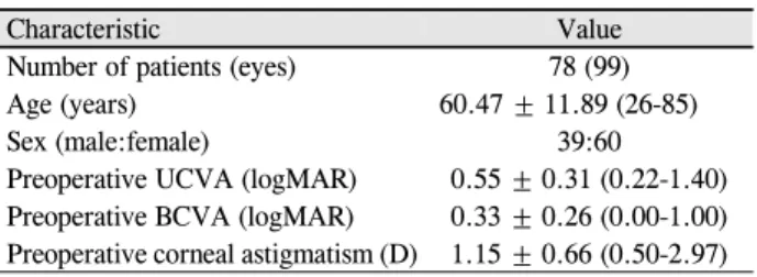
Figure 2. Preoperative and postoperative corneal astigmatism
(diopter) with standard deviation. Significant improvement was shown at 1-2 days and maintained without significant change for 2 years. W ilcoxon signed rank test was used to examine statistical difference. Preop = preoperative; mo = month; yr= year; yrs = years; cyl = corneal astigmatism. *Significant difference between preoperation and postoperation 1-2 days.
Figure 1. Preoperative and postoperative UCVA (logarithm of
minimal angle of resolution, logMAR) and BCVA (logMAR) with standard deviation. Large improvement was shown at 1-2 days and maintained without significant change for 2 years.UCVA = uncorrected visual acuity; BCVA = best corrected visual acuity; Preop. = preoperative; mo = months; yr(s) = year(s).
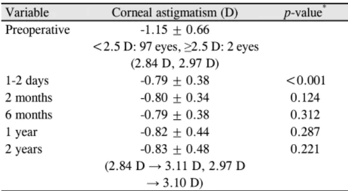
Variable Corneal astigmatism (D) p-value* Preoperative -1.15 ± 0.66
<2.5 D: 97 eyes, ≥2.5 D: 2 eyes (2.84 D, 2.97 D)
1-2 days -0.79 ± 0.38 <0.001
2 months -0.80 ± 0.34 0.124
6 months -0.79 ± 0.38 0.312
1 year -0.82 ± 0.44 0.287
2 years -0.83 ± 0.48
(2.84 D → 3.11 D, 2.97 D
→ 3.10 D)
0.221
Values are presented as mean ± standard deviation unless other- wise indicated.
D = diopter; SD = standard deviation.
*Wilcoxon signed rank test.
Table 3. Preoperative and postoperative corneal astigmatism
2년 후 0.13 ± 0.19 logMAR와 0.07 ± 0.09 logMAR로 유의한 시력상승을 보였다(p<0.001, p<0.001) (Fig. 1, Table 2).
수술 1, 2일 후 각막난시가 -1.15 ± 0.66D에서 -0.79 ± 0.38D로 개선된 후(p<0.001) 2년의 기간 동안 유의한 변화 없이 안정적으로 유지되었고(Fig. 2, Table 3), 수술 전 71.5%였던 0.50D 이상의 각막난시 비율이 수술 2년 후 44.7%로 감소하는 결과를 통해 난시교정 효과를 확인할 수 있었다(Fig. 3). 각막난시의 변화 양상을 산포도로 표시한 Fig. 4에서는 수술 전 0.5D 밖으로 광범위하게 산재되어 있 던 난시들이 수술 2개월 후 0.5D 내외로 수렴하는 양상을 볼 수 있고, Fig. 5에서는 2개월째 감소된 난시가 2년 후에 도 대부분 큰 변화 없이 유지되고 있었으나, 수술 전 2.5D 이상의 2안에서는 오히려 난시가 증가하는 양상을 보이기 도 하였다(Table 3). 수술 전 TIA는 평균 1.15 ± 0.66D였고, 수술 2년 후 SIA와 DV는 각각 0.69 ± 0.43D, 0.83 ± 0.48D
였으며, CI는 0.68 ± 0.45였고 1.0D 이상의 환자군에 비해 1.0D 이하의 군에서 유의하게 낮은 결과를 보였다(Table 4).
고위 수차는 동공 중심에서 4 mm와 6 mm 직경에서의 총 고위수차, 트레포일수차, 코마수차, 구면수차값을 측정 하였으며 수술 전과 수술 후 2개월 및 수술 후 2개월과 수 술 후 2년을 비교 분석하였다(Table 5, 6). 수술 전과 수술 2개월 후, 4 mm 총 고위수차에서는 0.18 ± 0.10에서 0.20
± 0.08로 유의하게 증가하였고, 6 mm 총 고위수차와 트레 포일수차에서도 각각 0.51 ± 0.30, 0.21 ± 0.19에서 0.57 ± 0.23, 0.27 ± 0.21로 유의한 상승을 보였다(p=0.001, p=0.001, p=0.004). 나머지 4 mm 트레포일수차는 수술 전 0.11 ± 0.07에서 수술 2개월 후 0.13 ± 0.08이었고(p=0.080), 4 mm 와 6 mm 코마수차는 각각 0.11 ± 0.07, 0.28 ± 0.21에서
Parameter Total <1.0 ≥1.0 p-value*
TIA (D) 1.15 ± 0.66 (0.50-2.97) 0.76 ± 0.21 1.65 ± 0.47 0.24
SIA (D) 0.69 ± 0.43 (0.04-1.80) 0.42 ± 0.29 0.92 ± 0.40 <0.01
DV (D) 0.83 ± 0.48 (0.04-2.24) 0.57 ± 0.29 1.06 ± 0.51 0.01
CI 0.68 ± 0.45 (0.05-1.89) 0.81 ± 0.58 0.57 ± 0.28 0.03
Values are presented as mean ± standard (range).
D = diopter; TIA = target induced astigmatism; SIA = surgically induced astigmatism; DV = difference vector; CI = correction index; SD
= standard deviation.
*Student’s t-test.
Table 4. Vector analysis using Alphins method of keratometric astigmatic correction at 2 years after femtosecond-assisted combined
phacoemulsification and arcuate keratotomyVariable 4 mm 6 mm
Preoperative Postoperative p-value* Preoperative Postoperative p-value*
tHOA 0.18 ± 0.10 0.20 ± 0.08 0.001 0.51 ± 0.30 0.57 ± 0.23 0.001
Trefoil 0.11 ± 0.07 0.13 ± 0.08 0.080 0.21 ± 0.19 0.27 ± 0.21 0.004
Coma 0.11 ± 0.07 0.11 ± 0.06 0.556 0.28 ± 0.21 0.26 ± 0.19 0.107
Spherical 0.06 ± 0.06 0.05 ± 0.03 0.260 0.28 ± 0.13 0.28 ± 0.11 0.284
Values are presented as mean ± standard deviation.
tHOA = total higher order abberations.
*Wilcoxon signed rank test.
Table 5. The comparison of higher order abberations between preoperation and postoperation (2 months) after femtosecond-assisted
combined phacoemulsification and arcuate keratotomyFigure 3. Distribution of corneal astigmatism at preoperative
and postoperative 2 years. Rate of patients below 1.0 diopter increased at 2 years after surgery. Preop. = preoperation;Postop. = postoperation; yrs = years; D = diopter.
Figure 4. Scatterplot of corneal astigmatism change. Scattered
dots gathered within 1.0 diopter at 2 months after surgery.Preop. = preoperation; cyl = corneal astigmatism; mon = months; D = diopter.
Figure 5. Scatterplot of corneal astigmatism change. Similar
distribution can be seen between 2 months and 2 years. mon = months; yr = years; cyl = corneal astigmatism; D = diopter.0.11 ± 0.06, 0.26 ± 0.19로 변화하였으며(p=0.556, p=0.107), 4 mm, 6 mm 구면수차에서는 각각 0.06 ± 0.06, 0.28 ± 0.13에서 0.05 ± 0.03, 0.28 ± 0.11 (p=0.260, p=0.284)로의 변화를 보였으나 유의성을 나타내지는 않았다. 수술 2개월 후와 2년 후의 비교 결과를 보면 4 mm, 6 mm 총 고위수차 는 0.20 ± 0.08, 0.27 ± 0.20에서 0.57 ± 0.23, 0.62 ± 0.38 (p=0.137, p=0.787)로 미세한 증가를 보였고, 4 mm, 6 mm 트레포일수차에서는 0.13 ± 0.08, 0.27 ± 0.21에서 0.14 ± 0.13, 0.27 ± 0.19 (p=0.914, p=0.830)로 거의 변화가 없었 다. 4 mm, 6 mm 코마수차는 0.11 ± 0.06, 0.26 ± 0.19에서 0.14 ± 0.14, 0.36 ± 0.35 (p=0.543, p=0.212)의 소폭 상승을 보였고, 4 mm, 6 mm 구면수차에서는 0.05 ± 0.03, 0.28 ± 0.11에서 0.05 ± 0.03, 0.25 ± 0.12 (p=0.892, p=0.433)로 변
Variable 4 mm 6 mm
2 months 2 years p-value* 2 months 2 years p-value*
tHOA 0.20 ± 0.08 0.27 ± 0.20 0.137 0.57 ± 0.23 0.62 ± 0.38 0.787
Trefoil 0.13 ± 0.08 0.14 ± 0.13 0.914 0.27 ± 0.21 0.27 ± 0.19 0.830
Coma 0.11 ± 0.06 0.14 ± 0.14 0.543 0.26 ± 0.19 0.36 ± 0.35 0.212
Spherical 0.05 ± 0.03 0.05 ± 0.03 0.892 0.28 ± 0.11 0.25 ± 0.12 0.433
Values are presented as mean ± standard deviation.
tHOA = total higher order abberations.
*Wilcoxon signed rank test.
Table 6. The comparison of higher order abberations between 2 months and 2 years after femtosecond-assisted combined phacoe-
mulsification and arcuate keratotomy화하였다. 수술 후 2개월과 2년의 고위수차에서는 전 영역 에 걸쳐 작은 변화들을 보이는 하지만 모두 정상 범위 내에 서의 변화였으며 유의성을 관찰할 수 없었다.
고 찰
백내장수술 후 발생하는 난시에 대해서는 여러 연구를 통해 보고된 바 있으며 절개창의 크기, 위치, 수, 창상치유 등이 난시 유발에 영향을 미치게 된다.13-17 수술 후 난시 발 생을 줄이기 위해 난시교정인공수정체를 삽입하거나, 각막 절개의 정확성을 높이기 위해 펨토초레이저를 백내장수술 에 적용하였으며, 난시각막절개술을 백내장수술 중 같이 시행하여 난시를 감소시키려는 노력을 하고 있다.17-20
펨토초레이저는 라식수술에서 각막절편을 만들기 위해 처음 사용된 이래 백내장수술에 도입되어 각막절개, 수정 체전낭절개, 수정체 분할에 이용되고 있으며 그 결과와 안 정성을 인정받고 있다.21-23 백내장수술 시 시행되는 투명각 막절개의 폭과 길이 및 시축으로부터의 거리는 수술 후 각 막난시 유발과 직접적으로 관련되어 있는데, 펨토초레이저 는 노모그램 수치와 정밀하게 근접한 절개를 가능하게 해 주고 결과의 예측성을 높여 고식적인 방법의 백내장수술보 다 난시감소를 비롯한 향상된 결과를 도출하고 있다.24 또 한 수정체유화술 중 사용되는 초음파는 유화기 첨단부의 진동으로 열을 발생시키고 전방의 관류액에 기포를 형성하 여 각막내피세포 손상을 유발하게 되는데, 펨토초레이저는 유효 초음파 시간을 단축시켜 이러한 손상을 감소시켜주고 수술 후 예후에도 좋은 영향을 미치게 된다.5-8,25
난시각막절개술을 백내장수술과 같이 시행받은 군과 난 시교정 인공수정체삽입술 시행군 및 대조군(단초점인공수 정체삽입)의 임상결과를 비교한 국내 한 보고에 따르면, 수 술 2개월째 각막난시의 감소율이 난시각막절개술군에서 30.6%로 세 군 중에서 가장 컸고, 난시교정 인공수정체삽 입술군과 대조군은 비슷한 결과를 보여 각막절개술이 직접 적으로 각막의 난시를 줄일 수 있는 좋은 방법임을 시사하
였다.2 또한 펨토초레이저를 이용해 백내장수술과 활모양 각막절개술을 동시에 시행받은 환자를 대상으로 한 국외 연구 결과에서도 수술 1달 후 각막난시가 0.71D가 감소하 는 결과를 보였으며, 동일 수술에 대해 2년의 장기간 경과 에 대해 연구한 또 다른 보고에서도 수술 전 TIA 1.35D 그 룹에서 수술 2년 후 1.23D의 각막난시가 감소하는 결과를 보여 장기간에도 효과가 유지됨을 보여준 바 있다.13,26
본 연구는 레이저 백내장수술 중 펨토초레이저를 이용한 활모양각막절개를 동시 시행한 후 여러 검사 결과값을 통 해 후향적으로 난시교정 효과를 확인하고 2년간의 장기간 에 걸친 임상경과를 알아보고자 하였다. 수술 후 나안시력 과 교정시력은 모두 향상된 후 2년간 안정적으로 유지되는 결과를 보였고, 이를 통해 2년의 장기간 경과 관찰에도 난 시 재발이 많지 않았고, 유의한 고위수차 및 불규칙난시 발 생 또한 없었음을 알 수 있었다. 본 연구의 대상군은 수술 전 평균 TIA가 1.15D였고, 수술 2년 후 DV 0.83D의 각막 난시가 남아있었다(Table 4). 이는 본 연구와 동일한 주제 로 연구한 기존의 보고26와 비교하였을 때 각 벡터값들의 표준편차가 크게 분포하고 있고 난시교정 효과가 상대적으 로 적은 결과이다. 원인을 생각해보면 먼저 대상자 선정의 차이를 들 수 있겠다. 기존 발표된 연구26에서는 수술 전 각 막난시 2.5D 미만의 환자를 대상으로 하였고 본 연구는 0.5D 이상의 난시 환자를 선별하였다. 본 연구에서 수술 전 각막난시 정도에 따른 Alphins method 분석 결과 수술 전 각막난시가 적을수록 상대적으로 수술 후 난시교정 효과가 컸음을 알 수 있으며, 수술 전 1.0D 미만의 환자에서 1.0D 이상의 환자에 비해 유의하게 높은 CI값을 확인할 수 있었 다(Table 4). 선정의 기준 차이로 인해 본 연구에서는 기존 에 발표되었던 연구에 비해 수술 전 큰 난시를 갖은 환자의 비율이 더 높았고, 작은 난시 환자의 비율이 더 낮았을 것 으로 생각되며 이러한 요인이 본 연구에서 비교적 적은 효 과의 결과로 이어졌을 것으로 보인다. 또 다른 원인으로는 레이저 수술에 사용한 노모그램과 수술 장비의 차이를 생 각해 볼 수 있다. 본 연구에서는 LenSx® (Alcon LenSx,
Fort Worth, TX, USA)와 Donnenfeld nomogram을 이용해 수술하였고, 기존의 연구26에서는 VICTUS (Bausch & Lomb Inc., Dornach, Germany) femtosecond-laser platform과 술 자가 수정한 노모그램(Modified from Wallace Limbal Relaxing Incision Nomogram)을 사용하였다. 노모그램은 각막절개창의 길이, 깊이, 수, 위치를 결정해 주고 그 정확 성에 따라 난시교정 정도가 달라질 수 있다.12 술자의 경험 치를 반영해 수정한 노모그램 결과값이, 수술 전 높은 각막 난시를 갖고 있었던 환자에게 더 큰 효과를 나타낸 것으로 보이며, 수술 장비에 따른 오차 정도가 결과의 차이를 유발 했을 것으로 생각된다.
각막절개술 후 고위수차를 연구한 결과 수술 후 의미 있 는 변화는 관찰되지 않았다는 보고들이 있다.26,28 본 연구 결과에서도 수술 후 고위수차 변화를 분석한 결과 수술 2개 월 후의 4 mm, 6 mm의 총 고위수차와 6 mm 트레포일수 차에서 미미한 증가를 보였지만 나머지 부분에서는 유의한 차이를 보이지 않았고, 2년 후의 고위수차에서도 모든 영역 에서 유의한 변화가 관찰되지 않았다. 이를 통해 수술이 고 위수차에 큰 영향을 미치지 않았고 장기간의 임상경과에도 안정적으로 유지됨을 확인할 수 있었으며, 고위수차의 변 화량 또한 시력의 질적 차이를 유발할 정도의 변화가 아님 을 알 수 있었다.
백내장수술 시 난시를 교정하는 데 있어, 난시교정 인공 수정체삽입술이 가장 큰 교정 효과를 보이고, 각막절개술 은 상대적으로 작은 효과 및 저교정의 경향을 보이나 중등 도 이하의 난시에서는 난시교정 인공수정체삽입술과 비슷 한 효과를 보인다는 보고가 있다.2 본 연구 결과에서도 CI 값이 0.1D 미만의 환자에서는 0.81, 0.1D 이상의 환자에서 는 0.57로 유의한 차이를 보여 경도의 난시에 대해 더 큰 효과를 확인할 수 있었다. 또한 전체적으로 저교정의 결과 를 보였으나 각막난시 변화를 나타낸 산포도상 수술 2달 후 0.5D 쪽으로 감소하는 양상으로 보였고 2년 후에도 비 교적 안정적으로 유지되는 것을 통해 수술의 안정성을 확 인할 수 있었다. 하지만 수술 전 2.84D와 2.97D였던 2안은 2년 후 각막난시가 오히려 증가하는 결과를 보여 2.50D 이 상의 큰 각막난시의 교정에 대한 추가적인 연구가 필요할 것으로 보인다.
앞서 언급한 한계점들에도 불구하고 본 연구에서도 펨토 초레이저 활모양각막절개술의 효과적인 난시감소 효과를 확인할 수 있었고, 기존의 보고들과 유사한 결과의 경향성 도 볼 수 있었다. 결론적으로 백내장수술 전 중등도 이상의 난시를 갖는 환자에서는 난시교정 인공수정체삽입술이 난 시를 교정하는 데 가장 효과적인 방법이지만,2 경도의 난시 에서는 펨토초레이저 백내장수술과 각막절개술의 동시 시
행이 또 하나의 대안으로 제시될 수 있을 것으로 보인다. 또한 수술의 숙련도 향상과 정확한 노모그램의 선택이 더 욱 향상된 수술 결과를 유도할 수 있을 것으로 기대된다.
REFERENCES
1) Vickers LA, Gupta PK. Femtosecond laser-assited keratotomy.
Curr Opin Ophthalmol 2016;27:277-84.
2) Park YY, Kim MS, Kim EC. Comparison of efficacies in treating astigmatism between toric intraocular lens implantation and limbal relaxing incision. J Korean Ophthalmol Soc 2017;58:1225-33.
3) Steinert RF. Cataract surgery: Technique, complication and management, 2nd ed. Philadelphia: WB Saunders, 2004;253-66.
4) Hossein M, Elham M, Goldis E, et al. Three methods for correction of astigmatism during phacoemulsification. J Ophthalmic Vis Res 2016;11:162-7.
5) Bahar I, Levinger E, Kaiserman I, et al. IntraLase-enabled astig- matic keratotomy for postkeratoplasty astigmatism. Am J Ophthalmol 2008;146:897-904.e1.
6) Hoffart L, Proust H, Matonti F, et al. Correction of postkeratoplasty astigmatism by femtosecond laser compared with mechanized as- tigmatic keratotomy. Am J Ophthalmol 2009;147:779-87.e1.
7) Takács AI, Kovács I, Miháltz K, et al. Central corneal volume and endothelial cell count following femtosecond laser-assisted refractive cataract surgery compared to conventional phacoemulsification. J Refract Surg 2012;28:387-91.
8) Conrad-Hengerer I, Al Juburi M, Schultz T, et al. Corneal endothe- lial cell loss and corneal thickness in conventional compared with femtosecond laser-assisted cataract surgery: three-month fol- low-up. J Cataract Refract Surg 2013;39:1307-13.
9) Abbey A, Ide T, Kymionis GD, Yoo SH. Femtosecond laser-assisted astigmatic keratotomy in naturally occurring high astigmatism. Br J Ophthalmol 2009;93:1566-9.
10) Chan T, Cheng G, Wang Z, et al. Vector analysis of corneal astig- matism after combined femtosecond-assisted phacoemulsification and arcuate keratotomy. Am J Ophthlamol 2015;160:250-5.e2.
11) Day AC, Lau NM, Stevens JD. Nonpenetrating femtosecond laser intrastromal astigmatic keratotomy in eyes having cataract surgery.
J Cataract Refract Surg 2016;42:102-9.
12) Baharozian CJ, Song C, Hatch KM, Talamo JH. A novel nomo- gram for the treatment of astigmatism with femtosecond-laser arc- uate incisions at the time of cataract surgery. Clin Ophthalmol 2017;11:1841-8.
13) Day AC, Stevens JD. Predictors of femtosecond laser intrastromal astigmatic keratotomy efficacy for astigmatism management in cataract surgery. J Cataract Refract Surg 2016;42:251-7.
14) Khan MI, Muhtaseb M. Prevalence of corneal astigmatism in pa- tients having routine cataract surgery at a teaching hospital in the united Kingdom. J Cataract Refract Surg 2011;37:1751-5.
15) Mayer WJ, Klaproth OK, Hengerer FH, et al. In vitro im- munohistochemical and morphological observations of penetrat- ing corneal incisions created by a femtosecond laser used for as- sisted intraocular lens surgery. J Cataract Refract Surg 2014;40:
632-8.
16) Richard J, Hoffart L, Chavane F, et al. Corneal endothelial cell loss
= 국문초록 =
펨토초레이저 백내장수술에서 활모양각막절개술 시행의 장기간 임상결과
목적: 펨토초레이저 백내장수술에서 활모양각막절개술을 시행받은 환자의 장기간 임상결과를 알아보고자 한다.
대상과 방법: 수술 전 각막난시 0.5 diopter (D) 이상의 78명 환자 99안을 대상으로 후향적으로 연구하였다. 수술 전과 수술 후 2년간 나안과 교정시력, 각막난시, 고위수차를 비교 분석하였고 Alpins method를 이용해 난시교정 효과를 분석하였다.
결과: 수술 전 평균 나안시력과 교정시력은 각각 0.55 ± 0.31 logarithm of minimal angle of resolution (logMAR), 0.33 ± 0.26 logMAR이었고, 수술 2년 후 0.13 ± 0.19 logMAR, 0.07 ± 0.09 logMAR로 유의한 시력 상승을 보였다(p<0.001, p<0.001). 각막난시 는 수술 전 -1.15 ± 0.66D에서 수술 1, 2일 후 -0.79 ± 0.38D로 유의하게 개선된 후(p<0.001) 2년간 유의한 변화 없이 유지되었다.
수술 전 평균 target induced astigmatism은 1.15 ± 0.66D였고, 수술 2년 후의 평균 surgically induced astigmatism과 difference vector는 각각 0.69 ± 0.43D, 0.83 ± 0.48D였으며 평균 correction index는 0.68 ± 0.45였다. 4 mm, 6 mm 총 고위수차와 6 mm 트레포일수차에서는 수술 전과 비교해 수술 2개월 후 유의한 변화를 보였지만(p=0.001, p=0.001, p=0.004), 나머지 영역에서는 유의 성 있는 차이를 관찰할 수 없었다.
결론: 펨토초레이저 백내장수술과 동시에 시행되는 활모양각막절개술은 중등도 미만의 난시를 교정할 수 있는 안전하고 효과적인 방법이다.
<대한안과학회지 2019;60(10):946-952>
방찬우 / Chan Woo Bang
성모안과병원 Sungmo Eye Hospital after cataract extraction by using ultrasound phacoemulsification
versus a fluid-based system. Cornea 2008;27:17-21.
17) Tejedor J, Murube J. Choosing the location of corneal incision based on preexisting astigmatism in phacoemulsification. Am J Ophthalmol 2005;139:767-76.
18) Khokhar S, Lohiya P, Murugiesan V, Panda A. Corneal astigma- tism correction with opposite clear corneal incisions or single clear corneal incision: comparative analysis. J Cataract Refract Surg 2006;32:1432-7.
19) Barequet IS, Yu E, Vitale S, et al. Astigmatism outcomes of hori- zontal temporal versus nasal clear corneal incision cataract surgery.
J Cataract Refract Surg 2004;30:418-23.
20) Amesbury EC, Miller KM. Correction of astigmatism at the time of cataract surgery. Curr Opin Ophthalmol 2009;20:19-24.
21) Hayashi K, Hayashi H, Nakao F, Hayashi F. Risk factors for cor- neal endothelial injury during pahcoemulsification. J Cataract Refract Surg 1996;22:1079-84.
22) Juhasz T, Loesel FH, Kurtz RM, et al. Corneal refractive surgery with femtosecond laser. IEEE J Sel Top Quantum Electron
1999;5:902-10.
23) Roverts TV, Lawless M, Bali SJ, et al. Surgical outcomes and safe- ty of femtosecond laser cataract surgery: a prospective study of 1,500 consecutive cases. Ophthalmology 2013;120:227-33.
24) Diakonis VF, Yesilirmak N, Cabot F, et al. Comparison of surgi- cally induced astigmatism between femtosecond laser and maual clear corneal incisions for cataract surgery. J Cataract Refract Surg 2015;41:2075-80.
25) Ahn DS, Han SY, Lee KH. Comparison of effective phacoemulsifi- cation time between femtosecond laser-assisted cataract surgery and conventional cataract surgery. J Korean Ophthalmol Soc 2016;57:236-42.
26) Chan TC, Ng AL, Cheng GP, et al. Corneal astigmatism and aberra- tions after combined femtosecond-assisted phacoemulsification and arcuated keratotomy: two-year results. Am J Ophthalmol 2016;170:83-90.
27) Eliwa TF, Abdellatif MK, Hamza II. Effect of limbal relaxing in- cisions on corneal aberrations. J Refract Surg 2016;32:156-62.