© 2016 The Korean Ophthalmological Society
This is an Open Access article distributed under the terms of the Creative Commons Attribution Non-Commercial License (http://creativecommons.org/licenses /by-nc/3.0/) which permits unrestricted non-commercial use, distribution, and reproduction in any medium, provided the original work is properly cited.
92
Original Article
Factors Affecting Long-term Myopic Regression after Laser In Situ Keratomileusis and Laser-assisted Subepithelial Keratectomy for
Moderate Myopia
Sung A Lim1, Yooyeon Park1, Yu Jin Cheong2, Kyung Sun Na3, Choun-Ki Joo1
1Department of Ophthalmology and Visual Science, Seoul St. Mary's Hospital, The Catholic University of Korea College of Medicine, Seoul, Korea
2Department of Ophthalmology, Ewha Mokdong Hospital, Ewha Womans University School of Medicine, Seoul, Korea
3Department of Ophthalmology, Yeouido St. Mary’s Hospital, The Catholic University of Korea College of Medicine, Seoul, Korea
Purpose: High myopia is known to be a risk factor for long-term regression after laser refractive surgery. There have been few studies about the correction of moderate myopias that did not need retreatment after long-term follow-up. We evaluated 10 years of change in visual acuity and refractive power in eyes with moderate myo- pia after laser refractive surgery.
Methods: We included patients that had undergone laser in situ keratomileusis (LASIK) or laser-assisted subep- ithelial keratectomy (LASEK) to correct their myopia and that had at least 10 years of follow-up. We evaluated the stability of visual acuity in terms of safety, efficacy, and refractive changes at examinations 6 months and 1, 2, 5, 7, and 10 years after surgery.
Results: The study evaluated 62 eyes (36 eyes in LASIK patients and 26 eyes in LASEK patients). In both groups, the efficacy index tended to decrease, and it was consistently higher in the LASEK group compared to the LASIK group over the 10 years of follow-up. The safety index improved over 10 years and was always higher than 0.9 in both groups. The difference between the spherical equivalent at 6 months postoperatively and later periods was statistically significant after 5, 7, and 10 years in both groups (LASIK, p = 0.036, p = 0.003, and p < 0.001, respectively; LASEK, p = 0.006, p = 0.002, and p = 0.001, respectively). Ten years after sur- gery, 26 eyes (66.7%) in the LASIK group and 19 eyes (73.1%) in the LASEK group had myopia greater than 1 diopter. In comparison with the thickness at 6 months postoperatively, central corneal thickness was signifi- cantly increased after 5, 7, and 10 years in both LASIK and LASEK groups (LASIK, p < 0.001, p < 0.001, and p
< 0.001, respectively; LASEK, p = 0.01, p < 0.001, and p < 0.001, respectively).
Conclusions: Moderately myopic eyes showed progressive myopic shifting and corneal thickening after LASIK and LASEK during 10 years of follow-up. We also found that early refractive regression may indicate the long-term refractive outcome.
Key Words: Laser-assisted subepithelial keratectomy, Laser in situ keratomileusis, Refractive regression
Received: May 19, 2015 Accepted: July 3, 2015
Corresponding Author: Choun-Ki Joo, MD, PhD. Department of Ophthalmology and Visual Science, Seoul St. Mary’s Hospital, The Catholic University of Korea College of Medicine, #222 Banpo-daero, Seocho-gu, Seoul 06591, Korea. Tel: 82-2-590-2615, Fax: 82-2-533-3801, E-mail: [email protected]
The prevalence of myopia has profoundly increased, and laser refractive surgery is a popular procedure for safe and effective myopic correction. Many studies have shown good visual acuity and stable refractive outcomes after myopic laser refractive surgery. Laser in situ keratomileu- sis (LASIK) is predominantly performed, due to its rapid visual outcome and little pain. In contrast, laser-assisted subepithelial keratectomy (LASEK) is preferred in cases with thin corneas and high myopia. However, the tendency toward myopia after laser refractive surgery has been re- ported frequently and is difficult for ophthalmologists to address.
Regression is the tendency of an eye to return to its orig- inal refraction and is dependent on the preoperative myo- pia and the amount of correction [1,2]. A previous study of LASIK reported that the myopic regression for moderate to high myopia was -1.66 ± 2.15 diopters (D) over 15 years, indicating a regression rate of -0.11 D per year [3]. LASIK performed with a mechanical microkeratome may be a factor affecting myopia regressions due to uneven flap thickness [4]. Also, a considerable regression of high myo- pia over time after laser refractive surgery is a well-known phenomenon [5,6]. Recently, LASEK has been used for the correction of high myopia and an increased incidence of regressions has been reported in LASEK patients.
Laser refractive surgery is frequently performed in mod- erate myopia, and most patients demonstrate successful outcomes; however, there is little knowledge about the general changes in visual acuity and the refractive tenden- cies of patients without postoperative problems. Most stud- ies have tried to identify risk factors in specific cases, such as those involving ectasia or retreatment, rather than in general cases. Patients may represent different degrees of regressions toward myopia, and the correlation between the amount of myopic regression and the visual acuity is not clear. Some patients are satisfied with a certain amount of myopia, but other patients complain of discomfort de- spite the existence of only a small degree of myopia. Pa- tient’s tolerances are variable, and thus, determining the need for retreatment is very difficult. It is essential to eval- uate the eyes with myopic regression in patients who did not require retreatment for a long period.
The purpose of this study was to identify general refrac- tive changes and to compare significant risk factors for re- gression between LASIK and LASEK for moderate myo- pia during a 10-year follow-up period.
Materials and Methods
Retrospective data were collected from February 1998 to February 2003 at Seoul St. Mary’s Hospital. The study protocol followed the guidelines of the Declaration of Hel- sinki and was approved by the hospital’s institutional re- view board. The charts of all patients who had LASIK or LASEK performed by the same surgeon (CKJ) were re- viewed. Among these patients, we included those that had a preoperative spherical error of less than -10 D and a cyl- inder error of less than -3 D and those that returned for ex- aminations annually, specifically at 6 months and 1, 2, 5, 7, and 10 years after surgery.
All surgery targeted distance vision. All patients were counseled on refractive surgery and provided informed consent before the surgery. The patients were examined with ophthalmologic tests preoperatively and at 6 months and 1, 2, 5, 7, and 10 years after surgery. The uncorrected visual acuity (UCVA), best spectacle corrected visual acui- ty (BSCVA), manifest refraction, slit lamp examination, intraocular pressure (IOP), ultrasound pachymetry, and corneal topography (Orbscan II; Bausch & Lomb, Roches- ter, NY, USA) were examined.
The inclusion criteria were the myopic refractive error and normal preoperative topography. The exclusion crite- ria included ocular pathology, retinal disorders, hyperopia (positive spherical equivalent), and insufficient follow-up.
Patients with ocular or general medical histories such as diabetes were also excluded. All patients had stable refrac- tion for at least 1 year before surgery. When patients were evaluated after surgery, we excluded those with topograph- ic signs suggesting corneal instability, those who needed retreatment, and those who had corneal haze or other com- plications.
We defined the regression group as patients having myo- pia of more than 1 D at 10 years postoperatively and the non-regression group as patients having myopia less than 1 D at 10 years postoperatively following LASIK or LASEK procedures.
Surgical technique
An experienced surgeon performed all of the proce- dures. The preoperative regimen was similar for all pa- tients, and anesthesia consisted of topical eyedrops of proparacaine (Alcaine; Alcon-Couvreur, Puur, Belgium).
94
LASIK and LASEK were performed using a VISX S4 ex- cimer laser system (VISX Inc., Santa Clara, CA, USA).
Refractive surgery was performed with a 6.5-mm optical zone with no wavefront-guided treatments.
In LASIK-treated eyes, a microkeratome (Moria, Ant- ony, France) was applied to create a nasal-hinged flap us- ing the manufacturer’s recommended parameters. Excimer laser stromal ablation was performed after the flap was re- flected nasally. Immediately after ablation, the stromal bed was irrigated with a balanced salt solution, and the flap was positioned. In eyes treated with LASEK, the corneal epithelium was removed with a 20% alcohol solution for 30 seconds and then irrigated with a balanced salt solution.
Laser surface ablation was performed using an excimer la- ser based on the surgeon’s criteria.
After LASIK and LASEK surgeries, drops of fluoro- quinolone antibiotics and fluorometholone 0.1% were in- stilled into the lower fornix. Topical antibiotic and cortico- steroid agents were applied four times daily for a minimum of four weeks, and gradual tapering was applied based on refraction, IOP, and corneal status.
Statistical analysis
The visual outcomes evaluated included the efficacy in- dex (postoperative UCVA / preoperative BSCVA) and the safety index (postoperative BSCVA / preoperative BSCVA).
The refractive outcome was evaluated as predictability, which was defined as the percentage of the eye within 1.0 D in a spherical equivalent (SE).
All data are expressed as the means ± standard devia- tions. Statistical analyses were performed using SPSS ver. 17.0 (SPSS Inc., Chicago, IL, USA). Demographic data were compared using chi-square tests. Paired t-tests were used to compare preoperative and postoperative visual and refractive outcomes. Comparisons between LASIK and LASEK were performed using unpaired t-tests. The p-values less than 0.05 were considered statistically signif- icant.
Results
Laser refractive surgery was performed on a total of 487 eyes between June 1, 1998 and December 31, 2003. Ac- cording to above inclusion criteria, 425 patients were ex-
cluded: 203 were lost to follow-up; 133 were not satisfied the inclusion of refraction (hyperopia, low myopia, high myopia, and severe astigmatism); 46 had corneal abnor- malities upon follow-up (topographic abnormality, corneal haze, ectasia, and infection); and 43 had systemic diseases (diabetes, autoimmune disease, and cancer). After these exclusions, 62 eyes were evaluated in annual follow-ups and were included in this study. Thirty-six eyes had LASIK, and 26 eyes had LASEK. The preoperative SEs ranged from -3.5 to -8.625 D (mean ± SE, -5.73 ± 2.76 D) in the LASIK group and from -3 to -8.625 D (mean ± SE, -5.45 ± 1.84 D) in the LASEK group. The patient’s preoper- ative data are shown in Table 1. Age, refraction, and visual acuity were similar in the LASIK and LASEK groups.
Central corneal thickness (CCT) and IOP were significant- ly different in the two groups.
Efficacy
UCVA showed significant improvements at all visits in both groups (p < 0.001). The efficacy index (postoperative UCVA / preoperative BSCVA) of LASEK was consistently higher than that of LASIK in moderate myopia. Between the LASIK and LASEK groups, significant differences were apparent at 2, 5, and 10 years after surgery.
Safety
Safety indices (postoperative BSCVA / preoperative BSCVA) were above 0.90 in both groups. The safety index of LASEK was higher than that of LASIK, but the differ- ences in the safety indices were not significant between the groups during follow-up. Table 2 shows the safety and effi- cacy indices of the LASIK and LASEK patients.
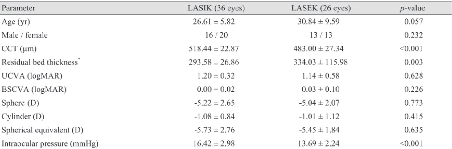
Predictability
The predictability of the refractive surgery is shown in Fig. 1A and 1B. In the LASIK group, postoperative mean refractive spherical equivalent (MRSE) within ±1.00 D was 93.3 % after 6 months and 68.3 % after 10 years. In the LASEK group, postoperative MRSE within ±1.00 D was 82.5 % after 6 months and 77.5 % after 10 years. The predictability was higher in the LASIK group than the LASEK group at 6 months after surgery. However, 10 years after surgery, the LASEK group showed superior
95 predictability.
Refractive change
The MRSEs were significantly improved for all fol- low-up periods. The difference between the SE 6 months
postoperatively and at later periods was statistically signifi- cant at 5, 7, and 10 years in both groups (LASIK, p = 0.036, p = 0.003, and p = <0.001, respectively; LASEK, p = 0.006, p = 0.002, and p = 0.001, respectively). The mean SEs were comparable between the LASIK and LASEK groups at all follow-up intervals in myopic eyes (p > 0.05). Myopic
Table 2. Efficacy and safety indices for 10 years after LASIK and LASEK
Follow-up period Efficacy index Safety index
LASIK LASEK p-value LASIK LASEK p-value
6 mon 0.83 0.92 0.204 0.96 0.99 0.574
1 yr 0.85 0.93 0.127 0.98 1.00 0.835
5 yr 0.77 0.99 0.002* 0.95 1.02 0.173
7 yr 0.77 0.89 0.070 0.98 1.02 0.461
10 yr 0.73 0.89 0.025* 0.99 1.00 0.936
LASIK = laser in situ keratomileusis; LASEK = laser-assisted subepithelial keratectomy.
*Statistically significant difference between groups.
80 60 40 20 0
6 mon 10 yr
Postoperative spherical equivalent refraction (D) Less than
-2 More than
-2 to -1 -1 to -0.5 -0.50 to 0 0 to 0.50 +0.5
Eyes (%)
80 60 40 20
6 mon 10 yr
Eyes (%)
80 60 40 20 0
6 mon 10 yr
Postoperative spherical equivalent refraction (D) Less than
-2 More than
-2 to -1 -1 to -0.5 -0.50 to 0 0 to 0.50 +0.5
Eyes (%)
80 60 40 20 0
6 mon 10 yr
Postoperative spherical equivalent refraction (D) Less than
-2 More than
-2 to -1 -1 to -0.5 -0.50 to 0 0 to 0.50 +0.5
Eyes (%)
Fig. 1. Predictability for 10 years after (A) laser in situ keratomileusis (n = 36 eyes) and (B) laser-assisted subepithelial keratectomy (n = 26 eyes) surgeries. D = diopter.
A B
Table 1. Preoperative data of 62 eyes treated with laser refractive surgery that were followed for up to 10 years after surgery
Parameter LASIK (36 eyes) LASEK (26 eyes) p-value
Age (yr) 26.61 ± 5.82 30.84 ± 9.59 0.057
Male / female 16 / 20 13 / 13 0.232
CCT (µm) 518.44 ± 22.87 483.00 ± 27.34 <0.001
Residual bed thickness* 293.58 ± 26.86 334.03 ± 115.98 0.003
UCVA (logMAR) 1.20 ± 0.32 1.14 ± 0.58 0.628
BSCVA (logMAR) 0.00 ± 0.02 0.03 ± 0.10 0.226
Sphere (D) -5.22 ± 2.65 -5.04 ± 2.07 0.773
Cylinder (D) -1.08 ± 0.84 -1.01 ± 1.12 0.415
Spherical equivalent (D) -5.73 ± 2.76 -5.45 ± 1.84 0.635
Intraocular pressure (mmHg) 16.42 ± 2.98 13.69 ± 2.24 <0.001
LASIK = laser in situ keratomileusis; LASEK = laser-assisted subepithelial keratectomy; CCT = central corneal thickness; UCVA = un- corrected visual acuity; logMAR = logarithm of the minimum angle of resolution; BSCVA = best spectacle corrected visual acuity; D = diopter.
*Calculated as the remainder from the whole corneal thickness subtracted by the corneal flap thickness.
96
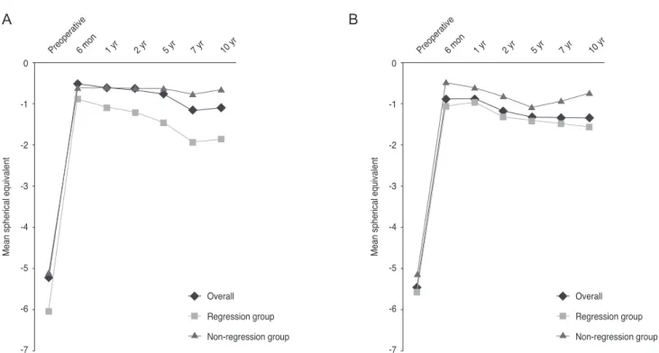
change was observed over time in both groups, even years after the initial surgical procedure. Fig. 2A and 2B shows the MRSE over time by surgery (LASIK or LASEK) and by degree of myopia 10 years postoperatively in all eyes over the duration of the study. The regression group (myo- pia of more than 1 D 10 years postoperatively) showed an early myopic tendency 1 year after surgery in the LASIK group and 6 months to 10 years after surgery, except at 5 years, in the LASEK group as compared with those in the non-regression group (myopia less than 1 D 10 years post- operatively). Preoperative characteristics of the regression and non-regression groups are shown in Table 3.
Central corneal thickness
Fig. 3A and 3B shows the mean CCT over time by sur- gery (LASIK or LASEK) and by degree of myopia 10 years postoperatively. There was an increase in CCT after myopic LASIK and LASEK in both the regression and non-regression groups. In comparison with the CCT at 6 months postoperatively, a significant increase was noted after 5, 7, and 10 years in both LASIK and LASEK pa- tients (LASIK, p < 0.001, p < 0.001, and p < 0.001, respec- tively; LASEK, p = 0.01, p < 0.001, and p < 0.001, respec-
tively). There were no significant differences between the regression and non-regression groups in either LASIK or LASEK patients.
Discussion
LASIK is the most popular refractive surgery, whereas LASEK is commonly used in patients with thin corneas or dry eyes [7,8]. This study evaluated the visual and refrac- tive outcomes of LASIK compared to LASEK. We focused on the differences and changes between patients having myopia of more than 1 D (regression group) and those hav- ing myopia of less than 1 D (non-regression group) 10 years postoperatively. Myopia of more than 1 D was seen in 76.6% of the LASIK group and 73.0% of the LASEK group 10 years after surgery. The mean myopic change throughout the 10-year follow-up period was -1.09 D in LASIK patients and -1.34 D in LASEK patients.
Previous studies showed variable results regarding the efficacies of LASIK and LASEK for the correction of my- opia. Tobaigy et al. [9] and Scerrati [10] suggested that vi- sual acuity and refractive outcomes were better in LASEK than in LASIK. Kim et al. [11] concluded that LASIK sur-
0 -1 -2 -3 -4 -5 -6 -7
6 mon
Preoperative 1 yr 2 yr 5 yr 7 yr 10 yr
Mean spherical equivalent
Overall Regression group Non-regression group
0 -1 -2 -3 -4 -5 -6 -7
6 mon
Preoperative 1 yr 2 yr 5 yr 7 yr 10 yr
Mean spherical equivalent
Overall Regression group Non-regression group
Fig. 2. Changes in mean spherical equivalents by surgery (A, laser in situ keratomileusis; B, laser-assisted subepithelial keratectomy) and by degree of myopia 10 years postoperatively.
A B
gery was superior to LASEK for high myopia. We retro- spectively reviewed 10-year follow-up charts and found that LASIK and LASEK had similar efficacies for moder- ate myopia within 2 years, with LASEK producing signifi- cantly superior efficacy 4 years postoperatively. Eyes treat-
ed with LASIK had a lower efficacy index of less than 0.8 at 5 years postoperatively and might show greater myopic regression. Safety indices were similar for LASIK and LASEK in our study.
Refractive changes after surgery were variable, although
Fig. 3. Changes in mean central corneal thickness (CCT) by surgery (A, laser in situ keratomileusis; B, laser-assisted subepithelial kera- tectomy) and by degree of myopia 10 years postoperatively.
530 520 510 500 490 480 470 460 450 440 430 420 410 400
6 mon Preoperative
1 yr 2 yr 5 yr 7 yr 10 yr
CCT (μm)
Overall Regression group Non-regression group
530 520 510 500 490 480 470 460 450 440 430 420 410 400
6 mon Preoperative
1 yr 2 yr 5 yr 7 yr 10 yr
CCT (μm)
Overall Regression group Non-regression group
A B
Table 3. Preoperative characteristics of regression and non-regression groups after laser refractive surgery Parameter
LASIK (n = 36 eyes) LASEK (n = 26 eyes)
Regression group
(n = 24) Non-regression
group (n = 12) p-value Regression group
(n = 19) Non-regression
group (n = 7) p-value
Age (yr) 26.66 ± 3.94 28.36 ± 7.57 0.328 31.42 ± 10.20 29.28 ± 8.22 0.592
IOP (mmHg) 17.08 ± 2.66 15.00 ± 3.25 0.081 13.05 ± 2.04 15.42 ± 1.90 0.017*
Mean keratometry 44.36 ± 44.17 44.17 ± 1.01 0.068 43.55 ± 1.22 44.07 ± 2.21 0.575
Sphere (D) -5.46 ± 3.16 -4.72 ± 1.05 0.308 -5.17 ± 2.07 -4.71 ± 2.21 0.645
Cylinder (D) -1.27 ± 0.79 -0.75 ± 0.85 0.095 -1.08 ± 1.37 -0.85 ± 0.24 0.549
Spherical equivalent -6.05 ± 3.23 -5.10 ± 1.33 0.223 -5.57 ± 1.71 -5.14 ± 2.29 0.663 CCT (µm) 515.45 ± 24.04 524.61 ± 19.94 0.247 475.63 ± 24.49 503.00 ± 26.03 0.035* Ablation depth (µm) 77.04 ± 20.38 69.75 ± 19.66 0.312 74.10 ± 22.12 60.28 ± 27.23 0.259 RBT (µm) 287.16 ± 28.27 306.41 ± 18.82 0.021* 330.17 ± 60.23 332.85 ± 38.95 0.898
UCVA (logMAR) 1.12 ± 0.35 1.29 ± 0.13 0.064 1.24 ± 0.57 0.88 ± 0.56 0.187
BSCVA (logMAR) 0.01 ± 0.03 0 >0.05 0.03 ± 0.11 0.02 ±0.07 >0.05
LASIK = laser in situ keratomileusis; LASEK = laser-assisted subepithelial keratectomy; IOP = intraocular pressure; D = diopter; CCT = central corneal thickness; RBT = residual bed thickness; UCVA = uncorrected visual acuity; logMAR = logarithm of the minimum angle of resolution; BSCVA = best spectacle corrected visual acuity.
*Statistically significant difference between groups.
98
we aimed to correct for refractive error [12]. A myopic shift after myopic laser refractive surgery is well known and is thought to result from undercorrection or regression [1,13]. The causes of the regression can be divided into cor- neal and non-corneal causes. Non-corneal causes include inaccurate preoperative refraction and inadequate laser energy delivery [1,14]. Corneal causes include biomechani- cal changes of the cornea after the keratectomy. Manifest SE was increased for up to 10 years after refractive sur- gery to correct myopia, and this tendency was shown over time in both groups, even years after the initial surgical procedure. The mean myopic change throughout the 10- year follow-up period was -1.09 D in LASIK patients and -1.34 D in LASEK patients. Our results showed more myo- pic change than did previous studies [15-17]. Because myo- pic regression is difficult to define with a specific value, we defined the regression group as patients having myopia of more than 1 D. According to Hirsch [18], a myopia of 1 D is approximately the 6 / 18 level of visual acuity, which de- fines the limitation of vision impairment according to the World Health Organization [19]. Between the regression and non-regression groups, the mean SEs were significant- ly different 1 year after surgery in the LASIK group and 6 months after surgery in the LASEK group.
We speculated that long-term myopic status was cor- related with preoperative refraction, CCT, and ablation profile. Compared with the non-regression group, the re- gression group had significantly lower residual bed thick- ness (RBT) preoperatively in the LASIK patients (p = 0.021) and lower CCT in the LASEK patients (p = 0.035).
Wang et al. [20] reported that an RBT below 250 µm in- creased the risk of ectasia after LASIK [20]. Erie et al. [21]
also found that an RBT of 250 µm does not safely prevent the development of keratectasia. Pan et al. [13] found that refractive regression after LASIK is mainly caused by cor- neal protrusion rather than postoperative central corneal thickening or RBT. Our study indicated that myopic re- gression after LASIK was correlated with a low RBT pre- operatively. In LASEK, patients with myopic regression had a mean preoperative CCT thinner than 500 µm. Some studies reported that surface ablation had stable visual and refractive outcomes in corneas thinner than 500 µm at long-term follow-up [22]. Ectasia after LASEK is rare and was correlated with abnormal topography and CCT preop- eratively [20,23]. Our results showed that regression in LASEK patients was correlated with low CCT and IOP,
but not with RBT.
Pan et al. [13] reported a progressive increase in CCT in the eyes in the regression group than in the non-regression group after LASIK in high myopic patients. Some studies also reported that the eyes with refractive regression after LASIK and photorefractive keratectomy showed a pro- gressive increase in CCT [1,24]. In our study, postoperative CCT increased significantly with time. CCT was signifi- cantly smaller at 6 months postoperatively than after 5, 7, or 10 years postoperatively in both surgeries. Some studies reported that an increase in central epithelial thickness af- ter refractive surgery is related to myopic regression [25- 27], but we did not detect a correlation between myopic re- gression and change in CCT. Our results are consistent with previous studies in that total corneal thickening may not completely explain the refractive change after laser re- fractive surgery [28]. It has been reported that the corneal epithelium has a tendency to restore its original contour and could be a nonuniform thickness during the wound healing process, leading to the development of refractive power [26,27,29]. Lohmann and Guell [27] reported that a difference in the epithelium of approximately 10 µm caused a refractive change of 1 D. Further study is needed to compare the epithelial thicknesses between the regres- sion and non-regression groups.
This study was a retrospective comparative case series, so we evaluated only the patients that came to our center regularly. The small number of patients was a limitation in this study, and a greater length of follow-up is needed to understand the natural course of laser refractive surgery.
Significant differences in CCT, RBT, and IOP were ob- served between the patients who had LASIK and those who had LASEK. Still, preoperative data were well con- trolled between the regression and non-regression patients.
Ten years ago, we performed blade-assisted flap creation rather than femtosecond laser-assisted flap creation and could not consider wave-front guided laser surgery.
In conclusion, our study with 10 years of follow-up demonstrated that both LASIK and LASEK performed to correct moderate myopia showed progressive myopic shifts and corneal thickening. Significant differences in SE between the regression and non-regression groups be- gan 1 year postoperatively in the LASIK group and 6 months postoperatively in the LASEK group. A larger sample size and more variable degrees of myopia are need- ed to support our study.
Conflict of Interest
No potential conflict of interest relevant to this article was reported.
References
1. Chayet AS, Assil KK, Montes M, et al. Regression and its mechanisms after laser in situ keratomileusis in moderate and high myopia. Ophthalmology 1998;105:1194-9.
2. Pang G, Zhan S, Li Y, et al. Myopic regression after photo- refractive keratectomy. Zhonghua Yan Ke Za Zhi 1998;34:
451-3.
3. Alio JL, Soria F, Abbouda A, Pena-Garcia P. Laser in situ keratomileusis for -6.00 to -18.00 diopters of myopia and up to -5.00 diopters of astigmatism: 15-year follow-up. J Cata- ract Refract Surg 2015;41:33-40.
4. Lin MY, Chang DC, Hsu WM, Wang IJ. Cox proportional hazards model of myopic regression for laser in situ ker- atomileusis flap creation with a femtosecond laser and with a mechanical microkeratome. J Cataract Refract Surg 2012;38:992-9.
5. Kim G, Christiansen SM, Moshirfar M. Change in ker- atometry after myopic laser in situ keratomileusis and pho- torefractive keratectomy. J Cataract Refract Surg 2014;
40:564-74.
6. Seiler T. Current evaluation of myopia correction with the excimer laser. Ophthalmologe 1995;92:379-84.
7. Sutton GL, Kim P. Laser in situ keratomileusis in 2010: a review. Clin Experiment Ophthalmol 2010;38:192-210.
8. Hammond MD, Madigan WP Jr, Bower KS. Refractive surgery in the United States Army, 2000-2003. Ophthal- mology 2005;112:184-90.
9. Tobaigy FM, Ghanem RC, Sayegh RR, et al. A con- trol-matched comparison of laser epithelial keratomileusis and laser in situ keratomileusis for low to moderate myo- pia. Am J Ophthalmol 2006;142:901-8.
10. Scerrati E. Laser in situ keratomileusis vs. laser epithelial keratomileusis (LASIK vs. LASEK). J Refract Surg 2001;
17(2 Suppl):S219-21.
11. Kim JK, Kim SS, Lee HK, et al. Laser in situ keratomileu- sis versus laser-assisted subepithelial keratectomy for the correction of high myopia. J Cataract Refract Surg 2004;
30:1405-11.
12. Liu HQ, Shi JP, Ma CR, et al. Multifactor analysis of the
reasons causing undercorrection after laser in situ ker- atomileusis. Zhonghua Liu Xing Bing Xue Za Zhi 2003;
24:307-9.
13. Pan Q, Gu YS, Wang J, et al. Differences between regres- sive eyes and non-regressive eyes after LASIK for myopia in the time course of corneal changes assessed with the Orbscan. Ophthalmologica 2004;218:96-101.
14. Netto MV, Mohan RR, Ambrosio R Jr, et al. Wound heal- ing in the cornea: a review of refractive surgery complica- tions and new prospects for therapy. Cornea 2005;24:509- 22.
15. O’Connor J, O’Keeffe M, Condon PI. Twelve-year fol- low-up of photorefractive keratectomy for low to moderate myopia. J Refract Surg 2006;22:871-7.
16. O’Doherty M, O’Keeffe M, Kelleher C. Five year follow up of laser in situ keratomileusis for all levels of myopia. Br J Ophthalmol 2006;90:20-3.
17. Reinstein DZ, Srivannaboon S, Gobbe M, et al. Epithelial thickness profile changes induced by myopic LASIK as measured by Artemis very high-frequency digital ultra- sound. J Refract Surg 2009;25:444-50.
18. Hirsch MJ. Relation of visual acuity to myopia. Arch Oph- thal 1945;34:418-21.
19. Wood PH. Appreciating the consequences of disease: the international classification of impairments, disabilities, and handicaps. WHO Chron 1980;34:376-80.
20. Wang Z, Chen J, Yang B. Posterior corneal surface topo- graphic changes after laser in situ keratomileusis are relat- ed to residual corneal bed thickness. Ophthalmology 1999;
106:406-9.
21. Erie JC, Patel SV, McLaren JW, et al. Effect of myopic la- ser in situ keratomileusis on epithelial and stromal thick- ness: a confocal microscopy study. Ophthalmology 2002;
109:1447-52.
22. Dirani M, Couper T, Yau J, et al. Long-term refractive out- comes and stability after excimer laser surgery for myopia.
J Cataract Refract Surg 2010;36:1709-17.
23. de Benito-Llopis L, Alio JL, Ortiz D, et al. Ten-year fol- low-up of excimer laser surface ablation for myopia in thin corneas. Am J Ophthalmol 2009;147:768-73.e1-2.
24. Randleman JB, Caster AI, Banning CS, Stulting RD. Cor- neal ectasia after photorefractive keratectomy. J Cataract Refract Surg 2006;32:1395-8.
25. Reinstein DZ, Ameline B, Puech M, et al. VHF digital ul- trasound three-dimensional scanning in the diagnosis of myopic regression after corneal refractive surgery. J Re-
100
fract Surg 2005;21:480-4.
26. Spadea L, Fasciani R, Necozione S, Balestrazzi E. Role of the corneal epithelium in refractive changes following laser in situ keratomileusis for high myopia. J Refract Surg 2000;16:133-9.
27. Lohmann CP, Guell JL. Regression after LASIK for the treatment of myopia: the role of the corneal epithelium. Se-
min Ophthalmol 1998;13:79-82.
28. Magallanes R, Shah S, Zadok D, et al. Stability after laser in situ keratomileusis in moderately and extremely myopic eyes. J Cataract Refract Surg 2001;27:1007-12.
29. Dierick HG, Missotten L. Is the corneal contour influenced by a tension in the superficial epithelial cells? A new hy- pothesis. Refract Corneal Surg 1992;8:54-9.