INTRODUCTION
Since first described by Quain in 1847 (1), hundreds of ab- dominal aortic stenosis have been reported with many varia- tions in anatomy. Though, the complete absence of infrarenal aorta, seen in presenting case, is extremely rare. Fitzpatrick reported similar case in a 15-yr-old boy (2). This represents the first case of complete segmental occlusion of infrarenal aorta in the old patient.
CASE REPORT
A 52-yr-old woman presented to the clinic with one-year history of bilateral claudication. Mild hypertension was diag- nosed one year earlier, but has been well without medication.
She complained right knee joint pain and frequent oral ulcers without genital ulcers. Physical examination revealed normal- ly developed female. Superficial arterial dilatations were seen on her abdomen. There were neither murmurs nor bruits in the chest and in the abdomen. Both dorsalis pedis arterial pulses were weak. Blood pressures were measured at 130/80 mmHg in the forearms and at 100/70 mmHg in the thighs, with ankle-brachial indexes of 0.77.
Urinalysis, blood cell counts, liver function tests and serum creatinine were normal. Serologic tests including C3, C4, vene- real disease research laboratory test (VDRL), and Immuno- globulin G were unremarkable except mild elevated erythro- cyte sedimentation rate (ESR) of 22 mm/hr and low tittered
anti-nuclear antibody of 1:80 with speckled pattern.
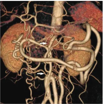
Computed tomographic angiography showed an abrupt segmental occlusion of infrarenal aorta (Fig. 1). Both renal arteries were intact. The abdominal aorta was reconstituted by multiple collaterals, such as marginal artery of Drummond, the anastomosis between superior mesenteric and inferior mesenteric artery, bilateral epigastric, lumbar, and abdomi- nal superficial arteries.
Operative findings revealed absence of infrarenal aorta repl- aced with fibrous band (Fig. 2). The pathology revealed fibr- omyxoid degeneration (Fig. 3). Polytetrafluoroethylene graft was interposed between the subdiaphragmatic thoracic aorta to the iliac bifurcation. The claudication resolved, and ankle- brachial index became 1.1. For her knee joint arthritis, she was started with methylprednisolon of 8 mg and non-steroid anti- inflammatory drug. After negative results of Pathergy test and human lymphocyte antigen B 51, we ruled out the Behcet’s disease and she tapered the medications to stop successfully.
She wanted the antihypertensive drug for mild hypertension and was taken amlodipine at 5 mg daily.
One year later, computer tomographic angiography showed patent bypass graft and decreased collateral vessels (Fig. 4).
DISCUSSION
The etiology of abdominal aortic stenosis is poorly under- stood. The young age of most of the patients suggests that the malformation may be congenital. Maycock first proposed an
950
Su-Ah Sung1, Young-Hwan Hwang1, So-Young Lee1, Young-Kwon Cho2, and Tae-Won Kwon3
Departments of Internal Medicine1, and Radiology2, Eulji General Hospital, Seoul; Division of Vascular Surgery3, Asan Medical Centre, Seoul, Korea
Address for Correspondence Su-Ah Sung, M.D.
Department of Internal Medicine, Eulji General Hospital, 14 Hangeulbiseok-gil, Nowon-gu, Seoul 139-872, Korea
Tel : +82.2-970-8205, Fax : +82.2-970-8621 E-mail : [email protected]
J Korean Med Sci 2010; 25: 950-2 ISSN 1011-8934
DOI: 10.3346/jkms.2010.25.6.950
An Infrarenal Aortic Hypoplasia Presented with Claudication
We describe a case of infrarenal aortic hypoplasia in a 52-yr-old woman who pre- sented with claudication. Computed tomographic angiography revealed an abrupt absence of the infrarenal aorta, with collateral flow reconstituting the iliofemoral sys- tems. After a polytetrafluoroethylene graft was interposed between the aortic stump and the iliac bifurcation, the patient’s claudication resolved.
Key Words : Aortic Coarctation; Intermittent Claudication
Received : 19 December 2008 Accepted : 30 March 2009
ⓒ 2010 The Korean Academy of Medical Sciences.
This is an Open Access article distributed under the terms of the Creative Commons Attribution Non-Commercial License (http://creativecommons.org/licenses/by-nc/3.0) which permits unrestricted non-commercial use, distribution, and reproduction in any medium, provided the original work is properly cited.
Infrarenal Aortic Hypoplasia 951
unequal fusion of embryonic aorta as a congenital mechani- sm (3). In first month of fetus, the paired primitive dorsal aortas fuse to form the abdominal aorta. Incomplete fusion due to infection or inflammation may lead of kinking of the aorta resulting to permanent constriction. Congenital rubel- la has been suggested for such vascular anomaly (4, 5). In Alagille syndrome which is congenital pulmonary artery stenosis transmitted in an autosomal dominant inheritance, deletion of the Jagged 1 gene has been suggested to relate with abdominal aortic coarctation (6). Acquired etiologies were also
suggested in fibromuscular dysplasia or Takayasu’s arthritis (7, 8). In our patients, several inflammatory presentations in- cluding the knee joint arthritis, recurrent oral ulcers, and ele- vation of ESR have suggested the possibility of Behcet’s dis- ease. Though, the pathologic study showed only fibromyxoid degeneration without any evidences of vasculitis. We ruled out the Behcet’s disease after the negative study of Pathergy test. Considering the complete absence of infrarenal aorta without any inflammatory reaction, congenital hypoplasia thought be most proposal cause in this patient.
In review of cases for abdominal aortic stenosis, coarctation, hypoplasia, and middle aortic syndrome were used diversely.
The term “coarctation”implies a congenital anomaly, and “hy- poplasia” is to longer segment of coarctation. Middle aortic syndrome is the clinico-anotomical entity involving long seg- mental narrowing of the abdominal aorta and various viscer-
Fig. 3. The microscopic image showed fibromyxoid degeneration (H&E stain; magnification×100,×400 in small box).
Fig. 4. Computed tomographic angiography showed a patent graft between thoracoabdominal junction to aortic bifurcation (arrows).
Fig. 1. Computed tomographic angiography showed an abrupt segmental occlusion of infrarenal aorta (white arrows). Both renal arteries were intact. The proximal and distal abdominal aorta were reconstituted by multiple collaterals, such as marginal artery of Drummond, the anastomosis between superior mesenteric and inferior mesenteric artery, bilateral epigastric, lumbar, and abdomi- nal superficial arteries.
Fig. 2. Operative findings revealed absence of infrarenal aorta replaced with fibrous band (white arrows). Inferior mesenteric artery (black arrow) reconstituted the abdominal aortic system.
Descending Thoracic Aorta
952 S.-A. Sung, Y.-H. Hwang, S.-Y. Lee, et al.
al arteries irrespective of its etiology. We described this case by “hypoplasia”, because 4 cm of infrarenal aorta was absent.
Presenting symptoms of abdominal aortic hypoplasia are related to the extent of the stenosis, and the collateral system.
In case involving renal artery, patients typically present with uncontrolled hypertension. Infrarenal aortic hypoplasia can give rise to claudication when the collateral circulation is not sufficient. An abdominal aortogram via angiography, ultra- sound, computed tomography, or magnetic resolution imag- ing can be used to diagnose and to determine the location and extent of disease. In case of renal artery stenosis, surgical inter- vention is aimed to treat severe hypertension and to preserve renal function. Medically treated patients have an average life expectancy of 32 yr (9). Claudication and aneurysmal dilata- tion also need surgical correction. In this case, as the subrenal aortic stump was too short, graft was interposed between the subdiaphragmatic thoracic aorta to the iliac bifurcation. Her hypertension seems not related to the aortic disease.
Claudication is not uncommon. Careful physical examina- tion for inspection of dilated superficial arteries and ausculta- tion of bruits can provide a clue for rare causes of ileofemoral insufficiency such as abdominal aortic stenosis. When suspect- ed, an angiography is mandatory to diagnose, and surgical in- tervention is necessary to improve severe claudication.
REFERENCES
1. Riemenschneider TA, Emmanouilides GC, Hirose F, Linde LM. Co- arctation of the abdominal aorta in children: report of three cases and review of the literature. Pediatrics 1969; 44: 716-26.
2. Fitzpatrick CM, Clouse WD, Eliason JL, Gage K, Podberesky DJ, Bu- sh DM. Infrarenal aortic coarctation in a 15-yr-old with claudication.
J Vasc Surg 2006; 44: 1117.
3. Maycock W. Congenital stenosis of the abdominal aorta. Am Heart J 1937; 13: 633.
4. Limbacher JP, Hill ME, Janicki PC. Hypoplasia of the abdominal ao- rta associated with rubella syndrome. South Med J 1979; 72: 617-9.
5. Siassi B, Klyman G, Emmanouilides GC. Hypoplasia of the abdomi- nal aorta associated with the rubella syndrome. Am J Dis Child 1970;
120: 476-9.
6. Raas-Rothschild A, Shteyer E, Lerer I, Nir A, Granot E, Rein AJ.
Jagged1 gene mutation for abdominal coarctation of the aorta in Alagille syndrome. Am J Med Genet 2002; 112: 75-8.
7. Vuong PN, Trinh AC, Lagneau P, Camilleri JP. Coarctation of the abdominal aorta and stenosis of the left renal artery with hypertension caused by fibrodysplasia. Arch Pathol Lab Med 1989; 113: 809-11.
8. Lande A. Takayasu’s arteritis and congenital coarctation of the des- cending thoracic and abdominal aorta: a critical review. AJR Am J Roentgenol 1976; 127: 227-33.
9. De Bakey ME, Garrett HE, Howell JF, Morris GC Jr. Coarctation of the abdominal aorta with renal arterial stenosis: surgical considera- tions. Ann Surg 1967; 165: 830-43.