INTRODUCTION
Tryptophan hydroxylase (TPH, EC 1.14. 16.4) is the first and the rate-limiting enzyme in 5-hydroxytryptamine (5- HT, serotonin) biosynthesis (1, 2). It catalyzes the conver- sion of L-tryptophan into 5-hydroxytryptophan in brain, pineal gland (PG), and enterochromaffin cells of gut. In the central nervous system serotonergic cell bodies are mainly localized in the raphe system and its terminals are spread throughout the central nervous system (3-6). Studies of TPH characteristics and regulation have been hampered partly because of the enzyme instability and partly due to the low abundance in animal tissues resulting in the low availability of purified enzyme protein. End product regula- tion of 5-HT biosynthesis at the rate-limiting step appeared to exist in vivo (7, 8) but even high concentrations of 5-HT did not inhibit TPH activity in vitro (9). Meanwhile TPH mRNA levels are greater in PG than in the dorsal raphe nucleus (DRN), while TPH activity is higher in DRN than in PG (10, 11). It has been also reported that brain TPH was immunochemically different from the mouse intestinal
TPH (peripheral enzyme) because antibodies against the mouse mastocytoma TPH immunoprecipitated TPH from intestine in a dose-dependent manner but did not signifi- cantly immunoprecipitate TPH from mouse brain (12). In addition, some biochemical characteristics of brain TPH have been reported to differ from pineal TPH in the previ- ous reports (11, 13). Although TPH was extensively puri- fied from rat brain and a mouse mastocytoma cell line, the quantity of purified enzyme protein was minimal (14-16).
Antiserum against rat brain TPH has been produced, but it also contained some antibodies to catalase (16). Therefore, extensive purification to remove contaminant catalase anti- bodies was required (16). The production of anti-peptide antibodies to rat TPH has also been reported (17) but these antibodies detect not only a TPH-like protein band on immunoblot but also three additional non-defined higher molecular weight bands from extracts of the rat raphe area.
Recently, recombinant mouse and rabbit TPH were suc- cessfully expressed in large quantities in bacteria, using bac- terial expression vectors and was shown to be enzymatically highly active (18, 19). Specific polyclonal antiserum to the
Young In Chung*, Dong Hwa Park, Myoungsoon Kim, Harriet Baker, Tong Hyup Joh
Department of Psychiatry*, Pusan National University Medical College, Pusan, Korea; Laboratory of Molecular Neurobiology, Cornell University Medical College, W.M. Burke Medical Research Institute, White Plains, New York 10605, U.S.A.
Address for correspondence Young In Chung, M.D.
Department of Psychiatry, Pusan National University Medical College, 1-10, Ami-dong, Seo-gu, Pusan 602-739, Korea
Tel : +82.51-240-7305, Fax : +82.51-248-3648 E-mail : [email protected]
489
Immunochemical Characterization of Brain and Pineal Tryptophan Hydroxylase
Recombinant mouse tryptophan hydroxylase (TPH) was expressed in Escheri- chia coli, using a bacterial expression vector and has been purified to homo- geneity by sonication followed by Sepharose 4B column chromatography and native slab gel electrophoresis. This purified enzymatically active TPH protein was used for production of a specific antiserum. This antiserum identified the predicted TPH band (molecular weight, 54 kDa) on Western blot of crude extracts from the rat and mouse dorsal raphe, and the rat pineal gland. Howev- er, this antiserum recognized an additional protein band of lower molecular weight (48 kDa) in pineal extract. It is not clear whether the 48 kDa TPH band represents an isozyme or a protease cleavage product of TPH. Since the pineal gland contains higher TPH mRNA and lower TPH activity when it is compared with dorsal raphe nucleus enzyme, this lower molecular weight TPH may partic- ipate in the reduced TPH specific activity. In addition, there are no specific TPH inhibitors in the pineal gland and this lower molecular weight TPH is inactive or has a very low specific activity. This antiserum specifically immunostained sero- tonergic cell bodies in the dorsal raphe nuclei, some large caliber serotonergic processes in the dorsal raphe area as well as terminals in the olfactory bulb. It also immunolabeled the pineal gland and immunoprecipitated equally well TPH protein from the dorsal raphe nucleus and the pineal gland in a concentration- dependent manner.
Key Words : Tryptophan Hydroxylase; Recombinant; Raphe Nuclei; Pineal Body; Immunochemistry
Received : 1 December 2000 Accepted : 26 March 2001
subunits of the recombinant mouse TPH (the holoenzyme consists of four identical subunits) has been produced in rabbit by injecting TPH band cut from SDS-polyacry- lamide gels (18). However, this antiserum, although specif- ic, had a major drawback for its use in immunocytochemi- cal localization of serotonergic neurons in brain due to some nonspecific immunostaining which perhaps resulted from usage of SDS-containing antigen for immunization.
TPH protein in rat brains is present in very small amounts (even in DRN) and very unstable due to the presence of small quantities of TPH protein during purification. The conventional many-step purification requires many rat brain tissues in addition to being difficult to maintain its enzyme activity. Also, the relative amount of TPH protein in brain tissue is minor compared to the other proteins pre- sent in the brain. Therefore, even if TPH preparation from one-step purification of Sepharose 4B column chromatogra- phy was applied to native gel electrophoresis, it is impossi- ble to detect TPH activity with a maximum load of pro- tein. On the other hand, the recombinant mouse TPH preparation after one-step purification of Sepharose 4B col- umn chromatography was enzymatically more active (434 nmol/mg protein/min at 37℃, 2.2 mg from 100 mL of culture) in our previous study (20) and contained more abundant TPH protein (one of the major protein bands is TPH protein in native slab gel electrophoresis) than puri- fied TPH (374 nmol/mg protein/min at 30℃, 19 g from 70 rat brain stems) reported by others (14). A large quanti- ty of enzymatically active recombinant mouse TPH prepa- ration can be easily produced using an expression vector containing the full coding region of the TPH cDNA just by growing culture (20).
In the present study, we describe a simple two-step proce- dure for purification of recombinant mouse TPH and pro- duction of immunocytochemically specific TPH antiserum to enzymatically active recombinant mouse TPH. Since we demonstrated that the coding sequence TPH cDNA from DRN and PG was identical, obviously the next question raised is whether TPH protein from both sources are the same or not. In order to examine the identity of TPH pro- tein from DRN and PG, we chose the commonly used techniques, i.e., immunocytochemical staining of central serotonergic neurons as well as pinealocytes, determination of molecular weight by Western blotting, and immunopre- cipitation of TPH protein by immunotitration with specific TPH antiserum, to immunochemically characterize TPH protein from both sources.
MATERIALS AND METHODS Expression of mouse TPH in Escherichia coli
We constructed a bacterial expression vector, pKSTPH,
containing the full coding sequence of mouse TPH (1.3 kb), as described elsewhere in detail (20). Briefly, the TPH cod- ing region was ligated into the EcoRI site of the plasmid, pKS6.8. pKS6.8, a vector containing a strong tac promoter, which can be regulated in bacteria by lac repressor, and which is inducible by isopropyl- -D-thiogalactoside (IPTG) (Gold Biotechnology, Inc., St. Louis, MO), was constructed by ligating a 1.5 kb DNA fragment from a PvuI, partial SalI digest of pKK233-3 (Pharmacia), with a PvuI/SalI digested 5.3 kb DNA fragment from PIN III A3 (21) gen- erously provided by Dr. M. Inouye (Department of Bio- chemistry, Rutgers University, NJ). This plasmid contains an intact lac I gene, which permits induction of the foreign gene by IPTG regardless of the genotype of the host E. coli strain. pKSTPH was transformed into E. coli strain MC 1061, inoculated into LB broth containing 100 g/mL ampicillin, and induced by IPTG. Cells were harvested by centrifuga- tion at 4,000 g for 20 min. The sediment was suspended in sonication buffer (5 mM Tris-HCl, pH 7.5, 10% glycerol, 50 M EDTA, 0.06% Tween 20, 2×10⁻⁵ M tryptophan, 1 mM phenylmethylsulfonyl fluoride, leupeptin (50 g/mL), 1% v/v aprotinin (Sigma Chemical Co., St. Louis, MO) and lysed by sonication for 30 sec in ice-water with the use of an ultrasonic model set (Vibra Cell, Sonics and Materials, Inc., Danbury, CT) at 60% of maximum output, followed by freeze-thawing twice and centrifuged at 16,000 g for 10 min.
The supernatant was used as a source of recombinant mouse TPH, unless otherwise specified. In case of antibody produc- tion the supernatant was partially purified by Sepha-rose 4B column (5×86 cm, equilibrated with 10 mM Tris-HCl buffer, pH 7.0, containing 2×10⁻⁵M tryptophan and 0.5 mM dithiothreitol) chromatography prior to subjection of TPH preparation to gel electrophoresis.
Enzyme and protein assay
TPH activity was determined by a minor modification of the method of Park et al. (18, 22), in which the conversion rate of L-[1-14C]tryptophan to 14CO2 was measured. Briefly, the assay mixture containing 25 mole of Tris-acetate buffer, pH 7.5, 112.5 nmol of DL-6-methyltetrahydropter- ine (Calbiochem., La Jolla, CA, U.S.A.), 600 nM of D,L- dithiothreitol, 5.1 nM of Fe (NH4)2(SO4)26H2O, 43 units of catalase (Sigma C-10), and 37.5 nM of L-[1-14C] trypto- phan (0.1 Ci) American Radiolabeled Chemicals, Inc., St.
Louis, MO in a total volume of 50 L was added to 100 L of enzyme solution. The mixture was incubated at 37℃for 15 min and then 50 L of solution containing partially purified hog kidney aromatic L-amino acid decarboxylase (30-50% ammonium sulfate fraction, the activity in 18 nM product formed/min at 37℃) and 50 nM of pyridoxal phosphate in H2O was added. The assay tube was then cov- ered by a rubber cap in which a plastic well containing a piece of filter paper soaked in 100 L of NCS II (Amersham)
was suspended. The mixture was further incubated at 37℃ for 30 min. The reaction was then stopped by injecting 100 L of 30% HClO4 and14CO2was trapped at 37℃for 30 min and counted with 15 mL of Econofluor (DuPont-NEN).
Activity was expressed as nmol of CO2formed per 15 min at 37℃. Protein concentration was measured according to Lowry et al. (23) using bovine serum albumin as standard.
Electrophoresis
Polyacrylamide gel electrophoresis was carried out by a minor modification to the standard procedure (24, 25) using 0.4 M glycine-Tris buffer, pH 8.2 in a 10% slab gel including 4% stacking gel (0.15 cm in thickness×14 cm in width×16 cm in length). Using partially purified recom- binant mouse TPH (2.78 mg protein loaded on each gel), electrophoresis was performed at 25 watts for 3 hr at 12℃, following 1 hr prerun. The gels were also stained for pro- teins with Coomassie blue R250. Following electrophoresis, the gels were cut consecutively (0.2 cm in length×0.15 cm in thickness×1 cm in width) and TPH activity measured as described above.
Production of Antiserum
Antibodies to recombinant mouse TPH were raised in rabbits by injecting the sections of the gels containing peak enzymatic activity. Active band containing about 200 g protein were cut and homogenized in 1 mL of 0.9% NaCl and an equal volume of complete Freund’s adjuvant. The resultant emulsion was injected subcutaneously at multiple sites on the back of rabbits. Booster injections of the anti- gen, using incomplete Freund’s adjuvant were given on days 43 and 64 and the animals were bled 1 week later.
Handling of rabbits, as well as rats and mice (see below) fol- lowed the guidelines of the Institutional Animal Care and Use Committee of the Cornell University Medical College.
Immunocytochemistry
For immunocytochemistry, rats and mice were anes- thetized with sodium pentobarbital (50 mg/kg, i.p.) and perfused transcardially with heparinized saline containing 0.5% sodium nitrite, followed by 0.1 M sodium phosphate buffer, pH 7.2, containing 4% formaldehyde generated from paraformaldehyde. The brain and pineal gland were post-fixed in 4% buffered formaldehyde for 1 hr, then rinsed in the buffer and placed in 30% sucrose at 4℃overnight.
Tissue sections of 30-40 m thickness were cut on a sliding microtome and collected in standard wells filled with 0.1 M sodium phosphate buffer. Tissue sections were incubated in 0.1 M sodium phosphate-buffered saline pH 7.4, contain- ing 0.2% Triton X-100 and 1% bovine serum albumin.
Tissues were washed in phosphate-buffered saline contain-
ing 0.5% bovine serum albumin and incubated overnight with a specific rabbit polyclonal antiserum to recombinant full-length TPH described above. The antiserum was used at a dilution of 1:10,000. A diluted 5-HT antiserum (1:30,000) was used for the purpose of comparison. Tissue was incubat- ed with biotinylated goat anti-rabbit IgG (Vector Laborato- ries, Burlingame, CA) for 1 hr and then treated with a Vecta Stain Elite Kit as per manufacturer’s direction. The antigen was visualized with 3,3′-diaminobenzidine-HCl (50 mg/
100 mL) and 0.005% hydrogen peroxide as a chromogen.
Sections were mounted onto gelatin-coated slides, dehydrat- ed through graded ethanols and coverslipped with Permount (Fisher Scientific).
Western blotting
Enzyme sources are 16,000 g supernatant of rat and mouse DRN and rat PG homogenate in 50 mM Tris-acetate buffer, pH 7.5, containing 0.5 mM dithiothreitol and recombinant mouse TPH preparation mentioned above. Western blotting was performed according to our minor modification (26) of the standard methods described by Towbin et al. (27) and Burnette (28). Briefly, proteins were subjected to gel elec- trophoresis using 10% SDS-polyacrylamide slab gels con- taining 4% stacking gel as described by Laemmli (29). Fol- lowing electrophoresis, the resolved proteins were trans- ferred at room temperature (200 mA for 3 hr) to Nitro- Screen West membrane (Dupont-New England Nuclear) in 192 mM glycine/25 mM Tris buffer, pH 8.3 containing 20%
methanol. The membrane was then incubated overnight in blocking buffer (150 mM NaCl, 50 mM Tris-HCl, pH 7.4 and 1% gelatin) to avoid nonspecific binding. The blot was rinsed with 150 mM NaCl, 50 mM Tris-HCl, pH 7.4, 5 mM EDTA, 0.25% gelatin and 5% normal goat serum and incubated for 2 hr with TPH antiserum described above, diluted (1:30,000 dilution for recombinant mouse TPH and 1:10,000 dilution for other sources of TPH) in rinsing buffer.
The blot was rinsed in the above buffer and then incubated for 2hr with a secondary antibody (peroxidase-conjugated goat anti-rabbit IgG (Kirkegaard and Perry Laboratories, Inc., Gaithersburg, MD) diluted (1:200) in rinsing buffer.
The antigen-antibody complexes were visualized with 0.05% 3,3′-diaminobenzidine containing 0.005% hydro- gen peroxide in 50 mM Tris-HCl buffer, pH 7.4.
Immunotitration
Increasing amounts of antiserum to recombinant mouse full-length TPH were added to the fixed quantity of enzyme to be titrated and the total volume was adjusted with preim- mune serum. The mixture was incubated for 1 hr at room temperature with occasional shaking and then for 16 hr at 4
℃. The antigen-antibody complex was removed by cen-
trifugation for 10 min at 16,000 g, and a 100- L aliquot of the supernatant was assayed for residual TPH activity (25).
RESULTS
Electrophoretic profile of recombinant mouse TPH
Crude recombinant mouse TPH (16,000 g supernatant, 103 mg protein) was subjected to Sepharose 4B column (5
×86 cm) chromatography, proteins were eluted with 10 mM Tris-HCl buffer, pH 7.0, containing 2×10⁻⁵M trypto- phan and 0.5 mM dithiothreitol, and the most enzymatical- ly active fractions were combined. Subsequently, the partial- ly purified recombinant mouse TPH subjected to slab gel electrophoresis with 10% gel at pH 8.2 showed a single enzymatically active peak located at 2.3 cm from the top of the separating gel (Fig. 1). This active peak overlapped with one major protein band stained with Coomassie blue. The distribution of enzyme activity (nM/15 min 37℃/section) and staining pattern of proteins on gel are shown in Fig. 1.
Western immunoblotting
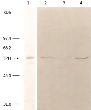
Western blot analysis was performed to determine whether the antiserum raised is specific and TPH proteins from the extracts of the rat and mouse DRN, the rat PG and recom- binant mouse TPH preparation are immunochemically identical. A specific rabbit polyclonal antiserum to recom- binant mouse TPH was used for this analysis. The TPH
antiserum recognizes an identical band with the same molecular weight (54 kDa) in all four TPH sources (Fig. 2).
This antiserum also detects an additional and equally abun- dant immunoreactive band with lower molecular weight in the extract of the rat PG. However, no immunoreactive band was detected with the preimmune serum (data not shown). Molecular weight was estimated by comparison to Coomassie blue stained marker proteins as indicated in the margin of Fig. 2. The main TPH band (MW 54 kDa) among these four sources is indicated by an arrow. Possibly because of the higher enzyme concentration of TPH in the recombinant enzyme preparations as compared to the ani- mal tissue extracts, a higher dilution of antiserum was ade- quate to detect the TPH band on Western blot.
Immunocytochemical localization of TPH
The new TPH antiserum localized TPH immunoreactive cells in the DRN. Cells were below the aqueduct and ven- tromedial area as well as in lateral portions of the DRN of rat and mouse (Fig. 3A and C). Additionally, moderate immunoreactivity was also observed in the medial raphe nuclei. Immunostaining in adjacent tissue sections of rat
TPH activity
5 4 3 2 1 0
0 1 2 3 4 5 6 7 8 9 10
Migration distance in centimeters
Fig. 1. Distribution of recombinant mouse TPH activity after native polyacrylamide gel electrophoresis. Partially purified enzyme preparation ( 2.78 mg protein) obtained from Sepharose 4B column chromatography was loaded on native slab gel (0.15 cm in thickness×14 cm in width×16 cm in length). Following electrophoresis as described in Materials and Methods, the gel was sliced into 0.2-cm sections (0.15 cm in thickness×1 cm in width×0.2 cm in length). The sections were assayed as described in Materials and Methods. The equivalent-sized gel section was stained for protein with Coomassie blue. Note that a single enzymatically active peak corresponding to a major stained protein band was observed. The activity was expressed in nM/15 min at 37℃/section.
1 2 3 4
kDa
97.4 66.2 TPH
45.0
31.0
Fig. 2. Western blots of rat and mouse brain, rat pineal and recombinant mouse TPH. Following SDS-polyacrylamide slab gel electrophoresis and subsequent transferring to a membrane, resolved proteins were analyzed as described in Materials and Methods with polyclonal antiserum to recombinant mouse TPH.
Note that an identical TPH band (indicated by an arrow) was detected with crude extract of recombinant mouse TPH (lane 1, 20 g), rat dorsal raphe nuclei (lane 2, 200 g), rat pineal gland (lane 3, 200 g), and mouse dorsal raphe nuclei (lane 4, 200 g).
An additional immunoreactive band with smaller molecular weight was also observed in the rat pineal gland.
DRN by 5-HT antiserum revealed a similar distribution of 5-HT immunoreactivity (Fig. 3B). Intense immunolabel- ing by TPH antiserum was also observed in the rat (Fig.
3D) and mouse PG (data not shown). Detailed immunos- taining patterns were illustrated in Fig. 3. As shown in the darkfield micrographs of TPH immunolabeling in Fig. 4, large caliber processes (Fig. 4A) can be seen in the DRN and only limited immunoreactive terminal fields (Fig. 4B) can be observed in the olfactory bulb.
Immunochemical crossreactivity
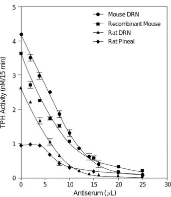
Antiserum directed against recombinant mouse TPH immunoprecipitated TPH protein from both rat and mouse DRN and recombinant mouse TPH preparations equally
well, judging from the nearly identical slopes of the immu- notitration curves (Fig. 5). Although a similar slope is obser- ved in the immunotitration curve for TPH from rat PG as compared to other sources, the low TPH activity in homo- genates from PG prevents a clear evaluation of the enzyme in this tissue. However, immunoprecipitation of TPH pro- teins from all four sources by the antiserum occurs in a con- centration-dependent fashion. TPH protein in all four cases was almost completely immunoprecipitated by addition of 25 L of the antiserum as shown in Fig. 5.
Effect of endogenous substances on TPH activity
In order to determine whether endogenous substances in the rat tissues inhibit TPH activity, assays of recombinant
A B C D
Fig. 3.Brightfield photomicrographs of TPH and serotonin immunostaining in dorsal raphe and pineal gland. Aand C, TPH immunore- activities in the rat and mouse dorsal raphe, respectively; B, serotonin immunoreactivity in the rat dorsal raphe; D, TPH immunoreac- tivity in the rat pineal gland. Note the similarity in the distribution of cells immunolabeled with antisera to TPH and serotonin (A, B, C).
Bar=300 m.
A B
Fig. 4. Darkfield photomicrographs of TPH immunostaining in the dorsal raphe nucleus (DRN) and the olfactory bulb (OB). In the DRN large caliber processes (A) can be demonstrated as they either course towards the ependyma or innervate blood vessels. In the termi- nal fields, such as the OB (B), only limited TPH immunoreactivity is apparent suggesting a low abundance of TPH protein. Bar=80 m.
mouse TPH were carried out in the presence of either DRN or PG extracts from rat. As indicated in Table 1, addition of PG extracts to recombinant mouse protein led to no alter- ation in TPH activity, while DRN extracts produced a small increase of recombinant mouse TPH activity. The data do not indicate that the PG contains a TPH inhibitor.
Effect of end-products on TPH activity
It has been reported that rat TPH is inhibited in vivo by an end-product, 5-HT (7, 8). In the present study using 7.5
×10-4M 6MPH4as the cofactor and 2.5×10⁻⁴M trypto- phan as the substrate, additions of various concentrations of end-products, either 5-HT, N-acetyl-5-hydroxytryptamine or melatonin were tested in our assay conditions for possible inhibition of enzyme activity in vitro. In contrast to the in vivo findings, TPH activity exhibited minimal inhibition in vitro with any of the above products, even when the con- centration was as high as 5×10⁻⁴M. The data are not shown here but all the experimental values were within a few per- centage of the control value.
DISCUSSION
TPH is highly labile during purification and storage, as a result of its very low tissue concentration (14-16, 20, 30).
Because of its instability and low abundance, previous puri- fication methods that involved numerous steps could not successfully produce large amounts of enzymatically active TPH protein from brain tissues (14, 16). Recently, we expressed abundant amounts of enzymatically active recom- binant mouse TPH in E. coli, using a bacterial expression vector, pKSTPH, containing the full coding region of mouse TPH. Subsequently, a specific antiserum to the sub- units of the recombinant mouse TPH was produced by injecting the TPH band cut from SDS-polyacrylamide slab gels (20). This antiserum detected a single band of predict- ed molecular weight for TPH from rat DRN, and PG by Western blotting. However, this antiserum immunostained not only TPH-containing neurons but also some nonspecif- ic staining was observed, perhaps because the antiserum was raised to TPH protein containing SDS. In the present study we described a simple procedure to produce large quantities of recombinant mouse TPH purified to an apparent homo- geneity by a two-step purification method, Sepharose 4B column chromatography and non-denaturing gel electro- phoresis. Subsequently, we produced a specific antibody to recombinant mouse TPH using this purified enzyme pro- tein. To our knowledge, this is the first report of the pro- duction and characterization of antibody to TPH using recombinant full-length mouse TPH, which immunochemi- cally as well as immunohistochemically recognizes native mouse and rat TPH from the brain as well as the PG. The specificity of this TPH antiserum was determined by three independent methods, Western blot analysis, immunostain- ing and immunoprecipitation.
In our Western blot analysis, this specific antiserum rec- ognized an identical protein band equivalent to molecular weight of 54,000 Dalton in crude extracts of rat and mouse DRN, rat PG as well as recombinant mouse TPH prepara- tions. This molecular weight corresponds to the one report- ed previously by others (14-16, 20) for native TPH from rat
Recombinant TPH 1.58±0.035
TPH from DRN 1.61±0.014
TPH from PG 3.22±0.028
Recombinant TPH+TPH from DRN 3.73±0.020 Recombinant TPH+TPH from PG 4.74±0.281
Enzyme Source TPH Activity
Table 1.Effect of rat DRN and PG extracts on recombinant mouse tryptophan hydroxylase activity
TPH activity (nM/15 min at 37℃±SEM) was determined in triplicate.
Enzyme source: Dorsal raphe nucleus and pineal gland extracts were 16,000 g supernatant and recombinant TPH was partially purified by Sepharose 4B column chromatography
Fig. 5.Immunochemical titration curves for TPH from rat and mouse DRN, recombinant mouse TPH and rat pineal gland using antiserum to recombinant mouse TPH. Immunotitration was performed as described in Materials and Methods. The slopes of immunotitration curves for TPH from all four sources are similar and immunoprecipitation of TPH proteins by the anti- serum occurs in a concentration-dependent manner. TPH activi- ty was measured in triplicate. Each point represents the mean of three separate experiments±SEM.
TPH Activity (nM/15 min)
5
4
3
2
1
0
0 5 10 15 20 25 30
Antiserum ( L) Mouse DRN Recombinant Mouse Rat DRN
Rat Pineal
brain, mouse mastocytoma, and recombinant mouse TPH.
However, an additional lower molecular weight band was also detected only in the crude extract of rat PG and the intensity of the two bands appeared almost equal. The pres- ence of this additional enzyme protein band in the PG extract was not detected using any of the previously report- ed TPH antisera. Detection of TPH on Western blot from rat and mouse DRN and rat PG extracts required loading of more protein and less dilution of the antiserum, com- pared with in the case of recombinant mouse TPH prepara- tion. The differences can be attributed to the very low level of TPH protein in the crude tissue extracts as compared with the recombinant TPH preparation.
Earlier reports described the production of antiserum to purified rat TPH but this antiserum also contained anti- catalase antibodies (10, 16) in addition to anti-TPH anti- bodies. Thus, prior to use this antiserum had to be purified to remove anti-catalase antibodies by column chromatogra- phy. In addition, their affinity-purified TPH antibody (10) reportedly recognized tyrosine hydroxylase protein in the PG extracts, but detected only one TPH band from extracts of rat raphe on Western blot. This antiserum was produced against native rat TPH protein which may fold differently from recombinant full-length mouse TPH. Thus, the spe- cific epitope recognizing the lower molecular weight band may be hidden. Recently, the production of anti-peptide antibodies to rat TPH was reported. These antibodies detected one principal (TPH protein band judging from its size) and three additional higher molecular weight bands from extracts of rat raphe areas on immunoblot (17) unlike our TPH antiserum used in this study, i.e., TPH antiserum directed against the subunits of recombinant mouse TPH (20) and the affinity-purified rat TPH antibodies (16).
Therefore, it seems that the peptide antiserum contains additional non-defined antibodies that interact with anti- gens other than TPH protein. They have not tested whether these anti-peptide antibodies to TPH crossreact with the pineal TPH. Unlike the new polyclonal antiserum to re- combinant full-length mouse TPH, our previously reported polyclonal antiserum to the subunits of the recombinant mouse TPH (20) also detected only a single predicted molec- ular weight (54 kDa) band for TPH from rat DRN as well as PG by Western blotting. Therefore, the antiserum to recombinant full-length mouse TPH likely contains an antibody to a different TPH epitope, which perhaps result- ed from the differences in protein folding. Thus, the follow- ing possibilities must be taken into consideration with respect to this tissue-specific presence of the lower molecu- lar weight band on Western blot. Firstly, it seems unlikely that this antiserum contains an antibody against a minor contaminant protein since the crude DRN extract does not show an equivalent lower molecular weight band. Also, the relative abundance of the two bands in the crude PG extract is equal and this antiserum is also immunohistochemically
specific as described below.
Secondly, TPH mRNA levels are greater in PG than DRN, while enzyme activity is higher in the DRN. This difference is possibly due to the presence of an endogenous activator or inhibitor in the DRN and the PG. In order to verify this possibility, TPH activity assays of partially purified recom- binant mouse TPH preparation were carried out in the pre- sence of a crude extract either from the rat DRN or from the rat PG. The results indicated the absence of either an activator or an inhibitor. Therefore, another explanation must be found for the differences in activity observed in these tissues. The in vivo inhibition of TPH activity by an end product, 5-HT has been reported (7, 8), but addition of up to 5×10⁻⁴M of end-products, either serotonin, N-acetyl- 5-hydroxytryptamine, or melatonin did not affect in vitro TPH activity in our assay conditions. These findings were consistent with the observations by Youdim et al. (9) with 5-HT in vitro. Therefore, the difference in the TPH activity of the DRN and the PG may not originate from end-prod- uct inhibition. If the lower molecular weight band from the PG represents another form of TPH, it is likely to be inac- tive, since only one enzymatically active peak is ob-served by chromatofocusing column chromatography of the crude PG extract (11, 20). As mentioned above, we are temp-ted to speculate that the lower molecular weight band, if it repre- sents a less or inactive form of TPH from the PG, may be responsible for the difference in the TPH activity. However, the nature of the additional band and its role in the lowered PG TPH activity remains to be resolved.
The TPH antiserum used in this study intensively immu- nostained serotonergic neurons located in dorsomedial, ven- tromedial as well as lateral portions of the rat and mouse DRN and the rat PG. These immunocytochemical distri- bution patterns of TPH-immunopositive cells in the DRN are in good agreement with the localization of serotonin- immunopositive cells in the same areas as illustrated in Fig.
3A, B, and C. The intensity of serotonin-immunoreactivity in the DRN appears stronger than that for TPH-immunos- taining. This difference may be due to the differences in the concentration of 5-HT and TPH protein present in the cells. These results are in good agreement with previous observations made by others, using affinity-purified TPH- antibodies (6, 10), anti-peptide TPH antibodies (17), sero- tonin-antibodies (4, 5, 11) or formaldehyde-induced fluo- rescence histochemistry (31). Our TPH antiserum also immunostained rat and mouse PG, consistent with previ- ous studies (10), while antibodies to peripheral TPH did not recognize brain TPH (12). The TPH antiserum used in this study also immunostained large caliber processes in the DRN and terminals in the olfactory bulb. The relative inef- fectiveness of this TPH antiserum for immunostaining sero- tonergic processes and terminals may be attributed to a lower concentration of TPH protein compared with the concentra- tion of 5-HT in processes and terminals.
The TPH antiserum described in the present study appears to equally immunoprecipitate TPH protein from all four sources used in a concentration-dependent manner. The immunocrossreactivity between species and tissues was expected from the almost identical amino acid sequences deduced from nucleotide sequences (11, 32-34).
In summary, we have purified large quantities of recom- binant mouse TPH by a simple two- step purification pro- cedure and subsequently produced a specific antiserum using this purified TPH. This antiserum recognizes one pre- dicted TPH protein band (54,000 Dalton) on immunoblot of crude extracts from rat and mouse dorsal raphe, and rat PG and also detect an additional lower molecular weight band only from the pineal extract. This lower band may be involved in the pineal low TPH activity despite its higher mRNA levels since there is no evidence for an endogenous inhibitor in PG or for end-product inhibition. Further stud- ies are needed to characterize the nature of the lower molec- ular weight band observed in the PG extract in view of its low TPH activity. This TPH antiserum specifically immu- nostained serotonergic neurons of rat and mouse dorsal raphe and some serotonergic processes in the DRN and ter- minals in the olfactory bulb as well as immunolabeling rat and mouse PG. It also immunoprecipitated TPH protein from the DRN and the PG in a concentration-dependent fashion. These data indicate that TPH protein from DRN and PG appears immunochemically identical as expected from the identical coding sequence of TPH cDNA from DRN and PG.
ACKNOWLEDGMENTS
This work was supported by NIH Grant MH 44043.
We would like to express our gratitude to Ms. Nan Min for her technical assistance in tryptophan hydroxylase immu- nostaining and Mr. Charles Carver for his outstanding art- work in preparing the figures and the photographs.
REFERENCES
1. Grahame-Smith DG. Tryptophan hydroxylation in brain. Biochem Biophys Res Commun 1964; 16: 586-92.
2. Jequier E, Lovenberg W, Sjoerdsma A. Tryptophan hydroxylase inhibition: The mechanism by which p-chlorophenylalanine depletes rat brain serotonin. Mol Pharmacol 1967; 3: 274-8.
3. Joh TH, Shikimi T, Pickel VM, Reis DJ. Brain tryptophan hydroxy- lase: Purification of, production of antibodies to, and cellular and ultrastructural localization in serotonergic neurons of rat midbrain.
Proc Natl Acad Sci USA 1975; 72: 3575-9.
4. Steinbusch HWM. Distribution of serotonin-immunoreactivity in the central nervous system of the rat cell bodies and terminals. Neu- roscience 1981; 6: 557-618.
5. Ishimura K, Takeuchi Y, Fujiwara K, Tominaga M, Yoshioka H, Sawada T. Quantitative analysis of the distribution of serotonin- immunoreactive cell bodies in the mouse brain. Neurosci Lett 1988;
91: 265-70.
6. Weissmann D, Belin MF, Aguera M, Meunier C, Maitre M, Cash CD, Ehret M, Mandel P, Pujol JF. Immunohistochemistry of trypto- phan hydroxylase in the rat brain. Neuroscience 1987; 23: 291-304.
7. Macon JB, Sokoloff L, Glowinski J. Feedback control of rat brain 5-hydroxytryptamine synthesis. J Neurochem 1971; 18: 323-31.
8. Hamon M, Bourgoin S, Morot-Gaudry Y, Glowinski J. End product inhibition of serotonin synthesis in the rat striatum. Nat New Biol 1972; 237: 184-7.
9. Youdim MBH, Hamon M, Bourgoin S. Properties of partially puri- fied pig brain stem tryptophan hydroxylase. J Neurochem 1975; 25:
407-14.
10. Dumas S, Darmon MC, Delort J, Mallet J. Differential control of tryptophan hydroxylase expression in raphe and in pineal gland:
Evidence for a role of translation efficiency. J Neurosci Res 1989;
24: 537-47.
11. Kim KS, Wessel TC, Stone DM, Carver CH, Joh TH, Park DH.
Molecular cloning and characterization of cDNA encoding trypto- phan hydroxylase from rat central serotonergic neurons. Mol Brain Res 1991; 9: 277-83.
12. Hasegawa H, Yanagisawa M, Inoue F, Yanaihara N, Ichiyama A.
Demonstration of non-neural tryptophan 5-mono-oxygenase in mouse intestinal mucosa. Biochem J 1987; 248: 501-9.
13. Jequier E, Robinson DS, Lovenberg W, Sjoerdsma A. Further stud- ies on tryptophan hydroxylase in rat brainstem and beef pineal.
Biochem Pharmacol 1969;18: 1071-81.
14. Nakata H, Fujisawa H. Purification and properties of tryptophan 5- monooxygenase from rat brain-stem. Eur J Biochem 1982; 122: 41-7.
15. Nakata H, Fujisawa H. Tryptophan 5-monooxygenase from mouse mastocytoma P815: A simple purification and general properties.
Eur J Biochem 1982; 124 : 595-601.
16. Cash CD, Vayer P, Mandel P, Maitre M. Tryptophan 5-hydroxy- lase: Rapid purification from whole rat brain and production of a specific antiserum. Eur J Biochem 1985; 149: 239-45.
17. Azmitia EC, Liao B, Chen Y. Increase of tryptophan hydroxylase enzyme protein by dexamethasone in adrenalectomized rat mid- brain. J Neurosci 1993; 13: 5041-55.
18. Park DH, Stone DM, Baker H, Kim KS, Joh TH. Early induction of rat brain tryptophan hydroxylase (TPH) mRNA following parachlo- rophenylalanine (PCPA) treatment. Mol Brain Res 1994; 22: 20-8.
19. Vrana KE, Rucker PJ, Kumer SC. Recombinant rabbit tryptophan hydroxylase is a substrate for cAMP-dependent protein kinase. Life Sci 1994; 55: 1045-52.
20. Park DH, Stone DM, Kim KS, Joh TH. Characterization of recom- binant mouse tryptophan hydroxylase expressed in Escherichia coli.
Mol Cell Neurosci 1994; 5: 87-93.
21. Ghrayeb J, Kimura H, Takahara M, Hsiung H, Masui Y, Inouye M.
Secretion cloning vectors in Escherichia coli. EMBO J 1984; 3:
2437-42.
22. Park DH, Park HS, Joh TH, Anwar M, Ruggiero DA. Strain differ- ences between albino and pigmented rats in monoamine-synthesiz-
ing enzyme activities of brain, retina and adrenal gland. Brain Res 1990; 508: 301-4.
23. Lowry OH, Rosebrough NJ, Farr AL, Randall RJ. Protein measure- ment with the Folin phenol reagent. J Biol Chem 1951; 193: 265-75.
24. Davis BJ. Disc electrophoresis II. Method and application to human serum proteins. Ann NY Acad Sci 1964; 121: 404-27.
25. Park DH, Baetge EE, Kaplan BB, Albert VR, Reis DJ, Joh TH. Dif- ferent forms of adrenal phenylethanolamine N-methyltransferase:
Species-specific posttranslational modification. J Neurochem 1982;
38: 410-4.
26. Park DH, Kim KT, Choi MU, Samanta H, Joh TH. Characteriza- tion of bovine aromatic L-amino acid decarboxylase expressed in a mouse cell line: Comparison with native enzyme. Mol Brain Res 1992; 16: 232-8.
27. Towbin H, Staehelin T, Gordon J. Electrophoretic transfer of pro- teins from polyacrylamide gels to nitrocellulose sheets: Procedure and some applications. Proc Natl Acad Sci USA 1979; 76: 4350-4.
28. Burnette WN.“Western blotting”: electrophoretic transfer of pro- teins from sodium dodecyl sulfate-polyacrylamide gels to unmodi- fied nitrocellulose and radiographic detection with antibody and
radioiodinated protein A. Anal Biochem 1981; 112: 195-203.
29. Laemmli UK. Cleavage of structural proteins during the assembly of the head of bacteriophage T4. Nature 1970; 227: 680-5.
30. Nukiwa T, Tohyama C, Okita C, Kataoka T, Ichiyama A. Purifica- tion and some properties of bovine pineal tryptophan 5-monooxyge- nase. Biochem Biophys Res Commun 1974; 60: 1029-35.
31. Dahlstrom A, Fuxe K. Evidence for the existence of monoamine- containing neurons in the central nervous system. I. Demonstration of monoamines in the cell bodies of brainstem neurons. Acta Physi- ol Scand 1964; 62: Suppl 232: 1-55.
32. Grenett HE, Ledley FD, Reed LL, Woo SLC. Full-length cDNA for rabbit tryptophan hydroxylase: Functional domains and evolution of aromatic amino acid hydroxylases. Proc Natl Acad Sci USA 1987; 84: 5530-4.
33. Darmon MC, Guibert B, Leviel V, Ehret M, Maitre M, Mallet J.
Sequence of two mRNAs encoding active rat tryptophan hydroxy- lase. J Neurochem 1988; 51: 312-6.
34. Stoll J, Kozak CA, Goldman D. Characterization and chromosomal mapping of a cDNA encoding tryptophan hydroxylase from a mouse mastocytoma cell line. Genomics 1990; 7: 88-96.