www.labmedonline.org 223 eISSN 2093-6338
mosome) 등이 포함된다[4-6]. 이로 인해 세포 증식, 분화 및 생존 등에 중요한 역할을 하는 유전자가 손상되면 악성종양이 발생할 수 있다[7, 8].
본 증례는 난소암 환자에서 화학요법 제제(특히 cisplatin, eto- poside) 투여로 인해 말초혈액 림프구의 염색체 또는 염색분체의 불안정성이 일시적으로 관찰되어 보고하고자 한다.
증 례
13세 여자가 한 달간 지속되는 복부팽만으로 내원하였다. 초음 파검사와 자기공명영상검사에서 우측 난소에 거대한 종양이 발견 되었다. 혈액검사에서는 AFP 수치가 34,000 ng/mL(참고치, 0-7 ng/mL), CA-125 수치가 50 U/mL (참고치, 0-35 U/mL)로 상승된 것 이외에는 특이소견이 없었다.
환자는 우측 난관난소절제술을 시행받았으며, 병리학적으로 악 성 혼합배아세포종양(95% 난황낭종양, 5% 미분화세포종)으로 판 명되었다. 화학요법은 5일간 매일 cisplatin 20 mg/m2, etoposide 100 mg/m2을 정맥주사하였고, 둘째날에만 추가로 bleomycin 15 mg을 근육주사하였다. 체질성 성염색체이상으로 인한 생식샘종양 등을 배제하기 위해 말초혈액을 이용한 핵형검사가 의뢰되었는데, cispl-
서 론
염색체불안정성(chromosomal instability)은 각종 암에서 나타 날 수 있는 현상이고, 체질적 염색체절단증후군이나 화학물질 및 전리 방사선 노출 후 등에서도 관찰될 수 있다[1-4]. 염색체 불안정 성의 종류에는 염색분체의 손상(break), 틈(gap) 또는 자매염색분 체교환(sister chromatid exchange), 4 방사상의 구조(tetraradial structures) 및 염색체의 손상, 틈, 고리염색체(ring chromosome), 표지염색체(marker chromosome), 두매듭염색체(dicentric chro-
난소암 환자에서 화학요법에 의한 말초혈액 림프구의 염색체 불안정성
Chromosomal Instability in the Peripheral Blood Lymphocytes of an Ovarian Cancer Patient Undergoing Chemotherapy
오세진1·문혜성2·허정원1
Se Jin Oh, M.D.
1, Hye-Sung Moon, M.D.
2, Jungwon Huh, M.D.
1이화여자대학교 의학전문대학원 진단검사의학과1, 산부인과2
Departments of Laboratory Medicine
1, Obstetrics and Gynecology
2, Ewha Womans University School of Medicine, Seoul, Korea
증례
Lab Med Online
Vol. 2, No. 4: 223-225, October 2012
http://dx.doi.org/10.3343/lmo.2012.2.4.223 진단유전학
Corresponding author: Jungwon Huh, M.D.
Department of Laboratory Medicine, Ewha Womans University School of Medicine, 1071 Anyangcheon-ro, Yangcheon-gu, Seoul 158-710, Korea Tel: +82-2-2650-5287, Fax: +82-2-2650-5091, E-mail: [email protected] Received: January 6, 2012
Revision received: February 1, 2012 Accepted: April 19, 2012
This article is available from http://www.labmedonline.org 2012, Laboratory Medicine Online
This is an Open Access article distributed under the terms of the Creative Commons Attribution Non-Commercial License (http://creativecommons.org/licenses/by-nc/3.0/) which permits unrestricted non-commercial use, distribution, and reproduction in any medium, provided the original work is properly cited.
Chemotherapy agents can induce chromosomal instability, including a variety of chromatid or chromosomal aberrations. However, only limited data is available on the effect of chemotherapy on the kinetics of chromosomal instability in peripheral blood lymphocytes. Here, we report the case of an ovarian cancer patient who showed chromosomal instability in peripheral blood lymphocytes while undergoing chemotherapy. Karyotypic analy- sis of peripheral blood 1 day after administration of cisplatin and etoposide showed chromosomal or chromatid aberrations, including gaps, breaks, and fragmentation. Chromosome study after completion of the first chemotherapy cycle showed normal karyotype. This finding suggests that che- motherapeutic agents can induce transient chromosomal instability in peripheral blood lymphocytes.
Key Words: Chromosome, Chemotherapy, Ovarian cancer, Peripheral blood, Instability
오세진 외: Chromosome Instability with Chemotherapy
http://dx.doi.org/10.3343/lmo.2012.2.4.223 224 www.labmedonline.org
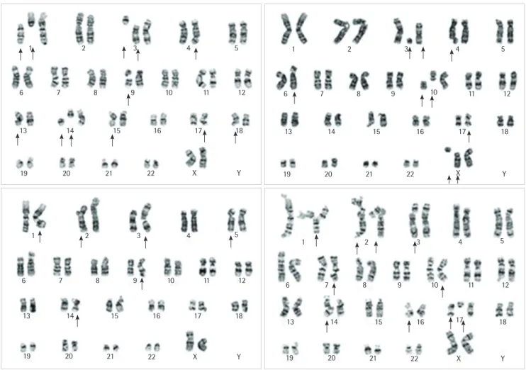
atin과 etoposide를 하루 투약한 후(bleomycin이 투약되기 전)에 시행되었다. 염색체검사는 전통적 단기배양(PHA 자극 72시간 배 양)법에 의해 시행되었다. 분석한 20개의 중기세포 중 90%에서 염 색분체 및 염색체의 손상, 틈, 조각 등을 포함한 이상이 관찰되었 다(Fig. 1). 이러한 이상은 특정한 염색체에 한정되지 않았고 특정 한 패턴의 이상에 국한되지 않았다. 첫 번째 화학요법이 끝난 19일 후(두 번째 화학요법이 시작되기 전), 핵형검사를 다시 시행하였는 데 결과는 정상으로 이전에 보였던 염색체이상은 발견되지 않았 다. 환자는 그 후 계속 치료를 받았으며 추적 관찰중이다.
고 찰
본 증례에서는 난소암 환자에서 화학요법에 의해 말초혈액 림프 구의 염색체 또는 염색분체의 이상이 일시적으로 관찰되었다. 본
환자의 경우 염색체절단증후군 환자에서 보이는 선천기형 등의 임 상양상이 나타나지 않았고 특정 염색체에 국한된 이상이 아닌 전 체 염색체에 걸친 무작위 이상이 관찰되었으므로 염색체절단증후 군을 배제할 수 있었다.
동물연구에 따르면, 쥐의 말초혈액 림프구에서도 약물 유도성 염색체이상이 보고되었다[9]. 염색체이상은 투여된 약물의 종류와 관계없이 약물 투여 후 3시간째에 가장 심하였고 치료 3일내에 정 상으로 회복되었다고 보고하였다[9]. 또한 유방암 환자들에서도 화학요법을 받는 중에 본 연구와 유사하게 다양한 염색분체 또는 염색체의 이상이 관찰되었다고 보고하였다[6]. 화학요법을 받기 전 에 비해 첫 번째와 네 번째 화학요법을 받는 사이에 염색체이상을 가진 세포수가 증가하였으며, 다섯 번째 화학요법 이후에는 정상 으로 회복되는 경향을 보였다고 하였다[6].
또한 폐암 환자와 난소암 환자에서, 소핵(micronuclei)이 첫 번째
Fig. 1. G-banded karyogram 1 day after chemotherapy (cisplatin and etoposide) showing chromosomal instability, including chromatid gap (chtg)
or break (chtb), chromosomal gap (chrg) or break (chrb), and fragmentation.Chtg, region of a single chromatid with minimal misalignment of the chromatid; chtb, discontinuity of a single chromatid with clear misalignment of one of the chromatids; chrg, a region at the same locus in both chromatids of a single chromosome with minimal misalignment of the chroma- tids; chrb, discontinuity at the same locus in both chromatids of a single chromosome, which gives rise to an acentric fragment and an abnormal monocentric chromosome.
1
1 1
2 1
2 2
2
6
6 6
6
13
13 13
14 13
14 14
15 14
15 15
16 15
16 16
17 16
17 17
18 17
18 18
18
19
19 19
20 19
20 20
21 20
21 21
22 21
22 22
X 22
X X
X Y
Y Y
Y 7
7 7
8 7
8 8
9 8
9 9
10 9
10 10
10 11
11 11
12 11
12 12
12 3
3 3
4 3
4 4
5 4
5 5
5
오세진 외: Chromosome Instability with Chemotherapy
http://dx.doi.org/10.3343/lmo.2012.2.4.223 www.labmedonline.org 225
화학요법 중 관찰되었고 두 번째 또는 세 번째 화학요법 때 가장 많 이 관찰되었으며 그 후에는 감소되었다는 연구가 있었다[10]. 소핵 의 존재는 체내 독성물질 노출 시 유전독성여부를 알 수 있는 유 용한 표지자 중 하나이다[10]. 본 증례에서는 화학요법 제제가 염색 체 또는 염색분체이상을 일으켰을 것으로 추정할 수 있는데, 이는 화학요법 제제 투여 후 하루 뒤 시행한 염색체 검사에서는 이상이 있었으나 화학요법 완료 후 시행한 염색체 검사에서는 정상소견을 보였기 때문이다. 이처럼 화학요법이 염색체의 이상을 초래할 수 있으므로[1, 5, 6] 염색체 검사는 반드시 화학요법 제제를 투여하기 전에 시행해야 할 것으로 생각된다. 염색체 또는 염색분체이상이 소멸된 요인으로는 염색체의 생리적 복구, 효과적인 DNA 복구기 전으로 인한 손상의 제거, 또는 치명적으로 파괴된 세포들의 자멸 등을 생각해 볼 수 있다[9].
염색체 불안정성의 발생기전은 DNA 복구, 복제 및 재조합 이상 등과 관련이 있을 것으로 생각된다[1, 6]. 염색체절단증후군에서는 DNA 복구에 결함이 있는데, 이로 인해 염색체 불안정성이 관찰되 고, 다양한 암 발생과 관련이 있다[11]. 또한 화학요법 제제가 염색 체 불안정성을 초래하는 기전은 확실히 밝혀져 있지 않으나 DNA 손상 및 DNA 복구능력의 감소를 유발해 염색체 불안정성을 야기 할 수 있을 것으로 생각된다. 염색체 불안정성이 중요한 유전자에 위치할 경우, 휴지기의 유전자 발현을 유도하거나 종양억제유전자 를 불활성화시켜서 암의 발생을 유도할 수 있다.
결론적으로, 본 증례는 화학요법 제제(cisplatin, etoposide)에 의 한해 일시적인 염색체 및 염색분체의 이상이 발생한 증례이다 이 는 화학요법 제제가 말초혈액 림프구에 끼치는 영향을 보여주었다.
요 약
화학요법 제제는 염색분체 및 염색체의 다양한 이상 등 염색체 의 불안정성을 야기할 수 있는데, 현재까지 화학요법이 말초혈액 림프구의 염색체불안정성을 초래하는 기전은 확실히 밝혀져 있지 않다.
본 증례에서는 난소암 환자에서 화학요법 치료 중 말초혈액 림 프구의 염색체불안정성이 관찰되었는데, cisplatin과 etoposide 투 여 후 하루 뒤 시행한 말초혈액의 핵형분석에서 손상, 틈, 조각 등 을 포함한 염색체 또는 염색분체의 이상이 관찰되었다. 첫 번째 화 학요법이 끝난 후 핵형분석을 다시 시행한 결과 정상소견이 관찰 되었다. 이러한 소견은 화학요법 제제로 인해 말초혈액 림프구의 일시적인 염색체 불안정성이 야기된 것으로 생각된다.
참고문헌
1. Martínez-López W, Folle GA, Cassina G, Méndez-Acuña L, Di-Tomaso MV, Obe G, et al. Distribution of breakpoints induced by etoposide and X-rays along the CHO X chromosome. Cytogenet Genome Res 2004;104:182-7.
2. Thompson SL, Bakhoum SF, Compton DA. Mechanisms of chromo- somal instability. Curr Biol 2010;20:285-95.
3. Lingle WL, Lukasiewicz K, Salisbury JL. Deregulation of the centro- some cycle and the origin of chromosomal instability in cancer. Adv Exp Med Biol 2005;570:393-421.
4. Fucic A, Jazbec A, Mijic A, Seso-Simic D, Tomek R. Cytogenetic conse- quences after occupational exposure to antineoplastic drugs. Mutat Res 1998;416:59-66.
5. Resende PA, Fidalgo C, Alves PM, Tavares-Murta BM, Murta EF, Dias FL. Analysis of the cytogenetic response in peripheral blood lympho- cytes from breast cancer patients following chemotherapy. Eur J Gyn- aecol Oncol 2010;31:75-9.
6. Sánchez-Suárez P, Ostrosky-Wegman P, Gallegos-Hernández F, Peñar- roja-Flores R, Toledo-García J, Bravo JL, et al. DNA damage in periph- eral blood lymphocytes in patients during combined chemotherapy for breast cancer. Mutat Res 2008;640:8-15.
7. Hagmar L, Brøgger A, Hansteen IL, Heim S, Högstedt B, Knudsen L, et al. Cancer risk in humans predicted by increased levels of chromo- somal aberrations in lymphocytes: Nordic study group on the health risk of chromosome damage. Cancer Res 1994;54:2919-22.
8. Rossner P, Boffetta P, Ceppi M, Bonassi S, Smerhovsky Z, Landa K, et al. Chromosomal aberrations in lymphocytes of healthy subjects and risk of cancer. Environ Health Perspect 2005;113:517-20.
9. Rosselli F, Zaccaro L, Venturi M, Rossi AM. Persistence of drug-induced chromosome aberrations in peripheral blood lymphocytes of the rat.
Mutat Res 1990;232:107-14.
10. Padjas A, Lesisz D, Lankoff A, Banasik A, Lisowska H, Bakalarz R, et al. Cytogenetic damage in lymphocytes of patients undergoing ther- apy for small cell lung cancer and ovarian carcinoma. Toxicol Appl Pharmacol 2005;209:183-91.
11. Stein CK. Applications of cytogenetics in modern pathology. In:
McPherson RA and Pincus MR, eds. Clinical diagnosis and manage- ment by laboratory methods. 22nd ed. Philadelphia: WB Saunders, 2011:1311-2.