Effects of Different Light Wavelengths on the Growth of Olive Flounder (Paralichthys olivaceus)
Ndada Regina Benedict1, Yeo-Reum Kim2 and Jong-Myoung Kim1,2*
1The World Fisheries Graduate School, Pukyong National University, Busan 48513, Korea
2Department of Fisheries Biology, Pukyong National University, Busan 48513, Korea Received January 30, 2019 /Revised February 7, 2019 /Accepted February 12, 2019
To investigate the effects of light on growth in fish, olive flounder (Paralichthys olivaceus) were reared under four kinds of monochromatic light-emitting diodes (LEDs) at violet (400 nm), blue (465 nm), green (508 nm), and red (635 nm) wavelengths, along with a white fluorescent lamp as control. The rearing experiments were carried out with 15 fish per tank under different wavelength illumination at the same intensity. After rearing the fish under a 12 hr:12 hr light:dark photoperiod for 60 days, percentage increases in weight gain of 269.92±13.02, 363.21±3.74, 433.22±4.83, 290.17±11.83, and 340.74±26.58% and increases in specific growth rates (SGR) of 2.18±0.06, 2.56±0.07, 2.79±0.01, 2.27±
0.05, and 2.47±0.10 were observed in fish grown under the illumination of red, blue, green, and vio- let LEDs and the white fluorescent light, respectively. The results show faster growth in fish reared under green LEDs, but slower growth in those reared under red light. Differences in most blood pa- rameters were minor, aside from an increased level of glutamic oxaloacetic transaminase in the fish grown under red LED illumination. Histological analysis of the retina showed few changes in the ra- tio of photoreceptor layer thickness to total retina thickness in fish reared under the green LEDs compared to those in other illumination groups. These results indicate that green LED light can fos- ter increased growth in olive flounder with no distinct harmful effects on their light-sensitive photo- receptor layers.
Key words : Growth, LED (Light-Emitting Diode), olive flounder, photoreceptor, wavelength
*Corresponding author
*Tel : +82-51-629-5919, Fax : +82-51-629-5908
*E-mail : [email protected]
This is an Open-Access article distributed under the terms of the Creative Commons Attribution Non-Commercial License (http://creativecommons.org/licenses/by-nc/3.0) which permits unrestricted non-commercial use, distribution, and reproduction in any medium, provided the original work is properly cited.
Journal of Life Science 2019 Vol. 29. No. 3. 311~317 DOI : https://doi.org/10.5352/JLS.2019.29.3.311
Introduction
The olive flounder (Paralichthys olivaceus) is a temperate coastal marine species native to the Northwestern Pacific Ocean. It belongs to the family Paralichthyidae, class Acti- nopterygii, phylum Chordata. Olive flounder usually in- habits offshore waters at depths of up to 100 m and is one of the most important cultured species in the aquaculture industry in South Korea. Its production was more than 40,000 tons, valued at over 473 million USD accounting for 51.1% of the total fish production in 2017 [16]. Thus, it is important to understand the physiology associated with growth to increase the productivity of olive flounder.
Growth in fish is controlled by various internal factors, including growth hormones, through the control of metabol-
ic activities [8]. External factors, such as light and temper- ature, are also known to affect growth-associated physio- logical processes such as stress responses, feeding, and re- production in fish [5]. Environmental light conditions in- cluding photoperiod, intensity, and spectrum are important for regulating behavioral and physiological changes that af- fect the growth and survival of fish [12, 25, 27]. These changes may be mediated by the primary effects of light on melatonin, which regulates the synthesis of growth hor- mone, affecting the feeding and survival of fish, or by its indirect effects on the homeostasis of various activities in the body [18, 23]. An adverse effect of light was also noted in some fish, a light-induced stress condition associated with impaired immune function, resulting in deterioration of growth, maturation, and survival [4, 14].
Primary perception of light in fish is mediated by two types of photoreceptor cells, rod and cone cells, which are located in the retina of the eye and are responsible for visual signal transduction [7, 17]. Visual pigments in the cells have a specific wavelength at which each photoreceptor type re- sponds best, indicating differences in the absorption of light and its effects on biological processes such as growth and
reproduction, which may lead to stressful conditions.
Therefore, it is important to understand the effects of wave- length and intensity of light to provide the optimal light con- ditions to promote the growth of fish.
LED technology has been widely adopted as an ecologi- cally friendly technology, replacing other light sources such as metal halide lamps. LEDs are manufactured to produce light of a desired wavelength with a low power requirement.
LEDs of various specific wavelengths have been tested for applicability as light sources in aquaculture. Overall, where- as green and blue light wavelengths were found to promote growth, red light seemed to inhibit growth in most cultured and studied fish [3, 9, 13, 20]. Light has also been shown to affect somatic growth in teleost fish, although different fish species may exhibit different responses to illumination conditions [19, 26]. For example, blue light was good for relieving stress in juvenile Nile tilapia Oreochromis niloticus together with a growth promoting effect of the green light [18, 28]. Red light was shown to inhibit growth in juvenile rotan, guppy, and crucian carp, but promoted weight gain in common carp [20, 27]. In this study, LED panels of differ- ent wavelengths were constructed and their effects on the growth of olive flounder were tested to identify optimal light conditions.
Materials and Methods
Experimental conditions
Four types of LEDs of different wavelengths were used in this study: violet (400 nm), blue (465 nm), green (508 nm), and red (635 nm), as well as a white fluorescent light for the control group. Two sets of replicated rearing experi- ments were carried out with 15 fish per glass tank (50 cm
× 50 cm × 50 cm). Olive flounder were obtained from Marineseed Co. (Yeosu, Korea). The mean weight and length of the fish subjected to the two sets of experiments were 7.52±0.74 g and 9.43±0.32 cm, and 11.75±0.15 g and 11.75±
0.06 cm (mean ± SD), respectively. Fish were fed commercial feed (3% of body weight) twice per day. Experiments were conducted for 60 days with a photoperiod of 12 hr: 12 hr light:dark. The intensity of light in each tank was controlled at the same level in both experiments. Water quality parame- ters such as temperature, pH, salinity, and dissolved oxygen (DO) were maintained at 20℃, pH 8, 32 psu, and 6-7 mg/L, respectively.
The length and weight of each fish in all five groups were
measured at the beginning and end of the experiment.
Growth parameters (weight gain and specific growth rate of each group) were calculated and statistically analyzed.
The growth parameters were calculated using the following equations:
Weight gain (%) = [(Wf- Wi)/Wi] ×100
Specific growth rate (SGR) (% day−1) = [(Ln Wf - Ln Wi)/days] ×100
where Wi and Wf are the weights (g) of the fish measured at the beginning and end of the experiment, respectively.
Hematological parameter analysis
Blood samples were collected from the fish using heparin to prevent clotting. Blood plasma was separated from whole blood using centrifugation (4℃, 1,500× g, 12 min), and the supernatants were stored at -80℃ until further analysis.
Levels of hematological parameters, including glutamic ox- aloacetic transaminase (GOT), glutamic pyruvic transaminase (GPT), total protein (TP), and glucose (GLU) were analyzed with a chemical analyzer (FUJI DRI-CHEM FDC NX500 V2.7).
Histological analysis
Eyes collected from the fish were fixed in 10% formalin and dehydrated in a series of ethanol concentrations, starting with 70% ethanol, then 80%, 90%, and 95% ethanol, and two washes in 100% ethanol with a 1 hr interval at each concen- tration. After two treatments with 100% xylene for 1 hr, sam- ples were subjected to 50:50 (paraffin: xylene) for 1 hr fol- lowed by 100% paraffin treatment overnight. Upon embed- ding in paraffin blocks, 3-μm sections were cut and mounted onto slides, followed by the removal of paraffin from the samples using 100% xylene and 100% ethanol, consecutively.
The slides were stained with hematoxylin and counter- stained with eosin for light microscopic examination. The thickness of the photoreceptor layer and the entire retina were measured using ImageJ 1.52a and Java 1.8 software.
Statistical analysis
Statistical analysis of all data obtained in this study was performed using the Statistical Package for Social Sciences (SPSS) and PASW base ver. 18 software (IBM Co. Ltd., Armonk, New York, USA). ANOVA was used for data evaluation. Significant differences among treatments were compared using the Duncan multiple range test (p<0.05) [10].
Table 1. Growth performance of olive flounder (Paralichthys olivaceus) reared under illumination of LEDs of four different wavelengths (red, blue, green, and violet) and white fluorescent light (control) for a period of 60 days
LED Red Blue Green Violet Control
Initial weight (g) Final weight (g) Initial length (cm) Final length (cm) Weight gain (%)
Specific growth rate (%/day)
7.59±0.76a 28.10±8.75a 9.52±0.33a 14.73±1.51a 269.92±13.02a 2.18±0.06a
7.65±1.04a 34.83±15.06b 9.46±0.40a 15.24±2.35ab 363.21±3.74b 2.56±0.07b
7.44±0.49a 40.05±12.17b 9.45±0.37a 16.06±2.13b 434.22±4.83c 2.79±0.01c
7.51±0.66a 29.32±10.26a 9.30±0.34a 14.60±1.74a 290.17±11.83a 2.27±0.05a
7.41±0.53a 32.65±8.59a 9.42±0.33a 15.33±1.35ab 340.74±26.58b 2.47±0.10b Values (mean ± SD, n=15) in the same row with different superscript letters are significantly different (p<0.05).
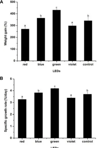
A
B
Fig. 1. (A) Weight gains (%) and (B) specific growth rates of olive flounder (Paralichthys olivaceus) grown under illumina- tion of four different LEDs (red, blue, green, and violet) and white fluorescent light (control) for a period of 60 days.
Results
Fish growth
To analyze the effects of different wavelengths of light on the growth of olive flounder, fish were reared under illu- mination from LEDs of different wavelengths (violet, 400 nm; blue, 465 nm; green, 508 nm, and red, 635 nm) for 60 days. Growth performance was calculated from the weights and lengths of fish measured at the beginning and end of the experimental period (Table 1). The specific growth rate and weight gain of fish grown under green LED illumination were significantly higher (p<0.05) than those of the other groups (Fig. 1). This finding was supported by weight gains (%) of 269.92±13.02, 363.21±3.74, 434.22±4.83, 290.17±11.83, and 340.74±26.58, and SGRs (%/day) of 2.18±0.06, 2.56±0.07, 2.79±0.01, 2.27±0.05, and 2.47±0.10 in fish grown under red, blue, green, and violet LEDs, and a white fluorescent light control, respectively. A similar result was obtained from the duplicate sets of rearing experiments. Overall, the growth performance of fish grown under blue light illumination was similar to that in the white fluorescent light control group.
The lowest specific growth rate was observed in fish grown under red LED illumination. Fish grown under violet LED illumination also showed a lower growth rate that did not differ significantly from that of fish grown under red LED illumination.
Hematological parameters
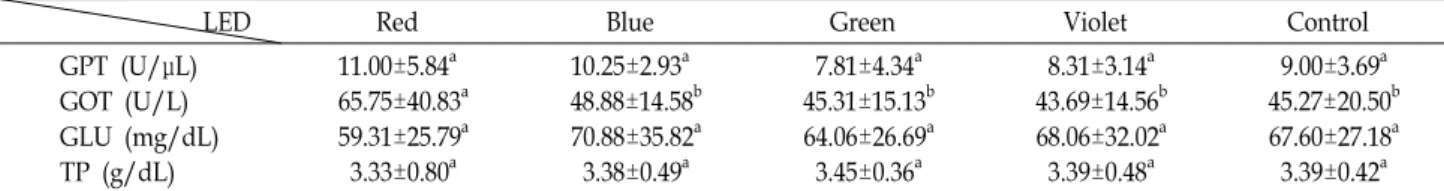
To assess the effects of light on fish physiology, blood samples were collected from fish grown under different light conditions. Levels of parameters including glucose (GLU), total protein (TP), glutamic oxaloacetic transaminase (GOT), and glutamic pyruvic transaminase (GPT) were analyzed in plasma separated from the blood collected from fish sub- jected to this experiment. The results showed no significant differences in the levels of GLU, TP, and GPT among fish
grown under different illumination conditions (Table 2).
This suggests that the levels of GLU, TP, and GPT are not affected by illumination at the different wavelengths em- ployed in this study. In contrast, fish grown under red LED illumination showed GOT levels that were significantly higher than those of other groups.
Table 2. Levels of glutamic pyruvic transaminase (GPT), glutamic oxaloacetic transaminase (GOT), glucose (GLU) and total proteins (TP) in the blood plasma. Blood samples were collected from olive flounder (Paralichthys olivaceus) grown under different LED wavelengths (red, blue, green, and violet) and white fluorescent light (control group) for a period of 60 days
LED Red Blue Green Violet Control
GPT (U/μL) GOT (U/L) GLU (mg/dL) TP (g/dL)
11.00±5.84a 65.75±40.83a 59.31±25.79a 3.33±0.80a
10.25±2.93a 48.88±14.58b 70.88±35.82a 3.38±0.49a
7.81±4.34a 45.31±15.13b 64.06±26.69a 3.45±0.36a
8.31±3.14a 43.69±14.56b 68.06±32.02a 3.39±0.48a
9.00±3.69a 45.27±20.50b 67.60±27.18a 3.39±0.42a Values (mean ± SD, n = 15) in the same row with different superscript letters are significantly different (p<0.05).
Table 3. Thickness (μm) of retina and photoreceptor layers in the eyes of olive flounder (Paralichthys olivaceus) reared under illumina- tion of LEDs at four different wavelengths (red, blue, green, and violet) and a white fluorescent light (control group).
Ratios of the thickness of the photoreceptor layer to that of the total retina (PR/RT) were calculated for each sample
LED Red Blue Green Violet Control
Retina layer thickness (RT) Photoreceptor layer thickness (PR) Ratio (PR/RT)
280.9±6.0a 94.1±3.8a 0.31±0.03
312.6±2.2b 97.7±1.4ab 0.31±0.01
301.8±5.8c 91.7±4.4ac 0.30±0.02
280.9±6.8a 93.7±1.0ac 0.31±0.02
291.5±4.7d 86.2±2.0d 0.30±0.01 Values (mean ± SD, n = 5) in the same row with different superscript letters are statistically significantly different (p<0.05).
Fig. 2. Representative photomicrographs of retina specimens pre- pared from the eyes of olive flounder (Paralichthys oliva- ceus) reared under illumination of four different LEDs (red, blue, green, and violet) and a white fluorescent lamp (control) for a period of 60 days. The photomicro- graph of a retina from a fish grown under blue light illustrates the regions measured for total retina (RT) and photoreceptor layer (PR) thickness. Prepared using H&E staining; 200× magnification; scale bar, 50 μm.
Histological analysis of the retina
To investigate the potential harmful effects of light of dif- ferent wavelengths, histological analysis was carried out on the retinas, which are the sites of the primary photoreceptors (Fig. 2). For this analysis, eyes were removed from fish grown under illumination of LEDs of different wavelengths.
The thickness of each layer was measured from microscopic images of the stained retina. The ratio of the thickness of
the photoreceptor layer to that of the entire retina was calcu- lated to determine effects on the layer of photoreceptors.
Although total retina thicknesses varied among the treat- ment groups, reflecting the sizes of the eyes, the ratio of the thickness of the photoreceptor layer to that of the total retina was similar among the treatment groups and the con- trol group (Table 3). In particular, the ratio of photoreceptor thickness to retinal thickness observed in the fish illumi- nated with green LEDs, which had a measurable effect on growth, did not differ from those in fish in other illumina- tion treatment groups, including the fluorescent light control.
Discussion
The light environment in aquatic ecosystems varies de- pending on water depth, due to differences in the pene- tration capacity of light at different wavelengths. Green and blue light penetrate more efficiently into deep water than light at longer wavelengths [5]. Fish have photoreceptor sys- tems adapted to the light environment of their habitats, lead- ing to different sensitivities to light of different wavelengths.
Wavelength-dependent effects of light on growth have been noted in various fish. To investigate the effects of light on the growth of olive flounder, one of the most important cul- tured species in Korea, fish were grown under illumination at different wavelengths, ranging from 400 to 635 nm.
Comparison of the sizes of fish after 60 days indicated that fish grown under green light illumination exhibited greater weight gain and growth rates than fish grown under illumi-
nation of LEDs of other wavelengths or white fluorescent light. The lowest weight gains and specific growth rates were observed in fish grown under red and violet illumination. These results were consistent with previous findings of higher growth rates under green light illumina- tion in various fishes, including rainbow trout Oncorhynchus mykiss, common carp Cyprinus carpio, and barfin flounder Verasper moseri [13, 15, 20, 28]. Furthermore, the growth of yellowtail clownfish Amphiprion clarkii was facilitated by green and blue light [22]. Overall, most studies examining the effects of light using various LEDs suggest that green and blue light are better light sources for promoting growth in fish, whereas red light negatively affects growth.
Fish have a photoreceptor system that is adapted to the light environment of their habitat. Two types of photo- receptor cells, rod and cone cells, contain visual pigments with distinct absorption profiles responsible for visual signal transduction [7, 17]. Differences in the growth of some fish under different illumination conditions may result from dif- ferences in their capacities to detect light of specific wave- lengths. Some fish may be able to detect specific wave- lengths more easily than others, enabling easier detection of food. A higher growth rate under green light illumination and slower growth under red light may be due to the ability of olive flounder to detect green light more efficiently with rhodopsin, which has an absorption maximum at 500 nm, close to the wavelength of the green LED. The influence of monochromatic light on energy metabolism and other phys- iological and biochemical activities of fish may also lead to the observed differences in growth performance. Upon illu- mination of Nile tilapia with blue light under stressful con- ditions, their plasma cortisol levels decreased, revealing that blue light can act as an anti-stress agent [25, 28]. When fish were exposed to chronic stress conditions caused by light that was hardly detectable, the growth rate was reduced due to energy utilization for tissue repair and osmoregulation to maintain normal homeostasis. In addition, yellow light has shown a negative effect on ATPase activity in Atlantic salmon Salmo salar [19].
Hematological parameters of fish grown under different LED illumination conditions were analyzed to determine the effects of light on fish physiology. GPT and GOT are known to link the metabolism of proteins and carbohydrates and to serve as an indicator of altered physiological or stress conditions. Under stress conditions, excess amounts of GPT or GOT leak into blood plasma. Although no significant dif-
ferences in the levels of hematological parameters (GPT, GLU, and TP) were detected among the experimental groups, a significantly higher level of GOT was observed in fish illu- minated with red light. This result suggests more stressful conditions for olive flounder under red light illumination.
A similar result was found in yellowtail clownfish, in which greater oxidative stress was observed under red light illumi- nation [21, 27]. Green light illumination also resulted in re- duced oxidative stress in fish that were already exposed to starvation, whereas oxidative stress induced by starvation increased in fish exposed to red light [9]. Stress has been suggested to inhibit growth in fish. Whereas green and blue light had favorable effects on fish growth performance, red light had a negative effect, possibly in response to hormonal imbalance, or to changes in energy metabolism or other physiological functions [2, 8, 9]. Stressors are known to neg- atively affect fish immunity, impairing their growth and ma- turation [11]. Higher levels of GOT in fish grown under red LED illumination compared to those in fish grown under violet LED illumination suggest that the former is more stressful for olive flounder than the latter illumination type.
Our results showed that excessive amounts of GOT may con- tribute to stress conditions or cause changes in the physio- logical activities of fish under red LED illumination, thus contributing to a lower growth rate.
The quality of light, including its wavelength and in- tensity, affect fish growth, as shown by the stimulatory ef- fects of green LED illumination on the growth of olive flounder. To widen its commercial application, it is important to assess any possible risk of harmful effects of green LED light on fish physiology. These effects can be tested by exam- ining changes in the retinal layer, which is one of the most light-sensitive tissues of the body and where primary per- ception of light occurs. Various methods have been used to assess changes in the retinal structure, such as the indirect fluorescence antibody technique and immunohistochemistry [1, 11, 24]. Adverse effects in the retina may be reflected in the ratio of the thickness of the photoreceptor layer to the thickness of the total retina. Our results suggest that the ratio of the photoreceptor layer thickness to that of the total retina layer in fish illuminated with green LEDs was similar to those in fish in the other experimental groups (Table 3).
Although such a change could represent an adaptive mecha- nism in fish, as expansion of photoreceptor layers or migra- tion of melanin granules may offer protection against un- favorable light conditions [6, 19], the lack of significant
changes observed among the ratios of photoreceptor thick- ness suggests little adverse effect of different wavelengths of light on the most light-sensitive tissues in fish. Therefore, we can conclude that green LED illumination can be used to facilitate growth in olive flounder without any distinct negative or adverse effects on their photoreceptors.
In summary, olive flounder growth was facilitated by illu- mination with green light as compared to illumination with LEDs of other wavelengths, but was retarded under illumi- nation with red and violet LEDs. The latter result appears to be due in part to a higher GOT level in fish grown under red light illumination. Green light does not show any harm- ful effects on the retinal layer containing primary photo- receptors. These results support the growth-promoting ef- fects of green LED illumination without any observable harmful effects on the retina, where the primary photo- response occurs.
Acknowledgement
This work was supported by a Research Grant of Pukyong National University (2017 year).
References
1. Allison, W. T., Hallows, T. E., Johnson, T., Hawryshyn, C.
W. and Allen, D. M. 2006. Photic history modifies suscepti- bility to retinal damage in albino trout. Vis. Neurosci. 23, 25-34.
2. Barton, B. A., Schreck, C. B. and Barton, L. D. 1987. Effects of chronic cortisol administration and daily acute stress on growth, physiological conditions, and stress responses in ju- venile rainbow trout. Dis. Aquat. Org. 2, 173-185.
3. Bayarri, M. J., Madrid, J. A. and Sánchez-Vázquez, F. J. 2002.
Influence of light intensity, spectrum and orientation on sea bass plasma and ocular melatonin. J. Pineal Res. 32, 34-40.
4. Biswas, A. K., Seoka, M., Inoue, Y., Takii, K. and Kumai, H. 2005. Photoperiod influences the growth, food intake, feed efficiency and digestibility of red sea bream (Pagrus major). Aquaculture 250, 666-673.
5. Boeuf, G. and Le Bail, P. Y. 1999. Does light have an influ- ence on fish growth? Aquaculture 177, 129-152.
6. Boulton, M., Rozanowska, M. and Rozanowska, B. 2001.
Retinal photodamage. J. Photochem. Photobiol. 64, 144-161.
7. Bowmaker, J. K. 1990. Retinal structure of fishes. In: Douglas, R. and Djamgoz, M. (Eds.), The Visual System of Fish.
Chapman & Hall, London.
8. Canosa, L. F., Chang, J. P. and Peter, R. E. 2007. Neuroendo- crine control of growth hormone in fish. Gen. Comp. Endocri- nol. 151, 1-26.
9. Choi, C. Y., Shin, H. S., Choi, J., Kim, N. N., Lee, J. and
Kil, G. S. 2012. Effects of LED light spectra on starvation-in- duced oxidative stress in the cinnamon clownfish, Amphpri- on melanopus, Comp. Biochem. Physiol. A 163, 357e363.
10. Duncan, D. B. 1955 Multiple range and multiple F test.
Biometrics 11, 1-42.
11. Harris, J. and Bird D. J. 2000. Modulation of the fish immune system by hormones. Vet. Immunol. Immunopathol. 77. 163e176.
12. Henne, J. P. and Watanabe, W. O. 2003. Effects of light in- tensity and salinity on growth, survival, and whole-body osmolality of larval southern flounder Paralichthys leth- ostigma. J. World Aquac. Soc. 34. 450-465.
13. Heydarnejad, M. S., Parto, M. and Pilevarian, A. A. 2013.
Influence of light colours on growth and stress response of rainbow trout (Oncorhynchus mykiss) under laboratory conditions. J. Anim. Physiol. Anim. Nutr. 97, 67-71.
14. Karakatsouli, N., Papoutsoglou S. E. and Manolessos, G.
2007. Combined effects of rearing density and tank color on the growth and welfare of juvenile white sea bream Diplodus sargus L. in a recirculating water system. Aquacul- ture 38, 1152-1160.
15. Karakatsouli, N., Papoutsoglou, E. S., Sotiropoulos, N., Mourtikas, D., Stigen-Martinsen, T. and Papoutsoglou, S. E.
2010. Effects of light spectrum, rearing density and light in- tensity on growth performance of scaled and mirror com- mon carp Cyprinus carpio reared under recirculating system conditions. Aquacult. Eng. 42, 121-127.
16. KOSTAT (Statistics KOREA). 2018. Agriculture & fishery products.
17. Levin, J. and McNicol, E. 1982. Color vision in fishes. Sci.
Am. 246, 108-117.
18. Luchiari, A. C. and Freire F. A. M. 2009. Effects of environ- mental colour on growth of Nile tilapia, Oreochromis niloticus (Linnaeus, 1758), maintained individually or in groups. J.
Appl. Ichthyol. 25, 162-167.
19. Migaud, H., Cowan, M., Taylor, J. and Ferguso, H. W. 2007.
The effect of spectral composition and light intensity on melatonin, stress and retinal damage in post-smolt Atlantic salmon, Salmo salar. Aquaculture 270, 390-404.
20. Ruchin, A. B. 2004. Influence of colored light on growth rate of juveniles of fish. Fish Physiol. Biochem. 30, 175-17.
21. Shin, H. S., Lee, J. and Choi, C. Y. 2012. Effects of LED light spectra on the growth of the yellowtail clownfish, Amphiprion clarkii. Fish Sci. 78, 549-556.
22. Takahashi, A., Kasagi, S., Murakami, N., Furufuji, S., Kikuchi, S., Mizusawa, K. and Andoh, T. 2016. Chronic ef- fects of light irradiated from LED on the growth perform- ance and endocrine properties of barfin flounder Verasper moseri. Gen. Comp. Endocrinol. 232, 101-108.
23. Taylor, J. F., Migaud, H., Porter, M. J. R. and Bromage, N.
R. 2005. Photoperiod influences growth rate and plasma in- sulin-like growth factor-I levels in juvenile rainbow trout, Oncorhynchus mykiss. Gen. Com. Endocrinol. 142, 169-185.
24. Vihtelic, T. S. and Hyde, D. R. 2000. Light-induced rod and cone cell death and regeneration in the adult albino zebra- fish (Danio rerio) retina. J. Neurobiol. 44, 289-307.
25. Villamizar, N., Blanco-Vives, B., Migaud, H., Davie, A.,
초록:빛의 파장이 넙치
Paralichthys olivaceus
의 성장에 미치는 영향엔다다 레지나 베네딕트1․김여름2․김종명1,2*
(1부경대학교 세계수산대학, 2부경대학교 수산생물학과)
빛의 파장이 넙치의 성장에 미치는 영향을 분석하기 위하여 보라색(400 nm), 청색(465 nm), 녹색(508 nm), 그리 고 적색(635 nm)의 LED와 백색 형광등하에서 성장 실험을 수행하였다. 수조당 15마리 넙치를 12:12 시간 각 파장 LED의 광주기하에서 60일 동안 사육한 결과, 각기 269.92±13.02%와 2.18±0.06(보라색), 363.21±3.74%와 2.56±0.07 (청색), 433.22±4.83%와 2.79±0.01(녹색), 290.17±11.83%와 2.27±0.05(적색), 그리고 340.74±26.58%와2.47±0.10(형광 등)의 체중 증가와 일간성장률(SGR: %/day)이 관찰되었다. 본 실험 결과 녹색 LED 하에서 넙치의 성장이 가장 빨랐으며, 적색 조명 하에서는 성장이 느린 것으로 나타났다. 대부분의 혈액 지표는 그룹별 차이가 없으나, 적색 LED조명 하에서 자란 넙치에서 높은 수준의 glutamic oxaloacetic transaminase (GOT)가 관찰되었다. 빛의 1차 감지 기관인 망막의 조직학적 분석 결과 광수용체 층의 상대적 두께에 별다른 영향이 없는 것으로 보아 녹색광이 무해함을 유추할 수 있다.
Carboni, S. and Sánchez-Vázquez, F. J. 2011. Effects of light during early larval development of some aquaculture tele- osts: a review. Aquaculture 315, 86-94.
26. Villamizar, N., García-Alcazar, A. and Sánchez-Vázquez, F.
J. 2009. Effect of light spectrum and photoperiod on the growth, development and survival of European sea bass (Dicentrarchus labrax) larvae. Aquaculture 292, 80-86.
27. Volpato, G. L., Bovi,T. S., de Freitas, R. H. A., da Sliva, D.
F., Delicio, H. C., Giaquinto, P. C. and Barreto, R. E. 2013.
Red light stimulates feeding motivation in fish but does not improve growth. PLoS One 8, e59134. doi: 10.1371/journal.
pone.0059134.
28. Yamanome, T., Mizusawa, K., Hasegawa, E. I. and Takahashi, A. 2009. Green light stimulates somatic growth in the barfin flounder Verasper moseri. J. Exp. Zool. A 311, 73-79.