Kor. J. Pharmacogn.
50(2) : 96∼ 101 (2019)
96
돼지감자꽃의 예쁜꼬마선충 내의 항산화 효과
김준형·이예빈·한선여·김수진·황인현·김대근*
우석대학교 약학대학
Antioxidant Activity of Helianthus tuberosus L. Flower in Caenorhabditis elegans
Jun Hyeong Kim, Ye Bin Lee, Seon Yeo Han, Su Jin Kim, In Hyun Hwang and Dae Keun Kim* College of Pharmacy, Woosuk University, Jeonju, Jeonbuk 55338, Korea
Abstract − Methanol extract of Helianthus tuberosus L. (Compositae) flower was investigated to research the anti-oxidative activity by using a Caenorhabditis elegans model system. Ethyl acetate soluble fraction of the methanol extract showed the most potent DPPH radical scavenging activity. The ethyl acetate fraction was measured on its activities of superoxide dismutase (SOD), catalase, and oxidative stress tolerance with reactive oxygen species (ROS) level in C. elegans. Furthermore, in order to verify if regulation of stress-response gene is responsible for the increased stress tolerance of C. elegans which treated by the ethyl acetate fraction, we checked SOD-3 expression using a transgenic strain. Consequently, the ethyl acetate fraction of H. tuberosus flower increased the catalase and SOD activities in a dose–dependent manner in C. elegans, reduced ROS accu- mulation dose-dependently. Besides, the ethyl acetate fraction-treated CF1553 worms showed higher SOD-3::GFP intensity than the control group.
Keywords − Helianthus tuberosus L., Caenorhabditis elegans, Anti-oxidative activity, Catalase, SOD
인간의 노화와 이에 따르는 질병을 일으키는 원인은 다양 하지만, 현재까지 가장 광범위하게 적용되는 원인으로 생체 내에서 생성되는 활성산소종(reactive oxygen species, ROS) 의 축적이다.1) ROS는 세포 내 정상적인 산소대사 과정과, 다양한 산화적 스트레스 및 인체 안에서 여러 가지 효소반 응에 의해 발생 한다.2) ROS는 세포 구성 성분들인 탄수화 물, 단백질, 지질 및 DNA와 반응하여 산화적 손상 및 효소 활성을 변화시켜 노화를 촉진시키고 뇌졸중 및 뇌 질환과 심장질환, 동맥경화, 암 등의 원인이 되기도 한다.3,4) 이러한 ROS에 의한 산화적 스트레스를 억제하기 위하여 항산화 효 능이 높으면서 부작용이 상대적으로 적은 천연 항산화제의 개발에 대한 필요성이 커지고 있다.5)
돼지감자(Helianthus tuberosus L., Compositae)의 잎이나 지상부에서 sesquiterpenes 화합물과 chlorogenic acid, quinic acid 등의 phenol성 화합물이 보고되어 있다.6,7) 약리 활성 연구로는 Kang 등이 보고한 돼지감자 괴근의 에탄올 추출물의 알러지성 피부에 함염증 효능8)및 항지방간 효
과9) 등이 알려져 있고, 돼지감자에서 분리된 methyl 2-(4'- methoxy-4'-oxobutanamide) benzoate의 항염증 효능 및 인 슐린 저항성에 관한 효능과 epoxysteroid 화합물의 항균작 용10) 등이 보고되어 있다.
본 연구는 돼지감자꽃 methanol 추출물의 항산화 효능을 예쁜꼬마선충(Caenorhabditis elegans)을 이용하여 확인해 보 고자 실험을 실시하였다. 돼지감자꽃 추출물의 용매분획으 로부터 DPPH free radical 소거능을 측정하여 가장 강한 항 산화능을 나타낸 ethyl acetate 분획에 대하여 선충 내의 항 산화 효소인 SOD와 catalase의 활성과 세포 내의 ROS의 축 적 억제 능력을 측정하였다. 또한, juglone으로 유도된 oxidative stress에 대한 저항능력을 확인하고 이와 관련된 선충 내의 oxidative stress에 저항하는 단백질의 증가여부를 SOD-3을 포함한 형질 전환 선충 CF1553을 이용하여 SOD- 3 발현 유전자의 증가 여부를 확인하였다.
재료 및 방법
실험재료 − 실험에 사용한 돼지감자꽃은 우석대학교에서
*교신저자(E-mail):[email protected] (Tel): +82-63-290-1574
채집하여 실험에 사용하였으며, 표준품은 우석대학교 약학 대학 생약표본실에 보관하고 있다(WS-18-005).
추출 및 분획 − 건조한 돼지감자꽃 300 g을 분쇄하여 methanol로 가끔 진탕하면서 5시간씩 50℃에서 3회 온침 추 출하였다. 그 추출액을 수욕상에서 감압농축하여 methanol 엑스 약 19.5g을 얻었으며, 이 methanol 엑스를 증류수로 현 탁시키고 상법에 따라 동량의 n-hexane(1.7 g), methylene chloride(3.92 g), ethyl acetate(1.36 g) 및 n-butanol(1.55 g)의 순으로 분획하여 각각의 분획물을 얻었다.
DPPH Free Radical 소거활성 측정11)− 96well plate에 시료를 ethanol을 용매로 각 농도별로 조제하여 넣고 0.2 mM 의 1,1-diphenyl-2-picrylhydrazyl(DPPH) (EtOH)을 일정량씩 가하였다. 10초간 진탕한 후 25oC에서 30분간 방치한 후 microplate reader를 이용하여 517 nm에서 흡광도를 측정하 였다. 대조약물은 L-ascorbic acid를 사용하였으며, 각 시료 에 대하여 3회 반복하여 측정하였다.
예쁜꼬마선충 배양12) − 예쁜꼬마선충(Caenorhabditis elegans, wild type)은 Escherichia coli OP50을 도말한 nematode growth medium(NGM) agar plate 상에서 배양하 였다(20℃). 시료는 DMSO를 용매로 한 stock solution 상태 로 멸균된 NGM plate(50oC)에 첨가되었으며, 최종 DMSO 농도는 0.1%(v/v)를 유지하였다.
선충 체내의 항산화 효소활성 측정13,14)− 돼지감자꽃 시 료를 농도별로 조제하여 plate에 넣고 성장 단계가 동일한 선충을 배양하였으며, 성체가 된 후 2일째의 선충을 모아 M9 buffer로 3회 세척 후 분쇄하여 효소활성 측정에 사용 하였다(homogenization buffer: 10 mM Tris-HCl, 150 mM NaCl, 0.1 mM EDTA, pH 7.5). SOD 활성은 Ibrahim등의 방법을 응용하여 측정하였다. 10 mM phosphate buffer(pH 8.0)를 용매로 반응혼합물(1.6 mM xanthine과 0.48 mM NBT) 0.49 mL를 만든 뒤 농도별 시료 10 μL와 37oC에서 5 분간 pre-incubation시켰다. 그 후 xanthine oxidase 100 μL (0.05 U/mL)을 첨가하고 37oC에서 20분 동안 incubation한 다음 69 mM SDS로 반응을 멈추고 570 nm에서 흡광도를 측정하였다. Catalase activity는 Aebi의 방법을 응용하여 25 mM H2O2에 농도별 시료 50 μL를 3분 동안 반응시키고 240 nm에서 흡광도를 측정하였다.
선충 세포 내 활성산소종(ROS) 분석15)− 선충 내 활성 산소종은 2',7'-dichlorodihydro fluorescein diacetate(H2DCF- DA)를 사용하여 측정하였다. 성장 단계가 동일한 N2 선충 을 돼지감자꽃 시료를 농도별로 제조한 plate에서 배양하여 성체가 된 후 4일째에 50 μM juglone을 함유한 M9 buffer 에 넣고 2시간 노출시킨 뒤 96 well plate에 담긴 50 μL M9 buffer에 5마리씩 옮겼다. 마지막으로 50 μM H2DCF-DA 50 μL를 첨가한 뒤 여기 485 nm, 방출 535 nm에서 흡광도 를 각각 측정하였다.
산화적 스트레스 저항성 평가14,17)− 돼지감자꽃 ethyl acetate 분획이 선충의 산화 스트레스 저항성에 미치는 영향 을 알아보기 위해 산화적 스트레스 조건하에서 선충을 배 양하여 생존율을 측정하는 실험을 수행하였다. 성체가 된 후 7일째 일시적으로 선충을 2 mM juglone이 함유된 M9 buffer가 담긴 96 well plate의 well에 옮기고 시간별로 생존 율을 확인하였다.
형질전환 선충 내 SOD-3::GFP 형광 측정 − 시료를 농 도별로 제조한 배지에서 형질 전환시켜 SOD-3::GFP를 포 함하고 있는 선충 CF1553을 배양하였다. 성체가 된 후 3일 째에 사용하였으며, 선충은 sodium azide(4%)로 마취시켰고 GFP 발현은 형광 실체 현미경(Olympus, Japan)으로 관찰하 였다. 발현 강도를 정량, 분석하기 위해 현미경을 이용한 사 진 촬영과 Image J 소프트웨어를 사용하여 분석하였다. 모 든 실험은 3회 반복하였다.
통계 분석 − 통계 자료의 값은 평균값±표준오차(mean±
S.E.M.)로 표시하였다. 그룹 간의 통계적 유의성 검정은 Student's t-test를 통해서 분석하였고 선충의 연속적인 생존 도는 Log-rank test분석 방법을 이용하였다. p값은 *p<0.05,
**p<0.01, ***p<0.001일 때 유의성이 있는 것으로 간주하 였다.
결 과
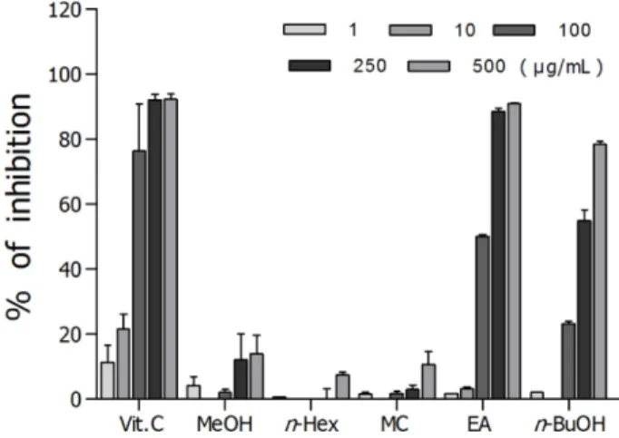
DPPH Radical 소거능 − 돼지감자꽃 분획물 중에서, DPPH radical 소거 효과는 Fig. 1에 나타난 바와 같이 분획 물들 중 ethyl acetate 분획(IC50 value, 95.9 μg)이 효과가 가 장 좋았으며, 대조군인 비타민 C는 IC50 값이 26.9 μg으로 측정되었다.
선충 체내의 항산화 효소 SOD 및 Catalase 활성증가 효능 − Xanthine을 기질로 xanthine oxidase의 효소반응 과
Fig. 1. DPPH radical scavenging effects of the methanol extract, and its fractions from H. tuberosus flower.
정 중에 생성되는 superoxide anion을 활용하여 SOD의 활 성을 측정한 결과, Fig. 2A에서와 같이 돼지감자꽃 ethyl acetate 분획 투여군은 SOD의 활성을 농도 의존적으로 증 가시켰으며, ethyl acetate 분획 500 µg/mL 투여군은 대조군 과 비교하여 SOD 활성을 약 32.5% 정도 증가시켰다 (***p<0.001). 활성산소종의 일종인 hydrogen peroxide를 체 내에서 대사시키는 catalase의 활성은 Fig. 2B에서 나타난 바와 같이 돼지감자꽃 ethyl acetate 분획 500 µg/mL 투여 군이 대조군에 비해 약 11.1% 정도 증가시켰다(*p<0.05)(Fig.
2).
선충 세포 내 ROS 감소 효능 − 돼지감자꽃 ethyl acetate 분획의 농도별 세포 내 활성산소종의 감소 효능을 알아보
기 위해 H2DCF-DA와 선충 내부의 활성산소종을 반응시켜 형광을 관찰하였다. 활성산소종으로 인한 120분까지의 평균 형광 감소폭은 대조군과 비교하여 ethyl acetate 분획 500 µg/
mL 투여군에서 약 12.7%(***p<0.001) 활성산소종을 감소 시켰으며, 120분 시점에서의 형광 감소폭은 대조군과 비교 하여 37.3%의 유의성 있는 감소폭은 나타냈다(Fig. 3).
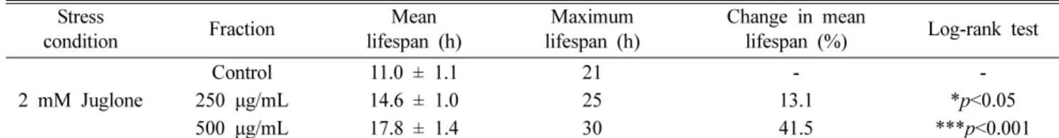
Oxidative stress 저항성 증가 효능 − 돼지감자꽃 ethyl acetate 분획이 선충의 산화적 스트레스 조건에서 선충의 생 존율에 미치는 영향을 확인하였다. 선충에 산화적 스트레스 를 유도하기 위해서 2 mM juglone이 함유된 M9 buffer가 담긴 96 well plate에서 배양한 대조군 선충의 최고 생존시 간은 21시간이었으나, ethyl acetate 분획 500 µg/mL 농도
Fig. 2. Effects of ethyl acetate fraction of the H. tuberosus flower on the antioxidant enzyme activity of C. elegans. (A) SOD activ- ity was showed as a percentage of superoxide-scavenged amount per control. (B) Catalase activity was expressed as a percentage of decrease in residual H2O2, measured by a spectrophotometric method. Differences compared to the control were considered sig- nificant at *p<0.05 and ***p<0.001 by one-way ANOVA.
Fig. 3. Effects of ethyl acetate fraction of the H. tuberosus flower on the intracellular ROS levels of wild-type N2 nematodes. Intra- cellular ROS accumulation was examined in a microplate fluorescence reader at 535 nm (emission) and 485 nm (excitation). (A) Plates were read for 120 min. (B) The average percentages of intracellular ROS accumulation were presented. Differences com- pared with the control were considered significant at **p<0.01 and ***p<0.001 by the one-way ANOVA.
에서는 생존시간을 30시간으로 증가시켰다. 대조군의 평균 생존시간이 11.0±1.1시간었으나 500 μg/mL 농도 처리군은 평균 생존 시간을 17.8±1.4시간으로 41.5%의 생존 시간을 향상시켰다(***p<0.001)(Fig. 4, Table I).
형질 전환 선충 내 SOD-3의 발현 증가 효능 − 돼지감 자꽃 ethyl acetate 분획이 선충 내에서 oxidative stress에 저 항하기 위한 단백질의 생성 여부를 확인하기 위해서 SOD 발현 유전자의 증가 여부를 확인하였다. SOD-3을 포함한 형질 전환 선충 CF1553을 사용하여 실험한 결과 돼지감자 꽃 ethyl acetate 분획 500 μg/mL 투여군이 처리되지 않은 선충군에 비해 상당히 높은 SOD-3::GFP 발현율(13.5%,
**p<0.01)을 보여주었다(Fig. 5).
고 찰
돼지감자꽃 ethyl acetate 분획은 DPPH radical 소거활성 시험에서 농도의존적으로 강한 항산화 활성을 보여 주었으 며, 이와 같은 결과는 기 보고된 돼지감자꽃 추출물의 항산 Fig. 4. Effects of ethyl acetate fraction of the H. tuberosus
flower on the stress tolerance of wild-type N2 nematodes. For the oxidative stress assays, worms were transferred to 96-well plate containing 2 mM of juglone liquid culture, and then their viability was scored. Statistical difference between the curves was analyzed by log-rank test.
Fig. 5. Effects of ethyl acetate fraction of the H. tuberosus flower on the expression of SOD-3 (CF1553) was determined using transgenic nematodes. (A) Images of SOD-3::GFP expressions of CF1553 nematodes in the presence or absence of the ethyl acetate fraction of the H. tuberosus flower. (B) The mean GFP-expressing intensity of CF1553 mutants was expressed as mean S.E.M. of values from 90 worms per each experiment (B). Data are expressed as the mean±standard deviation of three independent exper- iments (N=3). Differences compared with the control were considered significant at *p<0.05 and **p<0.01 by one-way ANOVA.
Table I. Effects of ethyl acetate fraction of H. tuberosus flower on the oxidative stress tolerance of C. elegans Stress
condition Fraction Mean
lifespan (h)
Maximum lifespan (h)
Change in mean
lifespan (%) Log-rank test 2 mM Juglone
Control 250 μg/mL 500 μg/mL
11.0 ± 1.1 14.6 ± 1.0 17.8 ± 1.4
21 25 30
- 13.1 41.5
-
*p<0.05
***p<0.001
Mean lifespan presented as mean±S.E.M data. Change in mean lifespan compared with control group (%). Statistical significance
of the difference between survival curves was determined by log-rank test using the Kaplan-Meier survival analysis. Differences
compared to the control were considered significant at *p<0.05 and ***p<0.001.
화 효능들과 잘 부합되는 결과이다.18)예쁜꼬마선충 내의 항 산화 효소인 SOD 및 catalase의 활성을 확인해 본 결과 돼 지감자꽃 ethyl acetate 분획은 농도 의존적으로 SOD 및 catalase의 활성을 높이는 것으로 확인되어 free radical에 의 해 야기되는 oxidative stress로부터 방어하는 기전에 영향이 있을 것으로 생각된다.19,20) Juglone으로 유도한 선충의 oxidative stress에 대한 저항능력을 측정한 실험에서는 같은 stress 조건 하의 대조군과 비교해 볼 때 돼지감자꽃 ethyl acetate 분획 처리군의 생존율이 농도 의존적으로 크게 증가 하였다. 이와 관련된 기전 중의 하나로써, 형질전환된 선충 인 GFP-fused transgenic strain CF1553을 이용한 oxidative stress 저항성 확인실험에서 저항성 지표인자로서 SOD- 3::GFP 형광 발현율을 측정해 본 결과, 형광 발현율이 농도 의존적으로 증가되는 것으로 관찰되어 oxidative stress에 저 항하는 단백질이 발현되었음을 확인하였다.21)이러한 결과 는 돼지감자꽃과 관련된 연구 중에 돼지감자꽃의 함유성분 으로 알려진 다수의 chlorogenic acid 유도체와 phenol성 화 합물들의 항산화 효능과 상당한 관련성이 있을 것으로 사 료된다.22) 체내에서 과잉 생성된 세포 내의 ROS와 각종 스 트레스, 세포 손상 등에 의해 생성되는 ROS는 체내의 항산 화 효소 등을 소멸시켜 oxidative stress를 야기시키며, 노화 관련 각종 퇴행성 질환을 비롯한 각종 암 발생이나 염증 생 성 등에 관련성이 있음이 알려져 있다.23) 돼지감자꽃 ethyl acetate 분획이 SOD나 catalase 등의 항산화 효소의 활성을 증가시키고, oxidative stress에 대한 저항력을 높여 주는 효 능은 암, 염증성 질환 등의 예방 및 치료에 도움을 줄 수 있 으며, 노화를 억제하여 수명을 연장시키는데 어느 정도 기 여할 수 있을 것으로 생각된다. 이후에 이어지는 연구로 돼 지감자꽃의 단일 물질 수준에서의 항산화 활성 및 기전 연 구가 필요할 것으로 사료된다.
결 론
돼지감자꽃 ethyl acetate 분획은 DPPH radical 소거활성 실험에서 강한 항산화력을 보여 주었으며, 예쁜꼬마선충 내 의 항산화 효소인 SOD 및 catalase의 활성이 돼지감자꽃 ethyl acetate 분획 투여에 의해 농도 의존적으로 높아졌다.
선충 세포 내 ROS의 축적량 억제실험에서 돼지감자꽃 ethyl acetate 분획은 ROS 발생을 유의성 있게 억제 하였으며, juglone을 처리한 oxidative stress 반응 관련 실험에서는 돼 지감자꽃 ethyl acetate 분획 투여군의 생존율이 농도 의존 적으로 증가하는 것을 확인하였다. 이와 관련하여 예쁜꼬마 선충의 mutant인 GFP-fused transgenic strain CF1553을 이 용한 oxidative stress 저항성을 확인한 결과, 농도 의존적으 로 형광 발현량을 증가시키는 것이 확인되어 oxidative stress 에 저항성이 있는 단백질의 발현이 증가되었음을 확인하였
다. 이러한 결과를 종합해 보면 돼지감자꽃 ethyl acetate 분 획은 항산화 및 이와 관련된 질병의 예방 및 치료를 위한 자원으로서의 가치가 있을 것으로 사료된다.
사 사
이 논문은 2017년도 교육부의 재원으로 한국연구재단의 지원을 받아 수행된 연구(2017R1D1A3B03032146)로 이에 감사 드립니다.
인용문헌
1. Labuschagne, C. F. and Brenkman, A. B. (2013) Current methods in quantifying ROS and oxidative damage in Caenorhabditis elegans and other model organism of aging.
Ageing Res. Rev. 12: 918-930.
2. Seifried, H. E. (2007) A review of the interaction among dietary antioxidants and reactive oxygen species. J. Nutr. Bio- chem. 18: 567-579.
3. Crack, P. J. and Taylor, J. M. (2005) Reactive oxygen species and the modulation of stroke. Free Radical Biology & Med- icine 38: 1433-1444.
4. Moskovitz, J., Yim, M. B. and Chock, P. B. (2002) Free rad- icals and disease. Archives of Biochemistry and Biophysics 397: 354-359.
5. Maarman, G. J. (2017) Natural antioxidants as potential ther- apy, and a promising role for melatonin against pulmonary hypertension. Adv. Exp. Med. Biol. 967: 161-178.
6. Yuan, X. and Yang, Q. (2017) Simultaneous quantitative determination of 11 sesquiterpene lactones in Jerusalem arti- choke (Helianthus tuberosus L.) leaves by ultra high per- formance liquid chromatography with quadrupole time-of- flight mass spectrometry. J. Sep. Sci. 40: 1457-1464.
7. Chen, F., Long, X., Liu, Z., Shao, H. and Liu, L. (2014) Anal- ysis of phenolic acids of Jerusalem artichoke (Helianthus tuberosus L.) responding to salt-stress by liquid chroma- tography/tandem mass spectrometry. The Scientific World Journal 2014: 568043. doi: 10.1155/2014/568043.
8. Kang, Y. M., Lee, K. Y. and An, H. J. (2018) Inhibitory effects of Helianthus tuberosus ethanol extract on dermato- phagoides farina body-induced atopic dermatitis mouse model and human keratinocytes. Nutrients 10: pii: E1657.
doi: 10.3390/nu10111657.
9. Okada, N., Kobayashi, S., Moriyama, K., Miyataka, K., Abe, S., Sato, C. and Kawazoe, K. (2017) Helianthus tuberosus (Jerusalem artichoke) tubers improve glucose tolerance and hepatic lipid profile in rats fed a high-fat diet. Asian Pac. J.
Trop Med. 10: 439-443.
10. Jung, Y. J., Kim, B. O., Kwak, J. H. and Pyo, S. (2016) Inhib- itory effect of methyl 2-(4'-methoxy-4'-oxobutanamide) ben- zoate from Jerusalem artichoke (Helianthus tuberosus) on
the inflammatory paracrine loop between macrophages and adipocytes. J. Agric. Food Chem. 64: 9317-9325.
11. Li, X. D., Miao, F. P. and Ji, N. Y. (2011) Two new epox- ysteroids from Helianthus tuberosus. Molecules 16: 8646- 8653.
12. Yoshida, T., Mori, K., Hatano, T., Okumura, T., Uehara, I., Komagoe, K., Fujita, Y. and Okuda, T. (1989) Studies on inhibition mechanism of autooxidation by tannins and fla- vonoids. V: Radical scavenging effects of tannins and related polyphenols on 1,1-diphenyl-2-picrylhydrazyl radical. Chem.
Pharm. Bull. 37: 1919-1921.
13. Brenner, S. (1974) The genetics of Caenorhabditis elegans.
Genetics 77: 71-94.
14. Mekheimer, R. A., Sayed, A. A. and Ahmed, E. A. (2012) Novel 1,2,4-triazolo[1,5-a]pyridines and their fused ring sys- tems attenuate oxidative stress and prolong lifespan of Caenorhabditis elegans. J. Med. Chem. 55: 4169-4177.
15. Aebi, H. (1984) Catalase in vitro. Method. Enzymol. 105:
121-126.
16. Kim, H. N., Seo, H. W., Kim, B. S., Lim H. J., Lee, H, N., Park, J. S., Yoon, Y. J., Oh, J. W., Oh, M. J., Kwon, J., Oh, C. H., Cha, D. S. and Jeon, H. (2015) Lindera obtusiloba extends lifespan of Caenorhabditis elegans. Nat. Prod. Sci.
21: 128-133.
17. Lee, E. Y., Shim, Y. H., Chitwood, D. J., Hwang, S. B., Lee, J. and Paik, Y. K. (2005) Cholesterol-producing transgenic Caenorhabditis elegans lives longer due to newly acquired enhanced stress resistance. Biochem. Biophys. Res. Commun.
328: 929-936.
18. Yuan, X., Gao, M., Xiao, H., Tan, C. and Du, Y. (2012) Free radical scavenging activities and bioactive substances of Jerusalem artichoke (Helianthus tuberosus L.) leaves. Food Chem. 133: 10-14.
19. Samarghandian, S., Azimi-Nezhad, M., Farkhondeh, T. and Samini, F. (2017) Anti-oxidative effects of curcumin on immobilization-induced oxidative stress in rat brain, liver and kidney. Biomed. Pharmacother. 87: 223-229.
20. Jiménez-Fernández, S., Gurpegui, M., Díaz-Atienza, .F, Pérez-Costillas, L., Gerstenberg, M. and Correll, C. U. (2015) Oxidative stress and antioxidant parameters in patients with major depressive disorder compared to healthy controls before and after antidepressant treatment: results from a meta-analysis. J. Clin. Psychiatry 76: 1658-1667.
21. Seo, H. W., Cheon, S. M., Lee, M.-H., Kim, H. J., Jeon, H.
and Cha D. S. (2015) Catalpol modulates lifespan via DAF- 16/FOXO and SKN-1/Nrf2 activation in Caenorhabditis ele- gans. Evidence-Based Complementary and Alternative Med- icine. doi 10.1155/2015/524878.
22. Wang, Y. M., Zhao, J.Q., Yang, J. L., Idong, P. T., Mei, L. J., Tao, Y. D. and Shi, Y. P. (2017) Antioxidant and α–glu- cosidase inhibitory ingredients identified from Jerusalem artichoke flowers. Nat. Prod. Res. 9: 1-5.
23. Moloney, J. N. and Cotter, T. G. (2018) ROS signalling in the biology of cancer. Semin. Cell Dev. Biol. 80: 50-64.
(2019. 3. 26 접수; 2019. 4. 27 심사; 2019. 5. 13 게재확정)