서 론 .
Ⅰ
(Spinal Cord Injury, SCI)
(paraplegia)
(tetraplegia) .
(American Spinal Injury Association,
ASIA) ‘
’ A
(B, C, D), E
.
, ,
, , ,
, ,
.
, , , ,
, , ,
[1].
.
.
손량희 손종상 황한정 임창환, , , , *김영호 , .
Steady-State Visual Evoked Potential
(SSVEP)-based Rehabilitation Training System with Functional Electrical Stimulation
R. H. Sohn, J. Son, H. J. Hwang, C. H. Im, *Y. H. Kim Dept of Biomed. Eng., Graduate School, Yonsei Univ.
Institute of Medical Eng., Yonsei Univ.
(Received March 22, 2010. Accepted October 13, 2010)
The purpose of the brain-computer (machine) interface (BCI or BMI) is to provide a method for people with damaged sensory and motor functions to use their brain to control artificial devices and restore lost ability via the devices. Functional electrical stimulation (FES) is a method of applying low level electrical currents to the body to restore or to improve motor function. The purpose of this study was to develop a SSVEP-based BCI rehabilitation training system with FES for spinal cord injured individuals. Six electrodes were attached on the subjects’
scalp (PO
Z, PO
3, PO
4, O
Z, O
1and O
2) according to the extended international 10-20 system, and reference electrodes placed at A1 and A2.
EEG signals were recorded at the sampling rate of 256Hz with 10-bit resolution using a BIOPAC system. Fast Fourier transform (FFT) based spectrum estimation method was applied to control the rehabilitation system. FES control signals were digitized and transferred from PC to the microcontroller using Bluetooth communication. This study showed that a rehabilitation training system based on BCI technique could make successfully muscle movements, inducing electrical stimulation of forearm muscles in healthy volunteers.
Brain-computer(machine) interface (BCI or BMI), Steady-state visual evoked potential (SSVEP), Functional electrical stimulation (FES)
Corresponding Author : 김영호 강원도 원주시 흥업면 매지리
(220-710) 234
연세대학교 첨단 의료기기 테크노타워204호
Tel : +82-33-760-2859 / Fax : +82-33-760-2859 E-mail : [email protected]
본 연구는 교육과학기술부와 한국산업기술재단의 지역혁신 인력양성사업으로 수행된 연구 결과임.
.
,
, ,
. Herbold J
[2].
,
.
. (Electroencephalography, EEG)
- (
) (Brain Computer (Machine) Interface, BCI or
BMI, ‘BCI’ ) 1970
[3,4]. ,
(Motor Imagery Training, MIT)
[5]
[6].
.
SSVEP(Steady State Visually Evoked Potentials) BCI . (Occipital Lobe)
, , . SSVEP
3.5Hz 75Hz
[7].
. (Functional Electrical Stimulation, FES)
(sensory awareness) ,
[8,9], (Bluetooth)
.
SSVEP BCI
FES
, .
연구방법 .
Ⅱ
피실험자 선정 및 실험 환경 구축 A.
5 .
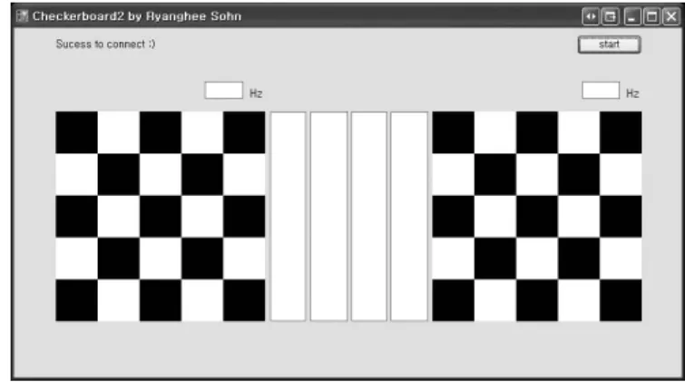
C# 7Hz 9Hz
, 1m
( 1).
.
PC (Bluetooth)
(Microcontroller: MSP430) .
시각자극 프로그램 B.
BCI
(Event Related Potential, ERP) P300 [10],
Application Control
BlueTooth Communication
EEG Signal
Acquisition DSP
그림1.시스템 흐름도 Fig. 1. System flow chart
μ
(Event Related Desynchronization, ERD) (Event Related Synchronization, ERS)
[11], (Steady State
Visually Evoked Potentials, SSVEP) [7].
SSVEP [12]. SSVEP
3.5Hz 75Hz
. SSVEP
2
( 2).
뇌파 계측 C.
BIOPAC Systems(MP150, EEG100C)
. 6 10-20
3 (a) PO
Z, PO
3,
PO
4, O
Z, O
1, O
2, A
1, A
2. (sampling rate) 256Hz .
PC
. 3 (b) .
무선 전기자극 시스템 D.
,
.
(implantable electrode method)
(percutaneous electrode method) [14-16].
(a) (b)
그림3.후두엽에서의 뇌파 계측(a)및 시스템 구성(b) Fig. 3. EEG acquisition of occipital lobe (a), and System configuration(b)
그림2. 시각 자극 프로그램 Fig. 2. Visual stimulation program
(Walking-Man , CyberMedic, KR)
. 18 (8
, 9 )
, 4 (a) 2~5
(flexor digitorum profundus)
, (extensor carpi radialis
longus) (motor point) .
, 10~20mA, 200us,
15~50Hz .
,
( 4 (b)).
E. 신호처리
5~30Hz
, (1) (Fast Fourier
Transform, FFT) (Frequency Series) (Power Spectrum)
[17].
(1)
7Hz 9Hz
. PC
, .
결 과 .
Ⅲ
, .
(Electrogoniometer)
, EN60601-1(IEC601-1) 93/42/EEC
SG110/A, 150(Biometrics Ltd, U.K.) .
동기화 시 뇌파 신호 및 손목관절각도 A. 7Hz
5 7Hz
. (a)
5Hz~30Hz , (b)
. (c)
7Hz (flexor
digitorum profundus) (flexion)
Lateral epicondyle of humerus Ulna
Radius Supinator Flexor digitorum profundus
Flexor pollicis longus
Pronator quadratus
Lumbricales
Medial epicondyle of humerus
Anconeus Extensor digiti minimi (cut) Extensor carpi
ulnaris (cut) Extensor carpi radialis brevis Extensor pollicis
longus
Extensor indicis Cut tendons
of extensor digitorum
Extensor carpi radialis longus Extensor digitorum
(cut and reflected)
Supinator (deep)
Abductor pollicis longus
Extensor pollicis
brevis
Ch1 Ch2 FES
Interrupt Port
External Port 3.7V
GND program Download
PC
(a) (b)
그림4. 기능적 전기자극기의 표면 전극 위치(a)및 무선 전기자극 시스템(b) Fig. 4. Surface electrode of FES locations (a) and Wireless electrical stimulation system (b)
, 10° 40°
.
동기화 시 뇌파 신호 및 손목관절각도 B. 9Hz
6 9Hz
, (a)
5Hz~30Hz , (b)
. (c) 9Hz
(extensor carpi radialis longus) (motor point) (extension)
, 13° -2° .
주파수 반응 결과 C.
(Power Spectrum) 7Hz
9Hz
, BCI
0 40
mV
20
0 -20
-40 0.3 0.6 0.9 1.2 1.5 1.8 2.1 2.4 2.7 3
Time (sec) (a)
0 Spectral density 10
8 6
2
0 3 6 9 12 15 18 21 24 27 30
Frequency (Hz) (b)
4
0 50
deg(°)
40 30
10
0 0.3 0.6 0.9 1.2 1.5 1.8 2.1 2.4 2.7 3
Time (sec) (c)
20
그림6. 9Hz동기화 시 뇌파 신호 및 손목관절각도 Fig. 6. EEG signal(a), Frequency spectrum(b), and Wrist joint angle(c)
0 40
mV
20
0 -20
-40 0.3 0.6 0.9 1.2 1.5 1.8 2.1 2.4 2.7 3
Time (sec) (a)
0 Spectral density 10
8 6
2
0 3 6 9 12 15 18 21 24 27 30
Frequency (Hz) (b)
4
0 50
deg(°)
40 30
10
0 0.3 0.6 0.9 1.2 1.5 1.8 2.1 2.4 2.7 3
Time (sec) (c)
20
그림5. 뇌파 신호 와 주파수 특성(a) (b)그리고 손목관절 각도(c) Fig. 5. EEG signal(a), Frequency spectrum(b), and Wrist joint angle(c)
. ,
5 10 . 1
, 50~80% .
결 론 .
Ⅳ
SSVEP
. Mueller-
Putz G [7] BCI
SSVEP ,
. 3.5Hz 75Hz
[7], 7Hz 9Hz
, 50~80%
.
,
.
참고문헌
[1] Delisa JA: Rehabilitation Medicine Principles and Practice, 3rd ed., pp 1259-1291, Lippincott-Raven, 1998.
[2] Herbold J., Walsh M. and Reding M., “Rehabilitation Hospital Versus Nursing Home Setting for Rehabilitation Following Stroke: A Case-Matched Controlled Study,” Archives of Physical Medicine and Rehabilitation, vol. 88, pp. e17-e18, 2007.
[3] Wolpaw JR, Birbaumer N, McFarland DJ, Pfurtscheller G, Vaughan TM.,“Brain-computer interfaces for communication and control,ˮ Clin Neurophysiol, 113(6):767-91, 2002.
[4] Janis J Daly, Jonathan R Wolpaw, “Brain computer interfaces
in neurological rehabilitation,ˮ The Lancet Neurology, vol 7, Issue 11, pp. 1032 - 1043, 2008.
[5] Dijkerman HC, Ietswaart M, Johnston M, MacWalter RS. Does motor imagery training improve hand function in chronic stroke patients? A pilot study. Clin Rehabil. 2004 Aug;18(5):538-49.
[6] Langhorne P, Coupar F, Pollock A. Motor recovery after stroke:
a systematic review. Lancet Neurol. 2009 Aug;8(8):741-54.
[7] Mueller-Putz G., Scherer G., Brauneis R.C. , and Pfurtscheller G.,: “Steady state visual evoked potential (SSVEP)-based communication: impact of harmonic frequency components,” J.
Neural Eng., vol. 2, no. 4, pp. 123 130, 2005.
[8] Robinson AJ, Snyder-Mackler, L. Clinical electrophysiology:
electrotherapy and electrophysi-ologic testing 3rd ed. Baltimore:
Lippincott Williams and Wilkins, 151-196, 198-237, 239-274, 2008.
[9] Alon G et al. Electrotherapeutic Terminology in Physical Therapy; Section on Clinical Electrophysiology. Alexandria, VA: American Physical Therapy Association, 2005.
[10] Emanuel Donchin, Kevin M. Spencer, and Ranjith Wijesinghe,
“The mental prosthesis: assessing the speed of a P300 based brain computer interface,ˮ IEEE Trans. on Rehab. Eng., vol. 8, no. 2, pp. 174~179, June 2000.
[11] J. R. Wolpaw, D. J. McFarland, and T. M. Vaughan,
“Brain-computer interface research at the wadsworth center,ˮ IEEE Trans. on Regab. Eng., vol. 8, no. 2, pp. 222-226, June 2000.
[12] J.R. Wolpaw , N.B., D.J. McFarland , G. Pfurtscheller , T.M.
Vaughan, “Brain-computer interfaces for communication and control,” Clinical Neurophysiology., 113: pp. 767-791, 2002.
[14] McNeal DR, Bowman BR., “Selective activation of muscles using peripheral nerve electrodes.” Med. Biol. Eng. Comput., 23:
249-253, 1985.
[15] Marsolais EB, Kobetic R. Functional walking in paralyzed patients by means of electrical stimulation. Clin.Orthop., 30-36, 1983.
[16] Cameron T, et al., “Micromodular implants to provide electrical stimulation of paralyzed muscles and limbs.” IEEE Trans.
Biomed. Eng., 44: 781-790, 1997.
[17] Walter, D.O., Leuchter, A.F. A Tourial on Classical Computer Analysis of EEGs: Spectra and Coherences in Analysis of the Electrical Activity of the Brain, ed. by Angeleri F., Butler S., Giaquinto S., Majkowski J. Wiley & Sons pp. 105-124. 1997.
피실험자 #1 #2 #3 #4 #5
성공률(%) 80% 77% 50% 68% 75%
표1. 주파수 반응 결과
Table 1. Result of frequency response