ISSN 1225-6552, eISSN 2287-7630 https://doi.org/10.7853/kjvs.2018.41.1.41
< Case Report >
Veterinary Service
Available online at http://kjves.org
*Corresponding author: Ji-Youl Jung, Tel. +82-54-912-0462, Fax. +82-54-912-0465, E-mail. jungjy1982@korea.kr
고양이 장에서 발생한 T 세포 림프종
정지연
1ㆍ이경현
1ㆍ최은진
1ㆍ김지현
1ㆍ소병재
1ㆍ이승희
2ㆍ신현호
3ㆍ정지열
1*
농림축산검역본부 질병진단과1, 리즈동물병원2, 다온동물병원3
Intestinal T cell lymphoma in a cat, Korea
Jiyeon Jeong
1, Kyunghyun Lee
1, Eun-Jin Choi
1, Ji-Hyeon Kim
1, ByungJae So
1, Seunghee Lee
2, Hyunho Shin
3, Ji-Youl Jung
1*
1
Animal Disease Diagnostic Research Division, Animal and Plant Quarantine Agency, Gimcheon 39660, Korea
2
Lee’s Animal Hospital, Seoul 07964, Korea
3
Daon Animal Hospital, Seoul 50441, Korea
(Received 30 October 2017; revised 22 December 2017; accepted 22 February 2018)
Abstract
An 11 year-old male Korean short-haired cat was presented to local animal hospital due to weight loss, vomiting, and intestinal hypomotility. After the cat was euthanized by poor clinical outcomes, necropsy was performed at Animal and Plant Quarantine Agency. At necropsy, the stomach was enlarged and had some nearly complete pellet food and the yellow mucous contents. The lumen of the middle and lower parts of the jejunum became narrow. Histopathologically, medium-sized lymphoid cells with hy- perchromatic nuclei enclosed by scant cytoplasm were diffusely proliferated from mucosa to serosa of the small intestine. These findings were mainly observed in the jejunum and slightly in the duodenum and ileum. The monomorphous lymphocytes were 1 to 1.5 times larger than red blood cells and had few mitotic figures. Metastasis of the tumor cells to other organs was not observed. In the result of immunohistochemical analysis for identifying the origin of tumor cells, CD3 was expressed, but CD79
was not detected in the infiltrated cells. This case was diagnosed as T cell intestinal lymphoma in a Korean short-haired cat based on the clinical signs, gross findings, histopathology, and immunohistochemistry.
Key words : Intestinal lymphoma, Cat, Immunohistochemistry, T lymphocyte, CD3
서 론
림프종(lymphoma)은 고양이에서 발생하는 종양의 33%를 차지할 정도로 가장 흔히 발생하는 종양이다 (Teske 등, 2002). 림프종은 병변 위치에 따라 위장관 림프종(gastrointestinal lymphoma), 종격동 림프종(me- diastinal lymphoma), 다중심성 림프종(multicentric lym- phoma), 결절외 림프종(extranodal lymphoma)의 네 가 지 형태로 분류하고, 조직학적 평가에 따라 고등급 (high grade), 중등급(intermediate grade), 저등급(low
grade)으로 분류하기도 한다(Grover, 2005; Waly 등, 2005).
1980년 전에는 7세령 미만의 어린 고양이에서 고 양이 백혈병 바이러스(Feline leukemia virus: FeLV) 감 염으로 인한 종격동 림프종이 가장 흔하게 발생하였 으나, 고양이 백혈병 바이러스 검사와 백신접종이 증 가하면서 고양이 백혈병 바이러스 음성인 10세령 이 상의 노령인 고양이에서 위장관 림프종이 가장 빈번 하게 발생하였다(Fox, 2003; Moore와 Ogilvie, 2001;
Okuda 등, 1994; Richter, 2003). 뉴잉글랜드의 경우 위 장관 림프종 발생률이 1983년 18%에서 1996년 32%
까지 증가하였고, 뉴욕은 1989년 27%에서 1995년 72%
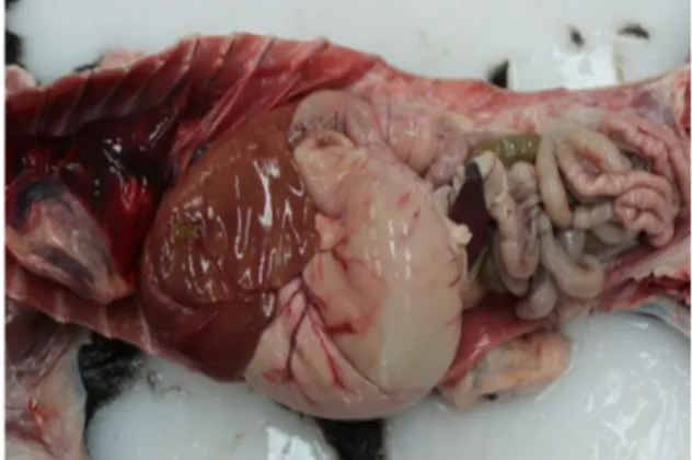
Fig. 1. Gross finding of the stomach. The stomach was abnormally enlarged.
로 증가하였다(Francis 등, 1979; Cotter, 1983; Mooney 등, 1989). 또한, T 세포 receptor gene rearrangement analysis 도입과 같은 진단기술의 발전으로 최근 위장 관 림프종의 발생 보고가 증가하고 있다(Grover, 2005).
위장관 림프종의 임상증상은 대부분 만성이며 발 병 후 1∼3개월 후에 증상이 나타난다(Moore와 Ogilvie, 2001). 주된 임상증상은 식욕부진과 체중감소이며 구 토는 50% 미만, 설사는 약 30% 정도의 개체에서 보 고되었다(Moore와 Ogilvie, 2001; Richter, 2003). 드물 게는 무기력, 허약, 갈증, 다뇨증, 그리고 복부팽만 등 이 나타난다(Grover, 2005).
위장관 림프종은 주로 소장에서 부분적 또는 전반 적인 비후를 나타낸다(Moore와 Ogilvie, 2001). 소장에 서 발병한 림프종은 내시경 검사로 진단하기 어렵기 때문에 개복술을 통한 전층 생체검사가 권장된다 (Okuda 등, 1994; Moore와 Ogilvie, 2001; Richter, 2003).
고양이 소장 림프종은 T 세포 림프종의 비율이 가장 높으며 특히 공장에서 T 세포 림프종이 다발한다 (Moore 등, 2012). B 세포와 T 세포 림프종은 형태학 적 특성으로 쉽게 진단할 수 있으며, CD3와 CD79
를 이용한 면역조직화학염색을 통해 유래 세포를 감 별할 수 있는 것으로 알려져 있다(Moore 등, 2012).
현재까지 고양이의 소장 림프종에 대한 국내 보고 사례가 드물기 때문에 본 증례를 통해 11세령의 수컷 잡종 고양이에서 발생한 소장 T 세포 림프종에 대한 육안 및 조직 병리학적 특징을 보고하고자 한다.
증 례
11세령 수컷 단모종 잡종 고양이가 지속적인 구토 와 체중감소를 보여 서울의 한 동물병원에 내원하였 다. 혈액검사결과 탈수가 확인되어 수액처치와 구토 에 대한 대증치료를 실시하였으며 초음파와 방사선 검사를 실시하여 위 확장과 장 운동이 거의 없음을 확인하였다. 다음 날, 조영촬영을 한 결과 물리적 장 폐색이 의심되어 탐색적 개복술을 실시하였으나 이 상소견은 확인되지 않았다. 수액처치, 항구토제, 위벽 보호제 등 치료를 실시한 결과 장 운동이 정상화되고 구토증상도 호전되었다. 그러나 두 달 후 다시 장 운 동이 멈추고 구토가 재발하였으며 병증의 속도가 매 우 빠르게 진행되었다. 심한 설사와 함께 결국 사지 마비가 진행되어 예후가 불량하다고 판단하였고 안 락사를 실시하였다. 고양이는 병리조직학적 검사를
위하여 농림축산검역본부 질병진단과에 부검 의뢰되 었다.
육안적으로 위는 팽창되어 있었다(Fig. 1). 위 내강 에는 원형 그대로의 펠렛 사료와 맑은 황색조의 끈끈 한 점액성 내용물이 존재하였다. 공장의 중하부부터 회장 상부까지 내강은 정상에 비해 좁아져 있었으며 대장 내용물은 정상이었다. 폐는 전반적으로 발적되 고 습윤하였으며 간은 정상에 비해 창백하였다. 췌장 에는 직경 1 mm 정도의 흰색 반점이 산재하였다. 기 타 장기에서는 특이 병변이 관찰되지 않았다.
병리조직학적 검사를 위하여 실질장기인 폐, 심장, 간, 비장, 신장, 위, 소장, 대장 등을 적출하여 10% 중 성 완충 포르말린에 고정하였다. 일반적인 조직 처리 과정을 거쳐 파라핀에 포매하고 4 m 두께로 조직절 편을 제작하여 Hematoxylin & Eosin (H&E) 염색을 실 시하였다. 또한, 종양세포의 기원을 확인하기 위하여 소장 조직에 대해 절편을 제작한 뒤 silane 코팅 슬라 이드 (Muto pure chemicals, Japan)에 부착시키고 Ventana discovery XT (Ventana Medical System, Tucson, AZ, USA)와 DAB Detection System (Ventana Medical System, Tucson, AZ, USA)을 이용하여 면역조직화학염색을 실시하였다. 1차 항체로는 CD3 (Dako, Denmark)와 CD79 (Dako, Denmark)를 각각 사용하였다.
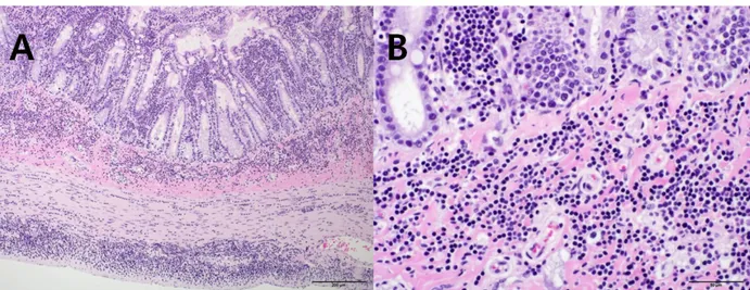
병리조직학적 검사결과, 소장의 점막, 점막밑, 근
육, 장막 층에 원형의 림프구양 세포가 광범위하게
관찰되었다(Fig. 2A). 이들은 균일한 형태로 적혈구의
1∼1.5배 정도 크기였으며 세포질은 거의 없고 핵은
진한 호염기성으로 염색되었으며 유사분열상은 관찰
되지 않았다(Fig. 2B). 이러한 소견은 공장에서 가장
두드러지게 관찰되었고 십이지장과 회장에서도 미약
하게 관찰되었다. 다른 실질장기에 대한 검사결과 종
Fig. 2. Histopathological findings in the jejunum. (A) Neoplastic cells were observed in the mucosa, submucosa, muscularis, and serosa of the jejunum. H&E stain, Bar=200 m. (B) Lymphoid neoplastic cells were round, with hyperchromatic nuclei and scant cytoplasm. H&E stain, Bar=50
m.
Fig. 3. Immunohistochemistry result of the jejunum. The neoplastic cells showed positive reactivity for CD3. IHC, Bar= 200 m.
양세포의 전이소견은 확인되지 않았다. 폐에서는 폐 수종과 폐포강 내 큰포식세포 침윤이 관찰되었고, 간 세포의 지방변성, 미약한 림프구성 췌장염이 확인되 었다. 면역조직화학염색 결과, 소장에 침윤된 세포는 T세포 마커로 사용되는 CD3에 강한 양성 반응을 보 였으나 B세포 마커로 사용되는 CD79에는 음성 반 응을 보여 종양 세포의 기원이 T 림프구임을 알 수 있었다(Fig. 3).
고 찰
본 증례는 소장에서 발생한 종양으로 소장의 점막, 점막밑, 근육, 장막 층에 림프구가 증식되어 있었고,
면역조직화학염색 결과, 대부분이 CD3에 양성 반응 을 보여 T세포 유래의 장 림프종으로 진단하였다.
고양이의 소장 림프종은 세계보건기구(World Health Organization: WHO)에서 분류한 장 병변 T 세포 림프 종(enteropathy-associated T cell lymphoma: EATL) 2형 분류와 일치하는 점막 T 세포 림프종이 가장 많이 나 타난다(Moore 등, 2012). EATL은 공장 또는 회장에서 가장 흔하게 발생하는 장상피 내 T 세포 악성 종양으 로 내시경 검사에서 다수의 궤양성 점막 결절 형태를 확인할 수 있으며 일부 사례의 경우 외적 형태로 관 찰할 수 있다(Isaacson 등, 2008). EATL은 셀리악 스 프루(celiac sprue)의 합병증으로 나타나는 1형 EATL 과 셀리악 스프루와 관련 없는 2형 EATL로 분류된다 (de Leval과 Gaulard, 2011). 1형 EATL은 다양한 크기 의 종양 림프구 집락이 관찰되며, 주로 불규칙한 수 포핵을 지닌 큰 림프구로 구성된다. 2형 EATL은 단 일형태인 중간 크기 림프구의 침윤이 관찰되며, 이들 은 세포질이 적고 과염색된 핵을 지닌 특징을 나타내 고 CD8, CD56에 함께 발현하거나 CD3와 TCR에 반 응을 나타낸다(Bautista-Quach 등, 2012). 본 증례는 림 프구의 조직학적 양상과 CD3에 양성을 보이는 특징 으로 2형 EATL인 것으로 추정된다. EATL의 두 유형 은 모두 5년 생존율이 8∼20%로 예후가 불량하고 발 병 개체는 장의 흡수불량이나 다른 합병증에 의해 폐 사한다(Isaacson 등, 2008; Van de Water 등, 2010).
고양이 소화기 림프종은 일반적으로 점막 내에서
발생하여 점막근육판과 점막밑층으로 침윤하는 것으
로 알려져 있다(Wilcock, 2012). 소형 림프구성 T 세
포 융모 림프종은 노령의 고양이에서 나타나며 융모 기저부에 소형 T 세포 림프구의 축적으로 질병이 진 행되기 시작한다. 이 세포들은 점차 점막 상피에서 고유층까지 퍼져나가며 장벽 전체로 침윤될 수 있다 (Carreras 등, 2003). 큰 세포(림프아구) 림프종은 연령 에 상관없이 나타나며 더 공격적이고 빠르게 진행되 고 장간막 림프절로 전이되는 특징을 보인다(Wilcock, 2012). 본 증례에서 또한 적혈구의 1∼1.5배 정도 크 기의 소형 T세포 림프구가 발견되었으며 장벽 전체 로 침윤된 양상을 관찰할 수 있었다.
고양이 소화기 림프종은 림프구형질세포성 염증 (lymphoplasmacytic enteritis)과 감별하여야 한다. 림프 구형질세포성 염증은 위장관에 림프구와 형질세포의 침윤을 나타내는 특발성 염증성 장질환(inflammatory bowel disease: IBD)의 한 형태이다(Hart 등, 1994). 올 바른 치료를 위해서 이들 질병의 감별은 매우 중요하 다(Briscoe 등, 2011). 소화기 림프종은 종양세포가 상 피세포에 침윤하는 정도로 염증성 병변과 구분할 수 있다. 소화기 림프종은 점막밑층보다 더 깊은 곳까지 림프구가 증식하거나 증상이 심할 경우 융모와 음와 의 구조적 변화를 초래한다. 반면, 림프구형질세포성 염증은 형질세포가 점막까지만 침윤된 양상을 보이 며 점막밑층보다 더 깊이 침윤된 경우는 오직 소화기 림프종 사례에서만 관찰되었다(Briscoe 등, 2011). 그 러므로 위장관 림프종을 정확히 진단하기 위해서는 소화기 전층 생검을 통한 조직학적 검사가 중요하다.
내시경을 통한 생검은 소장의 점막과 점막밑층 일부 만 채취하는 한계가 있으므로 염증성 장질환과의 감 별이 어려울 수 있다. 본 증례는 림프구 단일 세포의 증식이 관찰되었고 점막에서부터 장막까지 전층에 걸친 분포가 확인되었기 때문에 염증성 장질환과의 감별이 가능하였다.
국내 고양이 림프종 사례는 12세령의 잡종 고양이 와 7세령의 러시안블루 고양이에서 위장관 림프종이 발병된 증례가 보고된 바 있으며, 이들은 주로 방사 선 비투과성 표지자(radiopaque marker) 검사와 복부 초음파(hydrogram) 검사에 대한 결과를 중심으로 서 술하였으나, 림프종의 면역표현형은 구분하지 않았다 (Choi 등, 2012; Moore 등, 2012). 본 증례는 림프종의 조직학적 특징과 면역조직화학염색 결과 등을 고려 하여 림프종의 종류를 확인할 수 있었다.
림프종은 종양세포의 면역표현형에 따라 예후가 달라질 수 있는데, 특히 개의 경우는 T 세포 림프종 이 B 세포 림프종보다 예후가 더 안 좋은 것으로 알
려져 있다. 그러나 고양이의 경우는 개체의 치료 예 후 또는 생존과 종양세포 면역표현형과는 상관관계 가 없는 것으로 보고되어 있다(Cotter, 1983; Teske 등, 1994; Teske 등, 2002; Grover, 2005).
본 증례 환축의 경우, 폐사된 이후 위장관 전층의 조직검사를 실시하였으며 병변 부위가 소장이라는 점과 전반적으로 단일 형태의 소형 혹은 중간 크기인 종양 림프구가 침윤했다는 점, 면역조직화학적 검사 결과 림프구 대부분이 T 세포인 점을 고려하여 소장 에서 발생한 T 세포 림프종으로 진단하였다.
결 론
11세령 수컷 단모종 잡종 고양이가 수개월간의 구 토와 체중감소, 장 운동 정지 증상을 나타내고 간헐 적이던 증상이 연속적으로 진행되어 부검 의뢰되었 다. 육안 및 조직 병리학적 검사, 면역조직화학적 검 사 결과 소장부 점막, 점막밑, 근육, 장막 층 부위에 림프구가 광범위하게 침윤하였고, 대부분의 세포가 T 림프구 유래로 판명되어 소장 림프종으로 진단하였 다. 환축은 소장부위에 림프구가 과도하게 증식하여 장 운동이 약화되었고, 이로 인해 식욕부진, 지속적 인 구토, 체중감소, 설사 등의 여러 임상증상이 나타 난 것으로 판단된다.
감사의 글
본 증례보고는 농림축산검역본부 농림축산검역검 사기술개발 시험연구비(N-1543069-2015-99-01)의 지 원을 받아 수행되었습니다.
REFERENCES
Bautista-Quach MA, Ake CD, Chen M, Wang J. 2012.
Gastrointestinal lymphomas: Morphology, immunophe- notype and molecular features. J Gastrointest Oncol 3:
209-225.
Briscoe KA, Krockenberger M, Beatty JA, Crowley A, Dennis MM, Canfield PJ, Dhand N, Lingard AE, Barrs VR.
2011. Histopathological and immunohistochemical evalu- ation of 53 cases of feline lymphoplasmacytic enteritis and low-grade alimentary lymphoma. J comp Path 145:
187-198.
Carreras JK, Goldschmidt M, Lamb M. 2003. Feline epithelio- tropic intestinal malignant lymphoma: 10 cases (1997- 2000). J Vet Intern Med 17: 326.
Choi JH, Lee JS, Jang JY, Choi HY, Seo JM, Lee MJ, Kim HW.
2012. Radiopaque markers and hydrogram in feline ali- mentary lymphoma. Korean J Vet Res 52: 147-151.
Cotter SM. 1983. Treatment of lymphoma and leukemia with cy- clophosphamide, vincristine, and prednisone. II. Treatment of cats. JAAHA 19: 166-172.
de Leval L, Gaulard P. 2011. Pathology and biology of peripheral T cell lymphomas. Histopathology 58: 49-68.
Fox LE. 2003. Therapeutic choices for the medical management of lymphoma. Waltham Feline Med Symp 1-8.
Francis DP, Cotter SM, Hardy WD, Essex M. 1979. Comparison of virus-positive and virus-negative cases of feline leu- kemia and lymphoma. Cancer Res 39: 3866-3870.
Grover S. 2005. Gastrointestinal Lymphoma in cats. Compendium Vet 741-750.
Hart JR, Sharker E, Patnaik E, Garvey MS. 1994. Lymphocytic- plasmacytic enterocolitis in cats: 60 cases (1988-1990).
JAAHA 30: 505-514.
Isaacson PG, Chott A, Ott G. 2008. Enteropathy associated T cell lymphoma. pp. 289-291. In: Swerdlow SH, Campo E, Harris NL, Jaffe ES, Pileri SA, Stein H, Thiele J, Vardiman JW (ed). WHO classification of tumors of haematopoietic and lymphoid tissues. 4th ed. IARC, Lyon.
Mooney SC, Hayes AA, MacEwen EG. 1989. Treatment and prognostic factors in lymphoma in cats: 103 cases (1977-1981). JAVMA 194: 696-699.
Moore AS, Ogilvie GK. 2001. Lymphoma. pp. 191-219. In:
Yardley, PA(ed). Feline oncology: A comprehensive guide to compassionate Care. Veterinary Learning Systems, Treton, NJ.
Moore PF, Rodriguez-Bertos A, Kass PH. 2012. Feline gastro- intestinal lymphoma: mucosal architecture, immuno- phenotype, and molecular clonality. Vet Pathol 49:
658-668.
Okuda M, Umeda A, Sakai T, Ohashi T, Momoi Y, Youn HY, Watari T, Goitsuka R, Tsujimoto H, Haseqawa A. 1994.
Cloning of the feline p53 tumor-suppressor gene and its aberration in hematopoietic tumors. Int J cancer 58:
602-607.
Teske E, van Heerde P, Rutteman GR, Kurzman ID,Moore PF, MacEwen EG. 1994. Prognostic factors for treatment of malignant lymphoma in dogs. JAVMA 205: 1722-1728.
Teske E, van Straten G, van Noort R, Rutteman GR. 2002.
Chemotherapy with cyclophosphamide, vincristine, and prednisolone (COP) in cats with malignant lymphoma:
new results with an old protocol. J Vet Intern Med 16:
179-186.
Van de Water JM, Cillessen SA, Visser OJ. 2010. Enteropathy associated T cell lymphoma and its precursor lesions.
Best Pract Res Clin Gastroenterol 24: 43-56.
Waly NE, Gruffydd-Jones TJ, Stokes CR, Day MJ. 2005.
Immunohistochemical diagnosis of alimentary lympho- mas and severe intestinal inflammation in cats. J Comp Pathol 133: 253-260.
Wilcock B. 2012. Histopathology. pp. 345-347. In: Washabau RJ, Day MJ (ed). Canine and Feline Gastroenterology.
Missouri, St. Louis.