ISSN 1225-6552, eISSN 2287-7630 http://dx.doi.org/10.7853/kjvs.2014.37.4.313
< Short Communication >
Veterinary Service
Available online at http://kjves.org
*Corresponding author: Okjin Kim, Tel. +82-63-850-6668, Fax. +82-63-850-7308, E-mail. [email protected]
An occurrence of mammary spindle cell carcinoma in a dog
Sunhwa Hong
1, Hyun-A Lee
1, Dong-Woo Kim
1, Tae-Wan Kim
2, Okjin Kim
1*
1
Center for Animal Resources Development, Wonkwang University, Iksan 570-749, Korea
2
College of Veterinary Medicine, Kyungpook National University, Daegu 702-701, Korea (Received 23 August 2014; revised 11 November 2014; accepted 17 November 2014)
Abstract
A bitch was presented for investigation of the mass in left 5th mammary gland. The partial mastectomy was performed and submitted for the histopathological diagnosis. The mammary mass was firm and white colored. The cut surface was separated with several lobules and developed vessels. The central area of the mass formed the cavity filled with inflammatory exudates. The dominant component of the tumor was the bundles of spindle-shaped cells. Some tumor cells possessed atypical nuclei and were arranged in a solid nest. Cysts were microscopically composed of hemorrhage, necrosis, and exudates, partially surrounded by tumor cells and granulation tissues. Histopathologically, the mammary mass re- vealed spindle cell carcinoma. The bitch made a complete recovery following the mastectomy. This case was a rare mammary spindle cell carcinoma in a dog.
Key words : Mammary gland, Tumor, Spindle cell, Carcinoma, Canine
INTRODUCTION
One of the great advantages of canine mammary gland tumor model is that it is spontaneous in this organ. As the clinical evolution of spontaneous canine breast cancers is natural, their genetic and morphophy- siological aspects may be very much informative com- pared with some aspects of the human species (Vail and MacEwen, 2000). In comparative pathology, canine mammary gland tumors have been given special interest because of their similarities with human breast cancer.
Malignant epithelial tumors of the canine mammary gland that are composed of solid masses of interlacing elongate cells are relatively rare and may closely re- semble fibrosarcomas (Kusewitt et al, 1992). Such tu- mors are generally classified as spindle cell carcinomas and are often considered a subtype of solid carcinoma (Misdorp et al, 1972). Some pathologists have used the term ‘malignant myoepithelioma’ to identify the tumor
(Fowler et al, 1974), although there is little evidence to suggest that the tumors originate from myoepithelial cells. Recently, the incidences of canine mammary tu- mors have been increased in Korean veterinary clinics (Cho et al, 2008). The mammary gland tumor ranks first as the epidemiological issue in oncology of both female dogs and humans (Vail and MacEwen, 2000). Here, we report a rare mammary spindle cell carcinoma in a fe- male dog.
CASE REPORT
This study describes a case of the mammary spindle cell carcinomas in a dog. A 13-year old female mongrel dog was 9 kg weights and presented for investigation of mammary mass on the left 5th udder. The mass was en- larged from 3 to 9 cm diameter during recent 3 months.
The bitch had menopause status from 2 years ago. The
dog had been submitted to mammary dissection. Staging
of the tumors were evaluated by the physical examina-
Table 1. Laboratory results of the female mongrel dog in this case
Blood count Differential count (10
3/L ) Blood chemistry
WBC (10
3/L) 11.0 (5.0∼14.1)* Neutrophils segment 6.16 (2.9∼12.0) ALP (IU/L) 198 (1∼114)
RBC (10
6/L) 7.0 (4.95∼7.87) Lymphocyte 4.73 (0.4∼2.9) ALT (IU/L) 71 (10∼109)
HB (g/dL) 16.2 (11.9∼18.9) Monocyte 1.1 (0.1∼1.4) BUN (mg/dL) 5.0 (8∼28)
PCV (%) 45.5 (35∼57) Eosinophil 0 (0∼1.3) Creatinine (mg/dL) 0.60 (0.5∼1.7)
MCV (fl) 66.0 (66∼77) Basophil 0 (0∼0.14)
MCHC (g/dL) 35.8 (32.0∼36.3)
*The reference ranges in blanks have been derived from the textbook ‘Ducan & Prasse’s Veterinary Laboratory Medicine - Clinical Pathology’.
Fig. 1. Gross photograph of the mammary mass. (A) Mammary mass on the left 5th udder, (B) 9 cm diameter-mass, (C) Firm and white colored dissected mass, (D) The central cavity filled with in- flammatory exudates.
Routine screening laboratory tests were also performed on the basis of hematology. Blood cell counting and se- rum chemistry were performed with Fully Automatic Hematology Analyzer for Multispecies (Hemavet 950 FS, Drew Scientific Inc, TX, USA) and 7150 automatic analyzer (Hitachi Co, Tokyo, Japan). After surgical mas- tectomy operation, the mammary mass was fixed in 10% formalin, and was submitted for histopathological examination. Following fixation, tissues were processed routinely and embedded into paraffin blocks. Four-mi- crometer-thick sections were cut and stained with hema- toxylin and eosin.
Blood test revealed lymphocytosis and mild increase
(ALP) (Table 1). The mastectomy was selected and mi- croscopic histopathological examination was performed for the final diagnosis. Expanding and effacing the mammary gland was a 3x9 cm, unencapsulated, in- filtrative, multilobulated neoplasm with multifocal areas of necrosis, hemorrhage, mineralization, and acicular clefts (Fig. 1A, B). The mastectomy was selected and microscopic histopathological examination was per- formed for the final diagnosis. The mammary mass was firm and white colored (Fig. 1C). The cut surface was separated with several lobules and developed vessels.
The central area of the mass formed the cavity filled
with inflammatory exudates (Fig. 1D).
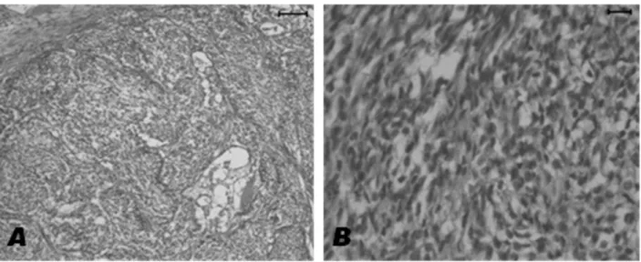
Fig. 2. Histopathological findings of the mammary mass. The dominant component of the tumor was the bundles of spindle-shaped cells.
Hematoxylin & Eosin stain. (A) ×100, (B) ×400.
Histopathologically, the neoplasm incorporates and compresses adjacent ducts and elevates the overlying epidermis. Lobules are separated by variably thick bands of collagen and are composed of polygonal neoplastic cells arranged in islands and solidly cellular areas sup- ported by fine fibrovascular stroma. The dominant com- ponent of the tumor was the bundles of spindle-shaped cells. The histological lesions were composed with bun- dles and whorls of fusiform cells without a lobular pat- tern and lack of glandular or tubular differentiation (Fig.
2A). Neoplastic cells resembled plump fibroblasts with round or elongated vesicular nuclei and frequently va- cuolated cytoplasm and were frequently embedded in or surrounded by a pink to light blue chrondromatous matrix. Some tumor cells possessed atypical nuclei and were arranged in a solid nest (Fig. 2B). Cysts were mi- croscopically composed of hemorrhage, necrosis, and exudates, partially surrounded by tumor cells and gran- ulation tissues. On the basis of the histopathognomic le- sions consisting of the bundles of spindle-shaped cells, the mammary mass revealed spindle cell carcinoma.
DISCUSSION
Dogs represent a remarkable incidence of neoplasia, usually associated with environmental exposure to vari- ous carcinogens which are important to humans
(Lindblad-Toh et al, 2005; Schneider et al, 1969).
Mammary tumors are the most common tumors in the female dog (Misdorp, 2002). Reports of the occurrence of malignant forms vary from 26 to 73% (Pérez Alenza et al, 1995), carcinoma being the most common malig- nant type (Misdorp et al, 1999). In dogs with high- grade simple carcinomas and metastasis to regional lymph nodes, the frequency of deaths was even worse.
According to many veterinary authors, lymph node in- volvement was related to prognosis (Hellmén et al, 1993; Yamagami et al, 1996; Misdorp, 2002), as in hu- man reports (Todd et al, 1987; Elston and Ellis, 1991).
ALP was abnormal in a high proportion of mammary
tumor cases with bone or liver metastases (Crivellari et
al, 1995). In this study, we observed elevated ALP
level. However, the metastasis to the regional lymph no-
des and other organs in the patient dog was not
occurred. We also detected the increased WBC numbers,
which may be induced by the inflammation of the mam-
mary tumor. The patient therefore had a good condition
and prognosis. In canine mammary gland tumor, the age
of the dog at mastectomy is considered by some authors
to be a factor of prognostic significance (Hellmén et al,
1993; Misdorp et al, 1999; Pérez Alenza et al, 1995). In
this study, the age of dog was very old. However, the
results of follow-up study revealed very healthy
recovery. Immunohistochemistry (IHC), in particular in
combination with the use of monoclonal antibodies
method in tumour evaluation, supplementary to histo- logical examination (Miettinen, 1990). But, IHC alone cannot decisively differentiate between tumours (Diaz et al, 1991; Tsubura et al, 1991). The current gold stand- ard for tumor diagnosis is histopathology (Nazir et al, 2010). In this study, we did not obtain a specific MoAb for canine mammary spindle cell carcinoma and could not conduct IHC for the differentiation of the tumor types. However, we diagnosed the case as a canine mammary spindle cell carcinoma based on the pathog- nomic histopathological lesions.
The bitch made a complete recovery following the mastectomy. This case was a rare mammary spindle cell carcinoma in a dog.
ACKNOWLEDGMENTS
This study was supported by the research fund of Wonkwang University in 2014. We wish to appreciate Yun-Seong Lee and Gi-Wook Oh, research assistants of Center for Animal Resources Development, Wonkwang University, for carrying out the histopathological work.
REFERENCES
Cho SJ, Son MW, Rho JR, Kim O. 2008. A case report of canine complex mammary gland tumor. Lab Anim Res 24:
457-460.
Crivellari D, Price KN, Hagen M, Goldhirsch A, Gelber RD, Castiglione M, Coates AS, Rudenstam CM, Collins J, Lindtner J, Cortes-Funes H, Gudgeon A, Simoncini E, Byrne M, Schniirch HG, Fey M, Tattersall MHN, Forbes JF, Cavalli F, Reed R, Senn HJ. 1995. Routine tests dur- ing follow-up of patients after primary treatment for op- erable breast cancer. International (Ludwig) Breast Cancer Study Group (IBCSG). Ann Oncol 6: 769-776.
Diaz NM, McDivit RW, Wick MR. 1991. Microglandular ad- enosis of the breast. An immunohistochemical compar- ison with tubular carcinoma. Arch Pathol Lab Med 115:
578-582.
Elston CW, Ellis IO. 1991. Pathological prognostic factors in breast cancer. I. The value of histological grade in breast cancer: experience from a large study with long-term follow-up. Histopathology 19: 403-410.
Fowler EH, Wilson GP, Koestner A. 1974. Biologic behavior of
classification. Vet Pathol 11: 212-229.
Hellmén E, Bergström R, Holmberg L, Spångberg IB, Hansson K, Lindgren A. 1993. Prognostic factors in canine mam- mary tumors: A multivariate study of 202 consecutive cases. Vet Pathol 30: 20-27.
Kusewitt DF, Hahn FF, Muggenburg BA. 1992. Ultrastructure of a spindle cell carcinoma in the mammary gland of a dog. Vet Pathol 29: 179-181.
Lindblad-Toh K, Wade CM, Mikkelsen TS, Karlsson EK, Jaffe DB, Kamal M, Clamp M, Chang JL, Kulbokas EJ 3rd, Zody MC, Mauceli E, Xie X, Breen M, Wayne RK, Ostrander EA, Ponting CP, Galibert F, Smith DR, DeJong PJ, Kirkness E, Alvarez P, Biagi T, Brockman W, Butler J, Chin CW, Cook A, Cuff J, Daly MJ, DeCaprio D, Gnerre S, Grabherr M, Kellis M, Kleber M, Bardeleben C, Goodstadt L, Heger A, Hitte C, Kim L, Koepfli KP, Parker HG, Pollinger JP, Searle SM, Sutter NB, Thomas R, Webber C, Baldwin J, Abebe A, Abouelleil A, Aftuck L, Ait-Zahra M, Aldredge T, Allen N, An P, Anderson S, Antoine C, Arachchi H, Aslam A, Ayotte L, Bachantsang P, Barry A, Bayul T, Benamara M, Berlin A, Bessette D, Blitshteyn B, Bloom T, Blye J, Boguslavskiy L, Bonnet C, Boukhgalter B, Brown A, Cahill P, Calixte N, Camarata J, Cheshatsang Y, Chu J, Citroen M, Collymore A, Cooke P, Dawoe T, Daza R, Decktor K, DeGray S, Dhargay N, Dooley K, Dooley K, Dorje P, Dorjee K, Dorris L, Duffey N, Dupes A, Egbiremolen O, Elong R, Falk J, Farina A, Faro S, Ferguson D, Ferreira P, Fisher S, FitzGerald M, Foley K, Foley C, Franke A, Friedrich D, Gage D, Garber M, Gearin G, Giannoukos G, Goode T, Goyette A, Graham J, Grandbois E, Gyaltsen K, Hafez N, Hagopian D, Hagos B, Hall J, Healy C, Hegarty R, Honan T, Horn A, Houde N, Hughes L, Hunnicutt L, Husby M, Jester B, Jones C, Kamat A, Kanga B, Kells C, Khazanovich D, Kieu AC, Kisner P, Kumar M, Lance K, Landers T, Lara M, Lee W, Leger JP, Lennon N, Leuper L, LeVine S, Liu J, Liu X, Lokyitsang Y, Lokyitsang T, Lui A, Macdonald J, Major J, Marabella R, Maru K, Matthews C, McDonough S, Mehta T, Meldrim J, Melnikov A, Meneus L, Mihalev A, Mihova T, Miller K, Mittelman R, Mlenga V, Mulrain L, Munson G, Navidi A, Naylor J, Nguyen T, Nguyen N, Nguyen C, Nguyen T, Nicol R, Norbu N, Norbu C, Novod N, Nyima T, Olandt P, O'Neill B, O'Neill K, Osman S, Oyono L, Patti C, Perrin D, Phunkhang P, Pierre F, Priest M, Rachupka A, Raghuraman S, Rameau R, Ray V, Raymond C, Rege F, Rise C, Rogers J, Rogov P, Sahalie J, Settipalli S, Sharpe T, Shea T, Sheehan M, Sherpa N, Shi J, Shih D, Sloan J, Smith C, Sparrow T, Stalker J, Stange-Thomann N, Stavropoulos S, Stone C, Stone S, Sykes S, Tchuinga P, Tenzing P, Tesfaye S, Thoulutsang D, Thoulutsang Y, Topham K, Topping I, Tsamla T, Vassiliev H, Venkataraman V, Vo A, Wangchuk T, Wangdi T,
Weiand M, Wilkinson J, Wilson A, Yadav S, Yang S, Yang X, Young G, Yu Q, Zainoun J, Zembek L, Zimmer A, Lander ES. 2005. Genome sequence, com- parative analysis and haplotype structure of the domestic dog. Nature 438: 803-819.
Miettinen M. 1990. Immunohistochemistry of solid tumors. Brief review of selected problems. APMIS 89: 191-199.
Misdorp W, Cotchin E, Hampe JF, Jabara AG, von Sandersleben J. 1972. Canine malignant mammary tumors. II.
Adenocarcinomas, solid carcinomas and spindle cell carcinomas. Vet Pathol 9: 447-470.
Misdorp W, Else RW, Hellmen E, Lipscomb TP. 1999.
Histological Classification of Mammary Tumors of the Dog and the Cat. Armed Forces Institute of Pathology and the American Registry of Pathology and The World Health Organization Collaborating Center for Worldwide Reference on Comparative Oncology, Washington, D.C., USA, vol. 7, pp. 11-29.
Misdorp W. 2002. Tumors of the mammary gland. In: Tumours in Domestic Animals, 4th Edit., D. J. Meuten, Ed., Iowa State Press, Iowa, pp. 575-606.
Nazir RT1, Sharif MA, Iqbal M, Amin MS. 2010. Diagnostic ac- curacy of fine needle aspiration cytology in hepatic
tumours. J Coll Physicians Surg Pak 20: 373-376.
Pérez Alenza MD, Rutteman GR, Kuipers-Dijkshoorn NJ, Peña L, Montoya A, Misdorp W, Cornelisse CJ. 1995. DNA flow cytometry of canine mammary tumours: the rela- tionship of DNA ploidy and S-phase fraction to clinical and histological features. Res Vet Sci 58: 238-243.
Schneider R, Dorn CR, Taylor DO. 1969. Factors influencing ca- nine mammary cancer development and postsurgical survival. J Natl Cancer Inst 43: 1249-1261.
Tsubura A, Okada H, Senzaki II, Hatano T, Morii S. 1991.
Keratin expression in the normal breast and in breast carcinoma. Histopathology 18: 517-522.
Todd JH, Dowle C, Williams MR, Elston CW, Ellis IO, Hinton CP, Blamey RW, Haybittle JL. 1987. Confirmation of a prognostic index in primary breast cancer. Br J Cancer 56: 489-492.
Vail DM, MacEwen EG. 2000. Spontaneously occurring tumors of companion animals as models for human cancer.
Cancer Invest 18: 781-792.
Yamagami T, Kobayashi T, Takahashi K, Sugiyama M. 1996.
Prognosis for canine malignant mammary tumors based on TNM and histologic classification. J Vet Med Sci 58:
1079-1083.