48(1) : 31∼ 37 (2017)
31
글루타메이트로 유발한 세포독성에 대한 삼채추출물의 뇌세포 보호 효과
김지윤1#·고원민2#·김애정1*
1경기대학교 일반대학원 대체의학과, 2원광대학교 약학대학
Effects of Allium hookeri Extracts on Glutamate-induced Neurotoxicity in HT22 Cells
Ji-Yun Kim1#, Wonmin Ko2# and Ae-Jung Kim1*
1The Graduate School of Alternative Medicine, Kyonggi University, Seoul 03752, Korea
2College of Pharmacy, Wonkwang University, Iksan 54538, Korea
Abstract − Glutamate-induced oxidative stress results in neuro-degenerative disorders in many central nervous system (CNS) such as Alzheimer’s disease, ischemia, Huntington’s disease, and Parkinson’s disease. Our study was performed to investigate neuroprotective effects of Allium hookeri extracts (leaf, root, and whole) on glutamate-induced HT22 cells. In this study, ethanol extract of A. hookeri showed the outstanding neuroprotective effect in HT22 cells. In addition, we found that ethanol extract of A. hookeri root increased heme oxygenase (HO)-1 in HT22 cells. Moreover, ethanol extract of A. hookeri root also upreg- ulated nuclear accumulation of nuclear factor E2-related factor 2 (Nrf2) in HT22 cells. These results demonstrate that ethanol extract of A. hookeri root contributes neuroprotective effects against glutamate-induced oxidative stress in HT22 cells, via Nrf2- mediated HO-1 expression. Our study suggests that ethanol extract of A. hookeri root could be the potential agent for the treat- ment of many neuro-degenerative diseases.
Keywords − Allium hookeri, HT22 cells, Glutamate, Neuroprotective effect, Heme oxygenase-1, ROS
최근 현대의학의 발달과 식생활 향상으로 인간의 평균 수 명이 증가함에 따라 노인성 치매와 같은 뇌질환에 대한 관 심이 고조되고 있다.1)
파킨슨병(Parkinsom’s diseas), 알츠하이머(Alzhheimer’s disease: AD), 뇌졸중(Stroke) 및 헌팅턴 증후군(Huntington’s disease)과 같은 뇌질환의 주요 원인으로 활성 산소종(reactive oxygen spesies, ROS)이 주목받고 있다.2,3)
활성 산소종은 산화질소, 흡연, 자외선, 오존 등 여러 가 지 외부 요인에 의해서도 생성 되며, 동식물 체내 산소대사 과정에서 자연스럽게 생성되는 물질이다.4) 그러나 과도하게 생성된 활성 산소종은 정상 세포의 사멸, DNA 변성, 단백 질 분해, 지질 산화 등을 초래하여 노화, 심혈관계 질환 및 암질환 발생의 주범이다.5)
글루타메이트(glutamate)는 central nervous system(CNS) 의 대표 흥분성 신경전달 물질로 시냅스에서의 신경전달,
생존 및 생장, 학습과 기억력, 행동 등에 중요한 생리학적 기능과 역할을 한다.6) 그러나 글루타메이트 수용체의 과다 흥분에 의한 독성은 산화적 스트레스를 일으키고 뇌 세포 를 손상시켜 뇌 질환을 일으키는 주된 요인으로 보고되고 있다.7,8)
생쥐의 해마유래 세포주인 HT22 세포는 글루타메이트 수 용체가 결여되어 있는 세포주로 글루타메이트 처리 시 세 포손상을 확인할 수 있어서 산화적 스트레스로 인한 뇌세 포 보호 물질을 찾는 연구에 유용하게 이용되고 있다.9)
Nuclear transcription factor-E2-related factor 2(Nrf2)는 외부 자극과 산화적 스트레스에 노출 되었을 때, 핵 내로 translocation하여 antioxidant responsive element(ARE)에 결 합하여 다른 해독 효소들과 heme oxygenase-1(HO-1)의 발 현을 유도하여 세포를 방어하는 기능을 한다고 알려져 있 다.10,11)
HO-1는 세포 항산화 시스템에서 중요한 구성요소로 HO 유도체중 하나로 세포 내 heme을 분해하여 부산물인 billiverdin, carbon monoxide(CO), free iron(Fe)을 만들며,
#
These authors contributed equally to this work.
*교신저자(E-mail):aj5249@naver.com
(Tel): 82-2-390-5044
그 부산물과 함께 활성 산소종 제거, 독성으로부터 세포보 호, 세포손상 및 사멸억제, 항산화 작용을 한다.12,13)
Allium속 식물은 백합과(Lilliaceae)로 500여 종으로 이루 어져 있으며 주요 화합물은 유황으로 항균활성과 심혈관계 질환 예방 등 여러 생리적 유용성이 밝혀지고 있다.14) 양파 (Allium cepa L), 부추(Allium tuberosum), 마늘(Allium sativum) 등은 대표적인 Allium속 식물들인데, 이 들 Allium 속 식물은 향신료로 수세기 동안 사용되어져 왔으며 현재 까지도 널리 활용되고 있다.15) 특히 Allium속 식물이 함유하 고 있는 유황화합물은 살균력이 강해 염증반응을 감소시켜 아토피, 건선 및 무좀 등과 같은 피부질환에 효과가 뛰어나 다고 알려져 있다.16) 특히 콜레스테롤 합성 억제와 혈전분 해 성질이 있어 혈전으로 인해 발생하는 당뇨병, 고혈압, 고 지혈증 등을 예방해 주는 효과도 있다.17)
삼채(Allium Hookeri) 역시 allium속 식물로 숲, 늪지, 해 발 1400~4200 m의 초원지대에 자생하며 동아시아의 중국 남부, 인도, 부탄, 스리랑카 등에 분포하고 있다.중국인들은 3000년 전 전부터 약용과 식용으로 이용해 왔으며 잎, 순, 뿌리 모두가 식용으로 이용 가능하다고 알려져 있다.18) 최 근 우리나라에서도 일반 파속 식물에 비해 유황화합물이 6 배 정도 많다고 알려져 있어 건강식품으로 삼채의 효능에 관심도가 높아지고 있다.19)
삼채에 대한 선행연구를 조사해 보면 Kim 등20)의 삼채 뿌리 메탄올 추출물에서 항염증 효과, Bae 등21)의 삼채 뿌 리 에탄올 추출물에서 항염증 효과, Won 등22)의 삼채의 이 화학적 성분, 항산화 작용, DPPH radical 소거능 및 NO 생 성 억제능, Cha 등23)의 항균 활성과 항산화 활성, 지질대사 및 당질에 미치는 효과의 연구24,25)가 있을 뿐 국내산 삼채 에 대한 연구는 매우 미비한 실정이다.
따라서 본 연구에서는 국내에서 재배되고 있는 삼채에 대 한 용매별(에탄올, 열수), 부위별(잎, 뿌리, 전체)로 나누어 생쥐의 해마유래 세포주인 HT22세포를 이용하여 HO-1 발 현에 대한 뇌세포 보호효과를 살펴보고자 하였다.
재료 및 방법
실험재료 − 본 연구에서 사용된 삼채는 강원도 대관령 삼 채농장(Gangwon-do, Korea)에서 11월에 재배 생산된 삼채 를 구입하여 사용하였으며, 경기대학교 대체의학대학원 김 애정 박사에 의한 형태학적 평가를 통하여 동정하였다. 삼 채는 흐르는 물에 수세 동결건조(NIHON freezer, Tokyo, Japan)한 후 분말화(Mixer HM-5000, Hyun Dae Household Appliances, Seoul, Korea)하였다.
시약 및 기기 − DMEM 배지와 trypsin-ethylenediamin- etetraacetic acid(EDTA)는 Gibco Laboratories사에서 구입하 였으며, fetal bovine serum(FBS)는 Hyclone Laboratories사에
서 구입하였다. L-glutamate, Trolox와 3'-(4,5-dimethylthiazol- 2-yl)-2,5-diphenyltetrazolium bromide(MTT)는 Sigma사에서 구입하였다.
96-well tissue culture plates와 기타 tissue culture dishes 는 Nunc사 제품을 이용하였다. 흡광도는 BioRad사의 Microplate Reader(Bio-Rad Laboratories, Philadelphia, PA) 를 이용하여 측정하였다. 모든 시약은 Sigma사(St, Louis, MO, USA)에서 구입하여 사용하였다.
시료의 제조 − 삼채 잎(A.H L EtOH)과 뿌리 동결건조 분 말(A.H R EtOH)을 각각 10 g씩 500 mL 증류수와 500 mL 99% 에탄올을 사용하여 2시간 동안 가열 환류추출(HEATING MANTLE, MS-EAM920400, Seoul, Korea)하였고, 여과 (No. 2, Whatman, Maidstone, England)한 다음 여액을 감압 농축(EYELA Digital water bath, SB-1000, Tokyo, Japan) 하여 -20oC에 보관하며 실험에 사용하였다.
세포배양 및 뇌 세포 보호활성 측정 − 마우스 해마유래 HT22 세포주는 묵인희 교수(Seoul National University, Seoul, Korea)로부터 분양 받아 사용하였으며, DMEM 배지 에 10% FBS와 1% antibiotics를 첨가하여 37oC의 5% CO2 incubator에서 1-2일 마다 계대 배양하면서 실험에 사용하였 다.
글루타메이트로 유도한 뇌세포 독성에 대한 보호 효과는 Jeong 등의 방법26)에 따라 실시하였다. 종양세포를 3×10⁴cells/
mL의 농도가 되도록 조절한 후 96 well plate에 1×10⁴cell/
well로 동일하게 분주하고 12시간 동안 배양하였다. 기존의 배지를 제거하고 새로운 배지를 넣어준 후 DMSO에 녹인 시료를 다양한 농도(100, 200, 400 µg/mL)로 DMEM 배지 에 희석하여 첨가하였다. DMSO의 처리 농도는 배지 대비 0.1% 이하가 되도록 하였다. 이를 다시 12시간 배양한 후에 배지를 제거하고 MTT시약(2.5 mg/ml)을 넣고, 3시간 동안 방치한 후 상등액을 제거하였다. 각 well에 형성된 formazan 에 DMSO 150 µl를 첨가한 후 orbital shaker를 이용하여 녹 이고, 30분 후 540 nm에서 흡광도(BioRad, Hercules, CA, USA)를 측정하였다. 실험은 3회 반복 실시하여 평균값을 구하였으며, Control의 흡광도 값을 기준으로 생존율을 비 교하였다. HT22세포를 96 well microplate에 1×10⁴cells/
well이 되게 분주한 후, 37oC의 5% CO2 incubator에서 12 시간 배양한 다음, 5 mM의 글루타메이트와 각 추출물과 분 획물(100, 200, 400 μg/ml)을 농도별로 처리하였다. 시료 처 리 12시간 후 세포 생존율을 MTT 방법으로 측정하였으며, Trolox 50 μM을 양성 대조군으로 하여 비교하였다. 모든 실 험은 대조군에 대한 세포 보효율을 mean±S.D.로 표시하였 으며, 각각 3회 반복 실험치를 이용하여 계산하였다.
ROS(Reactive Oxygen Species) 측정 − 배양된 세포를 PBS로 세척한 후, 10 μM 2',7'-dichlofluorescein diacetate (DCFDA, 35845)를 포함하는 Hank' balanced salt 용액에서
30분 동안 암실에서 반응시킨 후 세포의 형광광도(excitation wave length: 490 nm; emission wave length: 525 nm)를 microplate reader(SpectramaxGemini XS, Molecular Devices, Sunnyvale, CA, U.S.A.)로 측정하였다.
Western Blot Analysis − HT22 세포를 60 mm dish에 2×10⁵cells/well 농도로 12시간 배양한 후 각각의 시료를 농 도별로 처리하였다. HT22 세포에 RIPA buffer를 첨가한 다 음, 4oC, 16,000 rpm 에서 15분간 원심분리하고 상등액을 튜 브에 옮겼다. 단백질 정량은 BSA 단백질 실험 키트를 이용 하였고 각각의 시료를 12% SDS-polyacrylamide gel에서 영 동하고 Nitrocellulose membrane(NC membrane)으로 전사 하였다. 전사된 NC membrane을 5% 무지방유가 포함된 신 선한 blocking buffer(0.1% Tween 20 in Tris-buggered saline)에서 blocking한후 HO-1 antibody를 1:1000으로 희석 하여 넣고 1시간 30분 동안 반응시켰다. 다시 2차 안티바디 (Anti-mouse IgG)를 1:1000으로 희석하여 넣고 1시간 동안 반응한 다음, ECL 용액(enhanced chemiluminescent kit, Amersham Co.)을 1:1로 잘 섞어서 NC membrane 위에 부 어서 발광시키고 암실에서 X선 필름에 감광한 후 현상하였 다. 같은 방법으로 Actin antibody를 이용하여 Actin을 측정 한다.
핵과 세포질 분획 − HT22 세포에 protease inhibitor cocktail I과 1 mM phenylmethylsulfonyl fluoride(PMSF)를 첨가한 PER-Mammalian Protein Extraction buffer를 첨가하 여 균질화하고 4oC에서 14,000×g로 원심분리 하였다. 10분 간 원심분리 후 상등액을 세포질 분획으로 사용하기 위하 여 -80oC에서 저장하였다. 나머지 침사물은 PBS로 세척한 후 RIPA buffer[150 mM NaCl, 1% NP-40, 0.5% sodium deoxycholate, 0.1% SDS, 50mM Tris-HCl(pH 7.4), 50mM glycerophosphate, 20 mM NaF, 20 mM ethylene glycol tetraacetic acid(EGTA), 1 mM dithiothreitol(DTT), 1 mM Na3VO4, protease inhibitors]를 첨가하고 40oC에서 15분간 혼합한 후 4oC, 16,000×g에서 15분간 원심분리한다. 이후의 과정은 상술된 western blotting 방법을 이용하였다.
통계 처리 − 본 실험의 통계처리는 GraphPad Prism, version 3.03(GraphPad Software Inc., San Diego, CA, USA)을 사용하였다. 각 실험군의 결과는 평균치와 표준오 차로 나타내었으며, 각 실험군 간의 결과는 ANOVA test를 사용하여 분석하고 유의적인 차이가 있는 항목에 대해서만 검정하였다. 실험군 간의 차이는 95% 수준(p<0.05)에서 유 의성 검정을 실시하였다.
결과 및 고찰
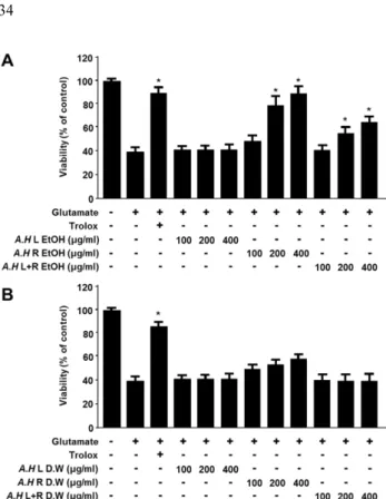
글루타메이트로 유발한 세포독성에 대한 뇌세포 보호효 과 − 용매(에탄올, 열수)에 따른 삼채 추출물 잎(A.H L), 뿌
리(A.H R), 그리고 전체(A.H L+R)에 대한 HT22 세포의 생 존율에 미친 영향을 알아보고자 농도별(100, 200, 및 400 µg/
mL)로 세포에 처리하여 생존율을 측정하였다(Fig. S1).
본 연구결과 HT22 세포에 처리된 삼채 잎과 뿌리, 전체 추출물은 400 µg/mL 이하의 농도에서 거의 100%의 생존율 을 보여 잎과 뿌리 모두 세포에 독성이 없는 것으로 나타났 다.
삼채 잎(A.H L), 뿌리(A.H R), 그리고 전체(A.H L+R) 추 출물(물과 에탄올)이 세포독성을 나타내지 않은 100, 200, 및 400 µg/mL농도에서 실험을 진행하였으며, 뇌세포 보호 효과를 측정하고자 글루타메이트를 처리하여 산화적 스트 레스를 유발시킨 생쥐의 해마 유래 HT22 세포의 생존율과 뇌세포 보호 효과를 검색하였다. 뇌세포 보호 효과 실험의 유의성 확인을 위한 양성 대조약물로는 항산화 물질로 알 려진 trolox(50 μM)을 사용하였다. 실험 결과, 처리 농도가 증가함에 따라서 세포 생존율 농도도 의존적으로 증가한 것 을 확인하였다(Fig. 1).
삼채 뿌리 에탄올추출물(A.H L EtOH) 400 μg/mL 농도 처리 시 81.3%의 생존율로 보호 효과가 가장 높았으며, 삼 Fig S1. Effects of A. hookeri EtOH (A) and distilled water (B) extracts on cell viability in HT22 cells. Cells were incubated for 24 h with various concentrations of A. hookeri extracts.
After 24 h, cell viability was measured by MTT assay as described in Materials and methods. Each bar represents the mean ± SD of three independent experiments.
채 뿌리 에탄올 추출물 200 μg/mL에서 68.5%, 증류수 용매 뿌리 400 μg/mL에서는 45.3%로 용매별, 부위별 다른 세포 생존율을 나타내었으며, 비교 결과 증류수 용매보다 에탄올 용매에서 높은 보호효과를 나타내었다.
유황에 관한 또 다른 선행연구 보고에 의하면 Munagami 등 쥐를 사용한 실험에서 S-allylcysteine 투여한 경우, 뇌경 색 감소, 운동능력 향상, 기억손상의 회복 등을 관찰하였 다.27) Nishiyama 등은 유황 유기화합물이 식이 내 2% 수준 으로 장기 투여하였을 때 노화촉진 마우스(SAM)에서 학습 능력과 전뇌 쇠퇴를 예방할 수 있다고 보고하였다.28)
본 연구 결과 삼채 뿌리가 잎에 비해 뇌세포 보호효과에 더 효과적으로 작용한 결과는 유황 함유량이 잎보다 뿌리 에 더 높게 나타났기 때문이라고 사료된다. 추후 삼채 부위 별 유황의 함유량에 따른 뇌세포 보호효과에 대한 실험이 좀 더 필요하다 생각된다.
Nrf2 핵내전사를 통한 Heme oxygenase(HO)-1 단백 질 발현 효과 − HO-1은 항산화 시스템에 주요한 역할을 하 는 단백질로서, 현재 많은 연구에 의하면 HO-1 단백질의 발 현에 대한 메커니즘은 nuclear factor-E2-related factor 2(Nrf2)의 핵 내 전사가 가장 직접적인 관련이 있는 것으로 알려져 있다. Nrf2는 전사인자로서 HO-1과 같은 항산화 단 백질의 유전자에 존재하는 ARE(Antioxidant Response Element)에 결합하여 이들 유전자의 발현을 조절하고 단백 질발현을 항진시킴으로써 산화적 스트레스에 대한 생체방 어기구에 중심적 역할을 담당하고 있다.10)
산화적 스트레스로부터의 뇌세포 보호 기전에 있어 중요 한 단백질인 Heme oxygenase-1(HO-1)의 발현 정도를 알아 Fig 1. Cytoprotective effect of A. hookeri EtOH (A) and dis-
tilled water (B) extracts on glutamate-induced neurotoxicity in HT22 cells. Cells were treated with various concentrations of A. hookeri extracts and then incubated for 12 h with glutamate (5 mM). Cell viability was determined by MTT assay as described in the Materials and methods. Each value represents the mean ± SD of three experiments. *p<0.05 compared with the group treated with glutamate alone. Trolox (50 µM) was used as a positive control.
Fig 2. Effects of A. hookeri root (EtOH) on the protein expression of HO-1 in HT22 cells. Cells were incubated for 12 h with the indicated concentration of A. hookeri root (EtOH) (A) and indicated periods with 400 µg/ml of A. hookeri root (EtOH) (B). Expres- sion of HO-1 was determined by Western blot analysis, and representative blots of three independent experiments are shown. Each bar represents the mean ± SD of three independent experiments. *p<0.05 vs. control. Band intensity was quantified by densitometry and normalized to actin; the values are presented at the bottom of each band.
보기 위해 뇌세포 보호 효과 결과에서 가장 높은 보호률을 나타낸 삼채 뿌리 에탄올 추출물을 이용하여 실험한 결과 농도 의존적으로 HO-1 단백질 발현을 증가시킨 것을 확인 하였다(Fig. 2A).
시간대 별로 관찰한 결과 3시간까지는 반응을 보이지 않 았으며, 6시간 이후부터 점차 반응을 나타내기 시작했다. 그 후 18시간에서 가장 높은 발현을 보였으며 24시간이 되었 을 때는 발현의 정도가 약간 감소하였다(Fig. 2B). 24시간 에서 HO-1의 발현이 약간 감소하는 경향이 나타나는 이유
로, 일반적으로 단백질의 발현은 자극에 의해 발현이 증가 되다가 정점에 이른 후 회복 단계에 이르러 발현이 감소하 는 형태를 나타내는데, HO-1의 발현 또한 정점에 이르는 시 간은 세포의 종류나 상태에 따라 차이가 나타나게 된다. 따 라서 우리의 결과와 같이 24시간에 이르기 전에 HO-1의 발 현량이 정점에 이른 후 감소하는 경우도 있고, 24시간 이후 까지 유지되는 경우도 나타난다.29-31) Bae 등은 생쥐 대식세 포주인 RAW264.7 세포에서 삼채 뿌리 에탄올추출물 500 μg/
mL 농도 처리 시 1시간 내 항염증에 관여하는 HO-1 발현
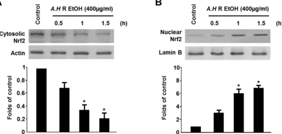
Fig. 3. Effects of A. hookeri root (EtOH) on Nrf2 nuclear translocation in HT22 cells. Cells were treated with 400 µg/ml of A.
hookeri root (EtOH) for 0.5, 1, and 1.5 h. The nuclei were fractionated from the cytosolic using PER-Mammalian Protein Extraction buffer as described in materials and methods. Cytosolic (A) and Nuclear (B) Nrf2 proteins were detected by Western blot analysis, and representative blots of three independent experiments are shown. Band intensity was quantified by densitometry and normalized to actin or lamin B; the values are presented at the bottom of each band.
Fig. 4. Cytoprotective effects of A. hookeri root (EtOH) on glutamate induced oxidative neurotoxicity and reactive oxygen species generation in HT22 cells. (A) Cells were treated with 400 µg/ml of A. hookeri (EtOH) in the presence or absence of SnPP (50 µM) and then exposed to glutamate (5 mM) for 12 h. (B) ROS production was assessed after 12 h incubation period with 100 and 200, 400 µg/ml of A. hookeri or SnPP (50 µM) and then exposed to glutamate (5 mM). Each bar represents the mean ± S.D. of three independent experiments, *p<0.05 vs. control. **p<0.05 vs. group treated with glutamate. #p<0.05 vs. same treatment plus SnPP.
이 나타났다고 보고한 바 있다.21)본 연구에서는 Bae 등의 결과와는 다르게 생쥐 해마세포에서 뇌세포 보호에 관여하 는 HO-1의 발현이 나타나는 것을 확인하였다. HO-1 발현 을 증가 시킨 삼채 뿌리 에탄올 추출물의 Nrf2 핵 내 이동 에 미치는 영향을 확인하기 위해, 삼채 뿌리 에탄올 추출물 (400 μg/mL)처리하여 시간별로 측정하였다(Fig. 3) 시간이 경과함에 따라 Nrf2가 핵 내로 전사 되면서(Fig. 3A) cytosol 의 Nrf2가 현저히 줄어드는 반면, (Fig. 3B) nuclear의 Nrf2 가 증가하는 양상을 보여, Nrf2의 핵 내로의 전사가 이루어 졌음을 확인 할 수 있었으며, HO-1 단백질의 발현에 대한 메커니즘 중 가장 직접적인 관련이 있는 것으로 알려진 nuclear factor-E2-related factor 2(Nrf2)의 핵 내 전사를 통 하여 HO-1 단백질의 발현을 조절하는 것을 실험을 통하여 다시 한번 확인하였다.
ROS 생성 억제 효과 − 세포 내에서 활성산소가 발생되 면 2',7'-dichlorofluorescein diacetate(DCFDA)가 esterase또 는 산화적 가수분해에 DCFH로 탈아세틸화 되고 비형광 성인 DCFH는 활성 산소에 의해 산화되어 2',7'-dichloro- fluorescein(DCF)로 전환되고 이는 강한 형광물질을 가진다.
이러한 원리를 이용하여 ROS생성 정도를 측정하였다. ROS 소거작용과 HO-1 발현 간의 직접적인 관계를 알아보기 위 하여, HO activity inhibitor로 알려진 Tin protoporphyrin (SnPP)를 처리하여 세포생존율 실험을 진행하였다(Fig. 4).
삼채의 뇌세포 보호효과와 ROS 소거 작용과 HO-1의 발 현간의 직접적인 관계를 알아보기 위하여, HO activity inhibitor로 알려진 Tin protoporphyrin(SnPP)를 처리하여 세 포생존율 실험을 진행한 결과, HO activity 억제제인 SnPP 를 3시간 전처리 후 삼채 뿌리 에탄올 추출물(A.H R EtOH) 과 함께 glutamtate 5mM 처리 시 농도별로 증가하였던 세 포 생존율과 ROS 소거 작용이 SnPP에 의해 역전된 것을 확인하였다. 이와 같은 결과를 통해 삼채의 ROS 생성 억제 능이 HO-1 단백질 발현과 관련 있음을 확인하였으며, 뇌세 포 보호활성에 기여함을 보여주었다.
결 론
최근 천연물을 이용한 산화적 스트레스를 저감시켜서 노 화를 지연시키거나, 억제하는 기능성 생리활성물질에 대한 연구가 활발히 진행되고 있다.32)
본 실험에서는 국내산 삼채 부위별(잎, 뿌리, 전체)과 용 매별(에탄올, 열수)로 그 분획물이 갖는 뇌세포 보호효과를 측정하였다. 삼채는 동결 건조한 잎과 뿌리, 전체 분말을 각 각 10 g 씩 500 mL 2시간동안 가열 환류추출 하였고, 여과 한 다음 여액을 다시 감압 농축하여 제조하였다.
글루타메이트로 유발한 세포 독성에 대한 삼채 추출물의 뇌세포 보호활성 효과를 알아보기 위해 독성이 검출되지 않
은 400 μg/mL 이하로 실험 농도를 정하였다. 그 결과 삼채 추출물 에탄올 용매뿌리 400 μg/mL에서는 81.3%에서 가장 높은 보호효과를 보였다.
뇌 세포 보호활성 효과가 가장 높은 삼채 추출물 뿌리부 위의 Nrf2의 핵 내 전사 유도를 통하여 HO-1 단백질을 발 현함으로써 뇌세포보호활성을 나타내는 것을 확인할 수 있 었다.
현대 의학의 발달로 수명이 길어진 지금 가장 두려워 하 는 질병 중 하나인 치매, 알츠하이머 등과 같은 뇌질환을 꼽 고 있는 현실에 맞춰 볼 때 이번 연구에서 사용된 삼채는 앞으로 뇌 질환 관련 증상들의 완화와 예방식품으로서 역 할 가능성이 높아 보이며, 이에 관한 연구는 계속되어야 한 다고 사료된다. 또한, 삼채의 효능이 잎에 비해서 뿌리에서 더 우수한 효과가 나타났는데 이를 확인하기 위해서는 HPLC profile 등의 분석이 필요할 것으로 보인다. 그리고 Allium속 의 대표적인 화합물인 유황 유래 화합물과의 비교 분석을 통해 추후 뇌세포 보호기전 연구와 활성 물질 분리, 구조규 명 및 동물실험 등에 관한 추가적인 연구가 필요할 것으로 생각된다.
인용문헌
1. Jin, D. Q., Lim, C. S., Hwang. J. K., Ha, I. H. and Han, J. S.
(2005) Anti-oxidant and anti-inflammatory activity of macel- ignan in murine hippocampal cell line and primary culture of rat microglial cell. Biochem. Biophys. Res. Commun. 331:
1264-1269.
2. Lim, C. S., Jin, D. Q., Sung, J. Y., Lee, J. H., Choi, H. G., Ha, I. H. and Han, J. S. (2006) Antioxidant and anti-inflammatory activities of the methanolic extract of Neorhodomela aculeate in hippocampal and microglial cells. Biol. Pharm. Bull. 29:
1212-1216.
3. Satoh, T., Enokido, Y., Kubo, K., Yamada, M. and Hatanaka, H (1999) Oxygen toxicity induces apoptosis in neuronal cells.
Cell Mol. Neurobiol. 18: 649-666.
4. Coyle, J. T. and Puttfarcken, P. (1993) Oxidative stress, glu- tamate, and neurodegenerative disorders. Science 262: 689- 695.
5. Frei, B. (1994) Natural antioxidants in human health and dis- ease. 25-55, Academic press, San Diego.
6. Neural science: Albright, T. D., Jessell, T. M., Kandel, E. R.
and Posner, M. I. (2000) A century of progress and the mys- teries that remain. Neuron 25: 209-216.
7. Halliwell B. (2006) Oxidative stress and neurodegeneration:
where are we now? J. Neurochem. 97: 1634-1658.
8. Packer, L. (1994) In methods in enzymology: Oxigen radicals in biological systems. Pare C, 23: 15-35. Academic press, San Diego.
9. Rossler, O. G., Bauer, I., Chung, H. Y. and Thiel, G. (2004) Glutamate-induced cell death of immortalized murine hip-
pocampal neurons: neuroprotective activity of heme oxy- genase-1, heat shock protein 70, and sodium selenite.
Neurosci. Lett. 362: 253-257.
10. Balogun, E., Hoque, M., Gong, P., Killeen, E., Green, C. J., Foresti, R., Alam, J. and Motterlini, R. (2003) Curcumin acti- vates the haem oxygenase-1 gene via regulation of Nrf2 and the antioxidant-responsive element. Biochem. J. 371: 887- 895.
11. Liu, X. M., Peyton, K. J., Shebib, A. R., Wang, H., Korthuis, R. J. and Durante, W. (2011) Activation of AMPK stimulates heme oxygenase-1 gene expression andhuman endothelial cell survival. Am. J. Physiol. Heart Circ. Physiol. 300: H84- 93.
12. Choi, B. M., Pae, H. O., Kim, Y. M. and Chung, H. T. (2003) Nitric oxide-mediated cytoprotection of hepatocytes from glucose deprivation-induced cytotoxicity: involvement of heme oxygenase-1. Hepatology 37: 810-823.
13. Choi, H. G., Lee, D. S., Li, B., Jun, K. Y., Jeong, G. S. and Kim, Y. C. (2011) Neuroprotective effect of the water-insol- uble fraction of root barks of Dictamnus dasycarpus 70%
ethanol extract on glutamate-induced oxidative damage in mouse hippocampal HT22 cells. Kor. J. Pharmacogn. 42:
175-181.
14. Lee, M. S., Lee, J., Kwon, D. Y. and Kim, M. S. (2006) Ondamtanggamibang protects neurons from oxidative stress with induction of heme oxygenase-1. J. Ethnopharmacol.
108: 294-298.
15. Rho, S. N. and Han, J. H. (2000) Cytotoxicity of garlic and onion methanol extract on human lung cancer cell lines. J.
Korean Soc. Food Soc. Nutr. 29: 870-874.
16. Sharma, G., Gohil, R. N. and Kaul, V. (2011) Cytological sta- tus of allium hookeri thwaites (2n=22) Genet. Resour. Crop.
Evol. 58: 1041-1050.
17. Kim, H. J. and Chun, H. S. (1999) Biological functions of organosulfur compounds in Allium vegetables. J. Korean Soc.
Food Sci. Nutr. 28: 1412-1423.
18. Ayam, V. S. (2011) Allium hookeri, Thw. Enum. A lesser known terrestrial perennial herb used as food its ethnobo- tanical relevance in Manipur. Afr. J. food Agric. Nutr. Dev. 11:
5389-5412.
19. Vazquez-Prieto, M. A. and Miatello, R. M. (2010) Orga- nosulfur compounds and cardiovascular disease. Mol.
Aspects Med. 31: 540-545.
20. Kim, C. H., Lee, M. A., Kim, T. W., Jang, J. Y. and Kim, H.
J. (2012) Anti-inflammatory effect of Allium hookeri root methanol extract in LPS-induced RAW264.7 cells. J. Korean Soc. Food Sci. Nutr. 41: 1645-1648.
21. Bae, G. C. and Bae, D. Y. (2012) The anti-inflammatory effects of ethanol extract of Allium hookeri cultivated in South Korea. Kor. J. Herbology 27: 55-61.
22. Won, J. Y., Yoo, Y. C., Kang, E. J., Yang, H., Kim, G. H., Seong, B. J., Kim, S. I., Han, S. H., Lee, S. S. and Lee, K. S.
(2013) Chemical components, DPPH radical scavenging activity and inhibitory effects on nitric oxide production in Aliium hookeri cultivated under open field and greenhouse conditions. J. Korean Soc. Food Sci. Nutr. 42: 1351-1356.
23. Cha, Y. S. (2014) Antimicrobial activity and antioxidative activity of the extract of Allium hookeri Thw. Enum. Grad- uate School of Joongbu University.
24. Kim, N. S., Choi, B. K., Lee, S. H., Jang, H. H., Kim, J. B., Kim, H. R., Kim, D. K., Kim, Y. S., Yang, J. H., Kim, H. J.
and Lee, S. H. (2015) Effect of Allium hookeri on glucose metabolism in type II diabetic mice. Kor. J. Pharmacogn. 46:
78-83.
25. Lee, S. H., Kim, N. S., Choi, B. K., Jang, H. H., Kim, J. B., Lee, Y. M., Kim, D. D., Lee, C. H., Kim, Y. S., Yang, J. H., Kim, Y. S., Kim, H. J. and Lee, S. H. (2015) Effects of Allium hookeri on lipid metabolism in type II diabetic mice. Kor. J.
Pharmacogn. 46: 148-153.
26. Jeong, G. S., Li, B., Lee, D.S., Byun, E., Kang, D. K., Lee, H. S. and Kim, Y. C. (2007) Cytoprotective constituents of Alpinia katsumadai seeds against glutamate-induced oxida- tive injury in HT22 cell. Nat. Prod. Sci. 13: 268-271.
27. Numagami, Y., Sato, S. and Ohnishi, S. T. (1996) Attenuation of rat ischemic brain damage by aged garlic extracts a pos- sible protecting mechanism as antioxidants. Neurochem. Int.
29: 135-143.
28. Nishiyama, N., Moriguchi, T. and Saito, H. (1997) Beneficial effects of aged garlic extract on learning and memory impair- ment in the senescence-accelerated mouse. Exp. Gerontol. 32:
149-160.
29. Yu, X., Song, N., Guo, X., Jiang, H., Zhang, H., and Xie, J.
(2016) Differences in vulnerability of neurons and astrocytes to heme oxygenase-1 modulation: Implications for mito- chondrial ferritin. Sci. Rep. 6: 24200.
30. Schwer, CI., Guerrero, A. M., Humar, M., Roesslein, M., Goebel, U., Stoll, P., Geiger, K. K., Pannen, B. H., Hoetzel, A., and Schmidt, R. (2008) Heme oxygenase-1 inhibits the proliferation of pancreatic stellate cells by repression of the extracellular signal-regulated kinase1/2 pathway. J. Phar- macol. Exp. Ther. 327: 863-871.
31. Park, S. Y., Jin, M. L., Kim, Y. H., Lee, S. J., and Park, G.
(2014) Sanguinarine inhibits invasiveness and the MMP-9 and COX-2 expression in TPA-induced breast cancer cells by inducing HO-1 expression. Oncol. Rep. 31: 497-504.
32. Masaki, H., Sakaki, S., Atsumi, T. and Sakurai, H. (1995) Active oxygen scavenging activity in plant extracts. Biol.
Pharm. Bull. 18: 162-166.
(2016. 11. 18 접수: 2016. 12. 8 심사; 2017. 3. 16 게재확정)