Identification of the Pig β-1,3-N-acetylglucosaminyltransferase 1 (pB3GNT1) that is Involved in Poly-N-acetyllactosamine (poly-LacNAc) Synthesis
Ji-Youn Kim1†, Hwan-Jin Hwang1†, Hak-Jae Chung2, Shinichi Hochi3, Mi-Ryung Park1, Sung June Byun1, Keon Bong Oh1, Hyeon-Yang1 and Kyung-Woon Kim1*
1Animal Biotechnology Division, National Institute of Animal Science, Rural Development Administration, Wanju-Gun 55365, Korea
2Swine Science Division, National Institute of Animal Science, Rural Development Administration, Cheonan 31000, Korea
3Faculty of Textile Science and Technology, Shinshu University, Ueda, Nagano 386-8567, Japan Received October 16, 2017 /Revised November 28, 2017 /Accepted March 9, 2018
The structure of glycan residues attached to glycoproteins can influence the biological activity, stabil- ity, and safety of pharmaceutical proteins delivered from transgenic pig milk. The production of ther- apeutic glycoprotein in transgenic livestock animals is limited, as the glycosylation of mammary gland cells and the production of glycoproteins with the desired homogeneous glycoform remain a challenge. The β-1,3-N-acetylglucosaminylatransferase1 (B3GNT1) gene is an important enzyme that attaches N-acetylglucosamine (GlcNAc) to galactose (Gal) residues for protein glycosylation; however, there is limited information about pig glycosyltransferases. Therefore, we cloned the pig B3GNT1 (pB3GNT1) and investigated its functional properties that could attach N-acetylglucosamine to gal- actose residue. Using several different primers, a partial pB3GNT1 mRNA sequence containing the full open reading frame (ORF) was isolated from liver tissue. The ORF of pB3GNT1 contained 1,248 nu- cleotides and encoded 415 amino acid residues. Organ-dependent expression of the pB3GNT1 gene was confirmed in various organs from adult and juvenile pigs. The pB3GNT1 mRNA expression level was high in the muscles of the heart and small intestine but was lower in the lungs. For functional characterization of pB3GNT1, we established a stable expression of the pB3GNT1 gene in the porcine kidney cell line (PK-15). As a result, it was suggested that the glycosylation pattern of pB3GNT1 ex- pression in PK-15 cells did not affect the total sialic acid level but increased the poly N-acetyllactos- amine level. The results of this study can be used to produce glycoproteins with improved properties and therapeutic potential for the generation of desired glycosylation using transgenic pigs as bioreactors.
Key words : N-acetylglucosamine (GlcNAc), N-acetyllactosamine (LacNAc), pig β-1,3-N-acetylglucosa- minyltransferase 1 (pB3GNT1), PK-15
†Authors contributed equally.
*Corresponding author
*Tel : +82-63-238-7264, Fax : +82-63-238-7297
*E-mail : [email protected]
This is an Open-Access article distributed under the terms of the Creative Commons Attribution Non-Commercial License (http://creativecommons.org/licenses/by-nc/3.0) which permits unrestricted non-commercial use, distribution, and reproduction in any medium, provided the original work is properly cited.
Journal of Life Science 2018 Vol. 28. No. 4. 389~397 DOI : https://doi.org/10.5352/JLS.2018.28.4.389
Introduction
Many physiologically active proteins contain various car- bohydrate structures whose formation is catalyzed by glyco- syltransferases [17, 19]. The glycosylation process is consid- ered to be important for intrinsic and extrinsic cellular signal transduction [12, 13], as various types of cancer avoid apop- tosis through glycan modifications. When Fas is hyper-α-2,6 sialylated by β-galactoside α2,6-sialyltransferase 1 (ST6Gal1),
apoptotic signaling is precluded, as both death-inducing sig- nal complex formation and Fas internalization are sup- pressed [27]. Conversely, proliferation is significantly in- hibited, and apoptosis is induced in human colorectal cancer cells treated with O-glycosylation inhibitors [22]. The ability of cells to adhere to the extracellular matrix is also altered by modifying glycan structures [16, 18, 25]. Poly N-ace- tyllactosamine (poly-LacNAc) is a chain of disaccharides composed of galactose (Gal) and N-acetylglucosamine (GlcNAc) that provides a unique structure for cell-type-spe- cific glycans when attached to glycoproteins and glycolipids [4, 6]. The linear form of poly-LacNAc is dominant in fetal erythrocytes, while the branched form is dominant in adult erythrocytes [7]. β-1,4-N-galactosyltransferase (B3GALT) of the β-1,3-N-acetylglucosaminyltransferase (B3GNT) family is a key enzyme in the synthesis of poly-LacNAc and the for- mation of i antigen, a human alloantigen [28]. Further mod-
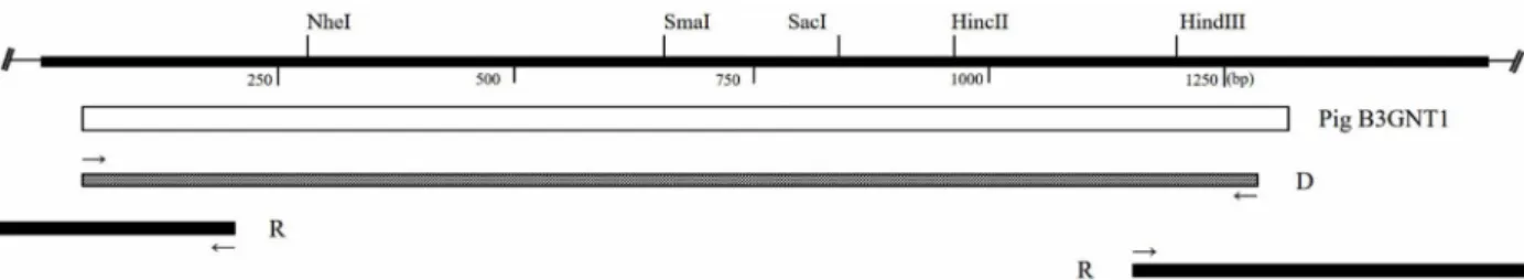
Fig. 1. Schematic diagram of pig B3GNT1 cDNA clones. The open bar represents the protein coding region, and thick lines indicate the noncoding regions of B3GNT1. D, Degenerate PCR clone, R, RACE clones. The small, thin arrows indicate the position and direction of primers.
ification of poly-LacNAc induces the display of various ter- minal epitopes [20]. Sialyl Lewis X, a terminal epitope, plays an important role in leukocyte adhesion [8, 23].
The B3GNT1 gene has been identified in many mamma- lian species, including humans, mice, rats and cows [24].
Functional analysis of B3GNT-/- genetic knock-out mice re- vealed inhibited neuronal migration, and the observed ab- normality was attributed to decreased terminal lactosamine levels [3]. Impaired sexual behavior has also been reported in male B3gnt-/- knock-out mice [2]. Conversely, over-ex- pression of the B3GNT gene appears to decrease tumor for- mation rate, probably due to increased synthesis of lam- inin-binding glycans that can interact with adhesion mole- cules in the epithelial cell basement membrane [1].
Pigs have received significant attention as animal bio- reactors in transgenic research as well as transplantable or- gan donors in regenerative medicine. The data on B3GNT1 in pigs is currently very limited but could be beneficial for understanding the equivalent enzymes in humans. There- fore, in the present study, we cloned the pig B3GNT1 gene (pB3GNT1), and its enzymatic activity was measured using CD39L3, which hydrolyzes phosphate groups from uridine diphosphate (UDP) [29]. The expression of pB3GNT1 in vari- ous organs from adult and juvenile pigs, the sialic acid and poly-LacNAc levels in pB3GNT1-overexpressing porcine kidney cells (PK-15) and the catalytic activity of this enzyme toward the O-glycan core were also investigated.
Materials and Methods
Ethics approval and consent to participate All experimental procedures were examined and ap- proved by the Animal Research Committee at National Institute of Animal Science (NIAS 2015-593).
RNA extraction and cDNA synthesis
Various tissues were homogenized in the presence of TRIzol (Invitrogen, San Diego, CA, USA), and RNA was isolated. The obtained RNA fragments were utilized for cDNA synthesis using a First-Strand cDNA Synthesis Kit (GE Healthcare, Pittsburgh, PA, USA).
Cloning of the pig B3GNT1 gene
Primers were designed for the untranslated region (UTR) through homology comparisons with equivalent sequences from other species obtained from NCBI, such as humans (NM 006876.2), mice (NM 175383.2), cows (NM 001034808.1), and rats (NM 001106324.1). The forward and reverse primers were 5‘-ATG CAR ATG TCS TAC GCC ATC-3’ and 5‘-TRG CCT TCA ACT CCT GYT TG-3’, respectively. Degenerate PCR was performed under the following conditions: 94°C for 3 min; 30 cycles of 94°C for 30 s, 42°C for 30 s, 72°C for 90 s; and 72°C for 5 min (Fig. 1).
Total RNA was extracted from pig liver tissue, and the GeneRacer Kit (Invitrogen, San Diego, CA, USA) was used for 5‘ and 3’ RACE. The 5‘ RACE primers consisted of a 5’-anchor primer: 5‘-GAA ATA TTG GTC TTG CTC CTC CTG C-3’, and the GeneRacer 5‘ primer. The 3’ RACE pri- mers consisted of a 3‘-anchor primer: 5’-ACG AAG GTT TCC TGG TTC ATA AAG G-3‘, and the GeneRacer 3’ primer (Fig. 1). The amplified PCR products were analyzed through agarose gel (Takara, Kyoto, Japan) electrophoresis and cloned using the TOPO-TA cloning kit (Invitrogen, San Diego, CA, USA). Finally, selected clones were identified via DNA sequence analysis (Macrogen, Seoul, Korea).
Quantitative real-time polymerase chain reaction (qPCR)
For quantitative analysis of the expression levels of pB3GNT1 and pig beta-actin gene were assessed in various organs from adult pigs (n=3) and a juvenile piglet (n=1).
qPCR was performed using LightCycler FastStart DNA Master SYBR Green I (Roche, Basel, Switzerland). The pri- mer sets used in these assays were as follows: for pB3GNT1, 5‘-AGC CCG GGA TCA ATT ATG CAC-3’ (forward) and 5’-ACC ATG TCC ACG TCG ATT ACC-3‘ (reverse); and for beta-actin, 5’-CAT CAC CAT CGG CAA CGA GC-3‘
(forward) and 5’-TAG AGG TCC TTG CGG ATG TC-3‘
(reverse). The thermal profile consisted of the following steps: pre-incubation at 95°C for 10 min and 45 cycles of amplification at 95°C for 10 s, 58°C for 5 s, and 72°C for 4 s. The 2-ΔΔCT method was used to analyze pB3GNT1 expression.
Construction of the pB3GNT1 expression vector For the cloning of the pB3GNT1 gene with a flag tag at the N-terminal region, the following primers were designed:
5‘-GAA TTC GCC GCC ACC ATG GAT TAC AAG GAT GAC GAC GAT AAG ATG CAG ATG TCG TAC GCC ATC-3’ and 5‘-GCG GCC GCT CAG CAG TGA CGT GGG GAG-3’ (the EcoRI and NotI sites are underlined, and the flag tag sequences are highlighted in bold). The amplified PCR products and the pcDNA3.1(+) vector (Invitrogen, San Diego, CA, USA) were sequentially digested with EcoRI (Takara, Kyoto, Japan) and NotI (Takara, Kyoto, Japan) and then ligated together with the Mighty Mix DNA Ligation Kit (Takara, Kyoto, Japan).
pB3GNT1 transfection into PK-15 cells
Porcine kidney cells (PK-15) were maintained in high-glu- cose Dulbecco’s Modified Eagle Medium (DMEM; Invitro- gen, San Diego, CA, USA) containing 10% fetal bovine serum (FBS; Invitrogen, San Diego, CA, USA), 50 U/ml penicillin, and 50 μg/ml streptomycin (Invitrogen). The cells were maintained at 37°C under 5% CO2 and 95% air. The pB3GNT1 expression vector was linearized with PvuI (Takara, Kyoto, Japan) and transfected into PK-15 cells using Lipofectamine (Invitrogen, San Diego, CA, USA). Transformed cells were selected via treatment with 800 μg/ml G418 (Sigma-Aldrich Corp., St. Louis, MO, USA) for 2 weeks.
Western blotting to detect FLAG-tagged pB3GNT1 Cells were harvested and lysed with M-PER buffer (Thermo Fisher Scientific, West Palm Beach, FL, USA).
Proteins were separated using SDS-PAGE and blotted onto 0.45 μm nitrocellulose membranes. The membranes were blocked in 5% non-fat dry milk in TBST buffer (Tris-buffered
saline, 0.05 M Tris, pH 7.4; 0.2 M NaCl; 0.1% Tween 20) for 1 h at room temperature (RT) with shaking and then incubated in 0.1% TBST containing a monoclonal FLAG anti- body (Sigma-Aldrich Corp., St. Louis, MO, USA) overnight at 4°C with shaking. On the following day, the membranes were washed 3 times with 0.1% TBST for 10 min and further incubated in 0.1% TBST containing an horseradish perox- idase (HRP)-conjugated anti-mouse secondary antibody (Invitrogen, San Diego, CA, USA) for 1 hr at RT. Finally, the membranes were washed 3 times with 0.1% TBST for 10 min and developed using an enhanced chem- iluminescence solution (GE Healthcare, Pittsburgh, PA, USA) to visualize the targeted proteins.
Immunofluorescence microscopy analysis
Cells were seeded onto Lab-Tek chamber slides and cul- tured for 24 hr. Subsequently, the cells were fixed with 3.7%
formaldehyde for 15 min, washed 3 times with phosphate- buffered saline (PBS) for 3 min, and then incubated for 10 min at -20°C with ice-cold MeOH. Next, the cells were wash- ed 3 times with PBS and incubated in PBS containing 1%
bovine serum albumin (BSA) and 0.2% Triton-X 100 (blocking buffer) for 1 hr. After 3 washes in PBS, the cells were in- cubated with a FLAG antibody (diluted 1:1,000 in dilution buffer consisting of PBS with 1% BSA and 0.05% Triton-X 100) overnight at 4°C. The cells were subsequently washed 3 times in PBS and incubated with fluorescein isothiocya- nate-conjugated mouse IgG (1:500 in dilution buffer) (Sigma- Aldrich Corp., St. Louis, MO, USA) for 1 hr in dark conditions. After 3 washes, cell nuclei and Golgi complexes were stained using 4′,6-diamidino-2-phenylindole (DAPI;
Roche, Basel, Switzerland) and BODIPY® TR ceramide (Invitrogen, San Diego, CA, USA), respectively. Finally, the cells were observed under a fluorescence microscope (Olympus, Tokyo, Japan).
Enzyme-linked immunosorbent assay (ELISA) ELISA was performed using the ELISA Starter Accessory Package (Bethyl Laboratories, Montgomery, TX, USA) ac- cording to the Vectorlabs method. Total protein lysates (10–
30 μg) from PK-control and PK-pB3 cells were coated onto 96-well plates with coating buffer (0.05 M carbonate bicar- bonate, pH 9.6) for 1 hr at 37°C. The plates were sub- sequently washed 3 times and filled with post-coat solution (50 mM Tris, 1% BSA, pH 8.0) for 30 min at RT. After 3 washes, each well was incubated with 10 μg/ml of bio-
A
B
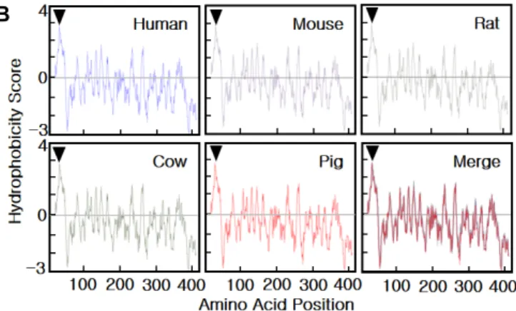
Fig. 2. Characterization of the pB3GNT1 gene. A - The cDNA sequence of the pB3GNT1 gene derived from pig liver cDNA and its deduced amino acid sequence. The underlined text indicates a putative transmembrane domain, and open circles indicates putative N-glycosylation sites. The sequence data have been submitted to the NCBI GenBank databases under accession No. JN975036. B - Hydrophobicity of B3GNT1 proteins from various species, calculated with the Kyte and Doolittle program (http://www.expasy.ch/cgi-bin/protscale.pl). High hydrophobicity in the N-terminal region (arrowhead) predicts that 1,3-N-acetylglucosaminyltransferase 1 protein shows a type II transmembrane topology.
tinylated Lycopersicon esculentum agglutinin (LEA; EY Laboratories, San Mateo, CA, USA) or Limax flavus ag- glutinin (LFA; EY Laboratories, San Mateo, CA, USA) for 1 hr at RT. The plates were next washed with Tris/Tween 20 buffered saline (pH 8.0) and incubated with streptavi- din-HRP (1:2,000; GE Healthcare, Pittsburgh, PA, USA).
Then, the plates were incubated for 30 min at RT and wash- ed 3 times and the 3,3′,5,5′-tetramethylbenzidine sub- strate was used as the chromogen. After the solution turned blue, stop solution (1 N H2SO4) was added to each well. The optical density (OD) value was finally determined at 450 nm.
Measurement of pB3GNT1 activity
pB3GNT1 activity was assessed using a Glycosyltransfer- ase Activity Kit (R&D Systems, Minneapolis, MN, USA) and 0.6 mM p-Nitrophenyl galacto-N-bioside (Gal-β1,3-GalNAc-
pNP; Sigma-Aldrich Corp., St. Louis, MO, USA), 200 μM UDP-GlcNAc (Sigma-Aldrich Corp., St. Louis, MO, USA), and 20 mg/ml of asialo-fetuin type I (Sigma-Aldrich Corp., St. Louis, MO, USA). To prepare the reaction, 10 μl of donor substrate solution (UDP-GlcNAc), 10 μl of acceptor substrate solution (Gal-β1,3-GalNAc-pNP or asialo-fetuin type I), and 5 μl of coupling phosphatase 1 solution were mixed in each well. Coupling phosphatase (ENTPD3/CD39L3) can remove a phosphate group from free-UDP. To initiate the reaction, glycosyltransferase solution (10 μg of cell protein lysate) was added to each well, and assay buffer (3.125 mM Tris HCl [pH 7.5], 1.25 mM CaCl2, 12.5 mM MnCl2) was then added to a total volume of 50 μl. As the negative control, assay buffer alone was used. The plate contents were mixed via gentle tapping and incubated at 37°C for 2 hr. Then, 30 μl of Malachite Green Reagent A was added to each well. The
A
B
C D
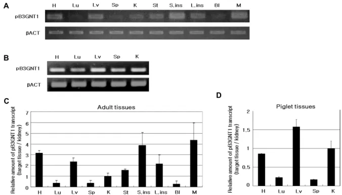
Fig. 3. qPCR analysis of pB3GNT1 mRNA levels in various pig organs. Data from duplicate samples of 3, 2-years-old adult pigs (A, C) and one 5-day-old piglet (B, D). H: heart, Lu: lung, Lv: liver, Sp: spleen, K: kidney, St: stomach, S.ins: small intestine, C: colon, Bl: bladder, M: muscle. The expression levels of pB3GNT1 were normalized to that of β-actin and subsequently normalized to kidney expression levels. The data represent the mean ± S.D.
plate contents were mixed by gentle tapping, and 100 μl of deionized water was added. Finally, 30 μl of Malachite Green Reagent B was added, followed by mixing by tapping.
The color reaction was allowed to develop for 20 min at RT, and the OD value was determined at 655 nm. A standard curve was generated through 2-fold serial dilution, where the highest concentration was 1 mM phosphate saline.
Results
Characterization of pB3GNT1 cDNA
Through degenerate PCR and RACE PCR targeting pB3GNT1, a partial pB3GNT1 sequence containing the full open reading frame (ORF) was identified (Fig. 1). The ORF consisted of 1,248 nucleotides (nt), encoding 415 amino acids (aa) (Fig. 2A). The pB3GNT1 gene sequence was highly ho- mologous to those of Homo sapiens (92% nt, 98% aa), Mus musculus (88% nt, 94% aa), Bos taurus (95% nt, 98% aa), and Rattus norvegicus (87% nt, 94% aa) (data not shown).
Moreover, the N-terminal region of pB3GNT1 exhibited high hydrophobicity (Fig. 2B). Comparison of the hydropathy plots of the hydrophobicity scores for B3GNT1 proteins from several species indicated high conservation across species
(Fig. 2B). These results confirm that the B3GNT1 gene is highly conserved in all investigated species.
pB3GNT1 expression in various organs
As B3GNT1 is known to be expressed in diverse tissues, we analyzed the relative expression levels of this gene in many organs through RT-PCR and quantitative PCR (Fig.
3A, Fig. 3B). In adult pig samples, pB3GNT1 mRNA levels were found high in the heart, small intestine and muscle, while lower in the lungs, stomach and bladder (Fig. 3C).
On the other hand, the piglet samples showed high level of pB3GNT1 mRNA in liver tissue, while low in lung and splenic tissues (Fig. 3D).
pB3GNT1 activity
In RT-PCR of PK-15 cells transfected with the pB3GNT1 expression vector, the expression of the introduced gene was confirmed (Fig. 4A), and qPCR indicated that the tran- scription and translation of pB3GNT1 were stronger in trans- fected cells (PK-pB3) than non-transfected control cells (PK-control) (Fig. 4B). Cells were double-labeled with a FLAG antibody and BODIPY® TR ceramide, and pB3GNT1 localization was identified within the Golgi complex (Fig.
A B
C
D E
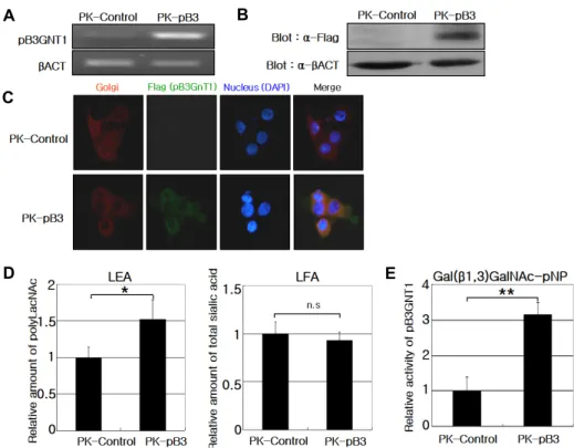
Fig. 4. Functional test of pB3GNT1 gene in PK-15 cell line. A - qPCR performed on PK-control and PK-pB3 cells to assess pB3GNT1 expression levels. B - Western blot analysis performed using a specific FLAG antibody. C - Immunofluorescence labeling for FLAG (green), the Golgi complex (red) and nuclei (blue), indicating pB3GNT1 localization within the Golgi complex. D - ELISA for poly-LacNAc and total sialic acid levels. The levels of poly-LacNAc structures, recognized by LEA, were higher in PK-pB3 than in PK-control cells. The total sialic acid levels, determined by LFA, were not significantly different between the PK-pB3 and PK-control cells. E - The specific activity of pB3GNT1 toward Gal(β1,3)GalNAc, a core structure of O-glycan, was analyzed using CD39L3. The activity was higher in PK-pB3 cells than in PK-control cells. The data represent the mean
± S.D. (*p<0.05, **p<0.005 by Student’s t-test, n.s: not significant).
4C).
As B3GNT1 plays an important role in the synthesis of poly-LacNAc, the intracellular poly-LacNAc content is in- dicative of B3GNT1 catalytic activity and can be measured using LEA lectin with a high affinity for LacNAc oligomers, rather than monomers or dimers. The poly-LacNAc level was approximately 1.5-fold higher in PK-pB3 cells than in PK-control cells (p<0.05). The sialic acid content, measured using a sialic acid-specific LFA lectin, was not significantly affected by pB3GNT1 expression in PK-15 cells (Fig. 4D).
p-Nitrophenyl galacto-N-bioside (Gal-β1,3-GalNAc-pNP) contains Gal residues and was employed as an acceptor substrate. As expected, the PK-pB3 cells showed approx- imately 3-fold higher catalytic activity than the PK-control cells (Fig. 4E). The specific activity of the PK-control cells was 0.325±0.158 pmol·min-1·μg-1, whereas, that of PK-pB3 was 1.194±0.149 pmol·min-1·μg-1 (data not shown).
Discussion
In this study, we cloned pig’s B3GNT1 gene and analyzed its function. pB3GNT1 exhibited a type II membrane top- ology, which is a common feature of many glycosyltransfer- ases [1, 5, 14, 24]. A 28-aa transmembrane domain in the N-terminal region was suggested to be a Golgi-anchoring region 253 based on sequence alignment with homologous proteins from other species, while the C-terminal region fa- cilitates catalytic activity in the lumen. As most glycosyl- transferases in the B3GNT family are glycoproteins, we searched for putative N-linked glycosylation sites in pB3GNT1. N-linked glycosylation usually occurs at the spe- cific peptide sequence Asn-X-Ser/Thr, where X can be any amino acid other than proline (Fig. 2A) [31]. The identified regions coincided with potential N-glycosylation sites in hu- man B3GNT1 [24].
qPCR analysis to detect pB3GNT1 mRNA levels in vari- ous pig organs indicated that the expression level in the liver
was higher than in the kidneys and spleen of pigs, whereas human B3GNT1 is more highly expressed in the kidneys and spleen than in the liver [24]. The pB3GNT1 activity of double labelled PK-15 cells was consistent with previous reports showing that enzymes in the B3GNT family are generally localized to the Golgi complex [17].
As sialic acids are added to Gal or GalNAc residues by various sialyltransferase families (ST3Gal, ST6Gal, ST6 GalNAc, ST8Sia, etc.), the donor substrates for B3GNT1 may be competitively used by dominant sialyltransferases. Over- expression of sialyltransferases in Chinese hamster ovary (CHO) cells has been shown to significantly decrease poly- LacNAc levels [30].
Therapeutic proteins are the fastest growing industry in the pharmaceutical. These proteins can be recombinantly produced in mammalian cells capable of glycosylation such as human. We have tried to produce safe and useful glyco- protein drugs using transgenic pigs that secrete recombinant drugs proteins such as von Willebrand factor (vWF) [15] and erythropoietin (EPO) [21], into their milk. However, there were some limitations that prevented the development of these production methods. Based on these studies, we found that the secreted proteins require proper glycan modification to function properly.
Modification of glycan structures has been attempted in the pharmaceutical industry [10, 11] to regulate the bio- logical activity and stability of glycoprotein biomedicines with the sialic acid content of N-glycan. When the sialic acid content of EPO is elevated, the biological activity and stabil- ity increase [9]. Some key enzymes in the B3GNT family that extend and diversify glycan structures could affect the stability of glycoproteins [26]. Therefore, efficient regulation of B3GNT family members and sialyltransferases in the mammary glands during lactation is critical to improve the biological activity of secreted recombinant proteins.
In conclusion, we characterized the molecular structure and function of pB3GNT1, an enzyme that attaches N-acetyl- glucosamine to galactose residues. The ORF contained 1,248 nt and encoded 415 aa. Furthermore, different expression levels of pB3GNT1 were confirmed via RT-PCR and qPCR in piglet organs and adult pig organs. We further concluded that the over-expression of the gene in a porcine kidney cell line (PK-15) did not affect the total sialic acid level.but in- crease the poly N-acetyllactosamine level. This enzyme ex- hibited catalytic activity toward the O-glycan core; however, the glycoprotein stability could be affected by poly-LacNAc
structures. These results will serve as a useful basis for the development of a new, efficient bioreactor in the pharma- ceutical industry.
Acknowledgments
This work was supported by "Cooperative Research Program for Agriculture Science & Technology Develop- ment (Project No. PJ01097601)", Rural Development Admi- nistration, Republic of Korea.
References
1. Bao, X., Kobayashi, M., Hatakeyama, S., Angata, K., Gullberg, D., Nakayama, J., Fukuda, M. N. and Fukuda, M. 2009.
Tumor suppressor function of laminin-binding alpha-dys- troglycan requires a distinct beta3-N-acetylglucosaminyl- transferase. Proc. Natl. Acad. Sci. USA. 106, 12109-12114.
2. Biellmann, F., Henion, T. R., Burki, K. and Hennet, T. 2008.
Impaired sexual behavior in male mice deficient for the be- ta1-3 N-acetylglucosaminyltransferase-I gene. Mol. Reprod.
Dev. 75, 699-706.
3. Bless, E., Raitcheva, D., Henion, T. R., Tobet, S. and Schwart- ing, G. A. 2006. Lactosamine modulates the rate of migration of GnRH neurons during mouse development. Eur. J.
Neurosci. 24, 654-660.
4. Dabrowski, U., Hanfland, P., Egge, H., Kuhn, S. and Dabrowski, J. 1984. Immunochemistry of I/i-active oligo- and polyglycosylceramides from rabbit erythrocyte mem- branes. Determination of branching patterns of a ceramide pentadecasaccharide by 1H nuclear magnetic resonance. J.
Biol. Chem. 259, 7648-7651.
5. Egan, S., Cohen, B., Sarkar, M., Ying, Y., Cohen, S., Singh, N., Wang, W., Flock, G., Goh, T. and Schachter, H. 2000.
Molecular cloning and expression analysis of a mouse UDP-GlcNAc:Gal(beta1-4)Glc(NAc)-R beta1,3-N-acetylglu- cosaminyltransferase homologous to Drosophila melanogaster Brainiac and the beta1,3-galactosyltransferase family. Glyco- conj. J. 17, 867-875.
6. Fukuda, M., Dell, A., Oates, J. E. and Fukuda, M. N. 1984.
Structure of branched lactosaminoglycan, the carbohydrate moiety of band 3 isolated from adult human erythrocytes.
J. Biol. Chem. 259, 8260-8273.
7. Fukuda, M., Fukuda, M. N. and Hakomori, S. 1979.
Developmental change and genetic defect in the carbohy- drate structure of band 3 glycoprotein of human erythrocyte membrane. J. Biol. Chem. 254, 3700-3703.
8. Fukuda, M., Spooncer, E., Oates, J. E., Dell, A. and Klock, J. C. 1984. Structure of sialylated fucosyl lactosaminoglycan isolated from human granulocytes. J. Biol. Chem. 259, 10925- 10935.
9. Fukuda, M. N., Sasaki, H., Lopez, L. and Fukuda, M. 1989.
Survival of recombinant erythropoietin in the circulation:
the role of carbohydrates. Blood 73, 84-89.
10. Fukuta, K., Abe, R., Yokomatsu, T., Kono, N., Asanagi, M., Omae, F., Minowa, M. T., Takeuchi, M. and Makino, T. 2000.
Remodeling of sugar chain structures of human interfer- on-gamma. Glycobiology 10, 421-430.
11. Fukuta, K., Yokomatsu, T., Abe, R., Asanagi, M. and Makino, T. 2000. Genetic engineering of CHO cells producing human interferon-gamma by transfection of sialyltransferases. Gly- coconj. J. 17, 895-904.
12. Hakomori, S. 2002. Glycosylation defining cancer malig- nancy: new wine in an old bottle. Proc. Natl. Acad. Sci. U SA. 99, 10231-10233.
13. Kitazume, S., Imamaki, R., Ogawa, K., Komi, Y., Futakawa, S., Kojima, S., Hashimoto, Y., Marth, J. D., Paulson, J. C.
and Taniguchi, N. 2010. Alpha2,6-sialic acid on platelet en- dothelial cell adhesion molecule (PECAM) regulates its ho- mophilic interactions and downstream antiapoptotic signal- ing. J. Biol. Chem. 285, 6515-6521.
14. Ko, H. K., Song, K. H., Jin, U. H., Seong, H. H., Chang, Y. C., Kim, N. H., Kim, D. S., Lee, Y. C. and Kim, C. H.
2010. Molecular characterization of pig alpha2,3-Gal-be- ta1,3-GalNAc-alpha2,6-sialyltransferase (pST6GalNAc IV) gene specific for Neu5Acalpha2-3Galbeta1-3GalNAc trisac- charide structure. Glycoconj. J. 27, 367-374.
15. Lee, H. G., Lee, H. C., Kim, S. W., Lee, P., Chung, H. J., Lee, Y. K., Han, J. H., Hwang, I. S., Yoo, J. I., Kim, Y. K., Kim, H. T., Lee, H. T., Chang, W. K. and Park, J. K. 2009.
Production of recombinant human von Willebrand factor in the milk of transgenic pigs. J. Reprod. Dev. 55, 484-490.
16. Lee, M., Park, J. J. and Lee, Y. S. 2010. Adhesion of ST6Gal I-mediated human colon cancer cells to fibronectin contrib- utes to cell survival by integrin beta1-mediated paxillin and AKT activation. Oncol. Rep. 23, 757-761.
17. Lee, P. L., Kohler, J. J. and Pfeffer, S. R. 2009. Association of beta-1,3-N-acetylglucosaminyltransferase 1 and beta-1,4- galactosyltransferase 1, trans-Golgi enzymes involved in coupled poly-N-acetyllactosamine synthesis. Glycobiology 19, 655-664.
18. Lin, J. T., Bhattacharyya, D. and Kecman, V. 2003. Multiple regression and neural networks analyses in composites machining. Compos. Sci. Technol. 63, 539-548.
19. Munro, S. 1998. Localization of proteins to the Golgi apparatus. Trends Cell Biol. 8, 11-15.
20. Nairn, A. V., York, W. S., Harris, K., Hall, E. M., Pierce, J. M. and Moremen, K. W. 2008. Regulation of glycan struc- tures in animal tissues: transcript profiling of glycan-related genes. J. Biol. Chem. 283, 17298-17313.
21. Park, J. K., Lee, Y. K., Lee, P., Chung, H. J., Kim, S., Lee,
H. G., Seo, M. K., Han, J. H., Park, C. G., Kim, H. T., Kim, Y. K., Min, K. S., Kim, J. H., Lee, H. T. and Chang, W. K.
2006. Recombinant human erythropoietin produced in milk of transgenic pigs. J. Biotechnol. 122, 362-371.
22. Patsos, G., Robbe-Masselot, C., Klein, A., Hebbe-Viton, V., Martin, R. S., Masselot, D., Graessmann, M., Paraskeva, C., Gallagher, T. and Corfield, A. 2007. O-glycan regulation of apoptosis and proliferation in colorectal cancer cell lines.
Biochem. Soc. Trans. 35, 1372-1374.
23. Rosen, S. D. and Bertozzi, C. R. 1996. Two selectins converge on sulphate. Leukocyte adhesion. Curr. Biol. 6, 261-264.
24. Sasaki, K., Kurata-Miura, K., Ujita, M., Angata, K., Nakaga- wa, S., Sekine, S., Nishi, T. and Fukuda, M. 1997. Expression cloning of cDNA encoding a human beta-1,3-N-acetylgluco- saminyltransferase that is essential for poly-N-acetyllactos- amine synthesis. Proc. Natl. Acad. Sci. USA. 94, 14294-14299.
25. Seales, E. C., Jurado, G. A., Brunson, B. A., Wakefield, J.
K., Frost, A. R. and Bellis, S. L. 2005. Hypersialylation of beta1 integrins, observed in colon adenocarcinoma, may contribute to cancer progression by up-regulating cell motility. Cancer Res. 65, 4645-4652.
26. Su, D., Zhao, H. and Xia, H. 2010. Glycosylation-modified erythropoietin with improved half-life and biological activity. Int. J. Hematol. 91, 238-244.
27. Swindall, A. F. and Bellis, S. L. 2011. Sialylation of the Fas death receptor by ST6Gal-I provides protection against Fas-mediated apoptosis in colon carcinoma cells. J. Biol.
Chem. 286, 22982-22990.
28. Ujita, M., McAuliffe, J., Hindsgaul, O., Sasaki, K., Fukuda, M. N. and Fukuda, M. 1999. Poly-N-acetyllactosamine syn- thesis in branched N-glycans is controlled by complemental branch specificity of I-extension enzyme and beta1,4-ga- lactosyltransferase I. J. Biol. Chem. 274, 16717-16726.
29. Wu, Z. L., Ethen, C. M., Prather, B., Machacek, M. and Jiang, W. 2011. Universal phosphatase-coupled glycosyltransferase assay. Glycobiology 21, 727-733.
30. Yang, Z., Wang, S., Halim, A., Schulz, M. A., Frodin, M., Rahman, S. H., Vester-Christensen, M. B., Behrens, C., Kristensen, C., Vakhrushev, S. Y., Bennett, E. P., Wandall, H. H. and Clausen, H. 2015. Engineered CHO cells for pro- duction of diverse, homogeneous glycoproteins. Nat. Bio- technol. 33, 842-844.
31. Yoshida, A., Minowa, M. T., Takamatsu, S., Hara, T., Oguri, S., Ikenaga, H. and Takeuchi, M. 1999. Tissue specific ex- pression and chromosomal mapping of a human UDP-N- acetylglucosamine: alpha1,3-d-mannoside beta1, 4-N-acetyl- glucosaminyltransferase. Glycobiology 9, 303-310.
초록:Poly-N-acetyllactosamine (poly-LacNAc) 합성에 관여하는 돼지 β-1,3-N-acetylgluco- saminyltransferase I (pB3GNT1) 유전자 동정
김지윤1†․황환진1†․정학재2․신이치 호치3․박미령1․변승준1․오건봉1․양 현1․김경운1*
(1국립축산과학원 동물바이오공학과, 2국립축산과학원 양돈과, 3신슈대학)
당 단백질에 붙어 있는 당사슬 구조는 형질전환 돼지 유즙으로 분비되는 의약용 단백질의 생물학적 활성, 안정 성 그리고 안전성에 영향을 줄 수 있다. 형질전환 동물을 이용한 치료용 당 단백질 생산은 유선 세포에서 이루어 지는 당사슬 부가능력에 의해 제한되며, 균일한 당사슬 형태를 가지는 당 단백질 생산은 도전 과제로 남아있다.
β-1,3-N-acetylglucosaminylatransferase1 (B3GNT1) 유전자는 N-아세틸글루코사민에 갈락토오스 잔기를 부착시키 는 단백질 당화기작에 중요한 효소이지만, 돼지 당 전이효소에 대한 정보는 매우 제한적이다. 따라서, 돼지 B3GNT1 (pB3GNT1) 유전자를 클로닝하고 N-아세틸글루코사민에 갈락토오스 잔기를 부착시키는 기능적 특성을 조사하였다. 몇가지 다른 프라이머를 사용하여 전체 전사영역(ORF)을 함유하는 부분적인 pB3GNT1 mRNA 염기 서열을 간 조직으로부터 분리하였다. 클로닝 된 pB3GNT1의 ORF는 1,248개의 뉴클레오티드를 가지며, 415개 아 미노산 잔기로 구성되어 있었다. pB3GNT1 유전자의 장기별 발현특성은 성돈 및 자돈의 여러 기관에서 분석하였 다. pB3GNT1 mRNA 발현 수준은 심장, 소장 보다는 근육에서 높았지만 폐에서는 낮았다. pB3GNT1의 기능적 특성 분석을 위해 돼지 신장 세포주(PK-15)에서 pB3GNT1 유전자의 안정적인 발현을 확립하였다. 그 결과, PK-15 세포에서 pB3GNT1 발현에 의한 당화 패턴은 총 시알산 증가에는 영향을 미치지 않지만, poly-N-아세틸글루코사 민은 증가하는 것으로 나타났다. 본 연구는 생물반응기로 형질전환 돼지를 이용할 때 희망하는 당사슬을 부가하 여 치료 가능성을 높이며 개선된 활성을 나타내는 당단백질 생산에 도움이 될 것이다.