간암 세포주에서 黃精의 주요 성분인 Kaempferol의 성장 억제 효과
주예진․정지천*
동국대학교 한의과대학 내과학교실
Anti-Growth Effect of Kaempferol, a Major Component of Polygonati Rhizoma , in Hepatocarcinoma Cells
Ye Jin Joo, Ji Cheon Jeong*
Department Internal Medicine, College of Korean Medicine, Dongguk University
Recently, herbal flavonoids have been implicated for anti-cancer therapy. Flavonoids as a commonly known for their anti-oxidant activity, are contained in the herbal medicine as well as root of plants, vegetables, fruits, grains, tea, and wine. Kaempferol, a component of
Polygonati rhizoma, a member of the herbal flavonoids, has been studied for anti-hypercholesterol, anti-hypertension and anti-diabetes. It is also known to be effective in anti-cancer therapy for breast, prostate and other type of cancers. However, the anti-cancer therapeutic mechanisms are pooly understood.
Here, we investigated the molecular mechanism underlying kaempferol-induced anti-cancer effects using the human liver cancer cell lines, Hep3B, HepG2, and Sk-Hep-1, and human Chang liver cell as a control. As shown by the FACS analysis, measurement of caspase activity, DAPI and trypan blue staining, and DNA fragmentation assay, kaempferol induced apoptosis in the liver cancer cells with the greater potential in Hep3B cells than other liver cancer cells. In addition, we performed microarray analysis to profile the genome-wide mRNA expression regulated by kaempferol.
Many of the apoptosis-related genes were significantly induced in kaempferol-treated Hep3B cells, in particular, the genes associated with MAPK cascade. Additionally, kaempferol induced the mRNA expression of genes involved in MKK7-JNK cascade, MKK3-p38 cascade, and caspase signaling pathway, which are all known to trigger apoptosis.
Overall, our data suggest that kaempferol has anti-liver cancer effects by inducing apoptosis through the MKK7-JNK cascade, MKK3-p38 cascade, and caspase signaling pathways.
Key words : Polygonati rhizoma, Kaempferol, Anti-liver cancer effects, MKK7-JNK cascade, MKK3-p38 cascade, Caspase signaling pathway
* 교신저자 : 정지천, 성남시 분당구 수내3동 동국대학교 분당한방병원
․E-mail : [email protected], ․Tel : 031-710-3727
․접수 : 2012/07/26 ․수정 : 2012/08/13 ․채택 : 2012/08/14
서 론
肝癌은 간에 발생하는 악성 신생물로서 아시아와 아프리카 지역에서 흔히 발생하는데, 한국에서는 2009년도 전체 암 중 5위 의 빈도로 발생한다. 간암은 다른 암에 비해 예후가 불량하고 치 사율이 높은 편이어서, 주요 암 사망분율이 전체 암 중 2위를 차 지하며, 5년 생존률이 25% 전후이다
1-3).
간암의 원인은 정확하게 알려져 있지는 않으나 B형, C형 간
염 유병 및 알코올 섭취와 관련성이 많은 것으로 보고 있으며, 그 치료법으로는 간 절제술, 간이식, 국소 치료술, 경동맥화학색 전술, 방사선치료, 항암화학요법 등이 쓰이고 있다
3-5). 최근 들어, 기존의 항암치료법 이외에 한약재 및 천연물질 들이 항암치료에 사용되고 있으며, 그 효과가 국내외 연구자들에 의해 보고되고 있다
6-9).
한의학에서 肝癌은 肝積, 癥積, 痞氣, 癖黃, 肝脹, 肝著, 黃疸,
積聚, 脇痛, 鼓脹, 蟲門 등과 그 증상이 유사한데
1,3), 《難經․五
十六難》에서는 “肝之積 名曰 肥氣 在左脇下 如腹背 有頭足 久
不愈 令人發咳逆 㾬瘧 連歲不已”라 하였고, 또한 “脾之積 名曰
痞氣 在胃脘 腹大如盤 久不愈則 令人四肢不隨 發黃疸 飮食不爲
肌膚”라 하여
10)肝癌과 유사한 병증을 기재하고 있다. 원인은 六 鬱, 七情傷, 過勞 등으로 인하여 氣血이 虛弱해진데다 飮食過度 로 인해 胃腸의 絡脈이 損傷되어 瘀血內溢하여 癥積痞塊가 형성 된다고 보았다
3).
黃精(Polygonati Rhizoma)은 둥굴레의 뿌리줄기로서 補氣養 陰, 健脾, 潤肺益腎 등의 효능이 있어 脾胃虛弱, 虛損, 肺虛咳嗽, 病後 虛弱 등을 치료하는데 사용되어 왔다
11-13). 또한 혈압과 혈당 을 낮추고, 동맥경화를 억제하며, 심장과 폐를 보호하여 강하게 하는 기능을 가지고 있는 것으로 알려져 있다
14-18). 黃精의 주요 성분인 kaempferol은 혈당 감소, 고지혈증 억제, 허혈성심질환 치료 등에 효과가 있다는 실험 결과들이 보고되어 있다
14,17,18). 또 한, 항산화, 항염증, 항균 작용이 있고, 암세포의 사멸 (apoptosis) 을 유도하여 항암작용에 관여하는 것으로 보고되어져 있다
19-24). 본 연구에서는 黃精의 주요 성분인 kaempferol이 간암 억제 에 미치는 영향을 분자생물학적 및 유전자 수준에서 비교 분석 하여, 유의한 결과를 얻었기에 이에 보고한다.
재료 및 방법
1. 재료 1) 약재
黃精을 시중(태원당약업사, 대구, 대한민국)에서 구입하고 정 선하여 사용하였다.
2) 시약
Kaempferol, 3-(4,5-dimethylthiazol-2-yl)-2,5-diphenyl tetrazolium bromide (MTT), propodium iodide (PI), Hoechst (1 μ g/ml), 7-amino-actinomycin D (7-AAD), DAPI, trypan blue, Dimethysulfoxide (DMSO)는 Sigma (ST. Louis, MO, USA)에서 구입하였다. Cell cultured media RPMI 1640 배지와 Fetal bovine serum (FBS)는 Invitrogen (Carlsbad, USA) 제품을 사용 하였다.
2. 방법
1) 검액의 조제
黃精 300 g을 잘게 분쇄하여 3배량의 95% methanol을 가하 여 60℃에서 중탕으로 24시간씩 3회 반복 추출하여 추출액을 얻 었다. 이 추출액을 실온으로 냉각시키고 여지로 여과한 다음 여 액을 회전 감압농축기를 사용하여 건조시켜 추출물 110.12 g (수 율 36.71%)을 얻어 실험에 필요한 농도로 DMSO에 희석하여 사 용하였다.
2) 세포 배양 및 약물 처리
간 세포주 (liver cell line)인 Chang 세포 (대조군)와 간암 세 포주 (liver cancer cell line)인 Hep3B (hepatocellular carcinoma), HepG2 (hepatocellular carcinoma) 및 Sk-Hep-1 (liver adenocarcinoma) 세포는 heat inactivation한 10% (v/v) FBS, antibiotics/antimyotics (Invitrogen)를 포함한 RPMI 1640 medium을 이용하여 5% CO
2, 37℃ 조건에서 배양하였다.
3) MTT assay
Chang, Sk-Hep-1, Hep3B 및 HepG2 세포 각각을 1,000 cells/well의 농도로 96 well plate에 분주한 후 黃精 추출물 또는 kaempferol을 농도 별로 처리하여 72시간 동안 배양하였다. PBS 에 녹인 MTT를 0.5 mg/ml이 되게 처리하여 4시간 배양 후 배지 를 제거하고, 150 ml의 DMSO를 well에 넣어준 후 540 nm에서 흡광도를 측정하였다. 또한, 세포 성장률을 측정하기 위하여, 50 μ M의 kaempferol을 각각의 세포에 시간별로 처리 후 배양하여 MTT assay를 수행하였다.
4) 유동세포분석법 (FACS)을 이용한 subG1 및 사멸세포 측정 subG1 및 사멸세포 측정을 위해, 유동세포분석법 (Fluorescence-activated cell sorting; FACS)을 실시하였다. 1 × 10
6개의 세포에 DMSO 또는 50 μM의 kaempferol을 처리한 후 1, 2, 4일 동안 배양하였다. 배양 후 각각의 세포들은 80% 에탄올 로 고정시킨 다음, propodium iodide (PI)을 이용하여 염색시킨 후, FACSCaliburTM Flowcytometer와 CellQuest software (BD Biosciences)를 이용하여 분석하였다. 또한, 살아있는 세포와 사 멸되고 있는 세포를 분석하기 위해서 1 × 10
6개의 세포에 DMSO 또는 50 μM의 kaempferol을 처리한 후 2일 동안 배양한 다음, hoechst (1 μg/ml)와 7-AAD (1 μg/ml)를 이용하여 세포를 염색 한 후 FACS를 통하여 분석하였다.
5) Western blot, DAPI 염색법, Trypan blue 염색법 및 DNA fragmentation법을 이용한 세포 사멸 분석
세포들은 DMSO 또는 50 μM kaempferol을 처리한 후 2일 동안 배양한 다음, 단백질을 추출하였다. 추출한 단백질들은 6-12
% SDS-polyacrylamide gel을 이용한 전기영동법으로 분리한 다 음, nitrocellulose membrane에 옮긴 후 caspase-8, poly ADP-ribose polymerase (PARP) 및 actin (대조군) 항체를 이용 하여 검출하였다. DAPI 및 trypan blue 염색을 위해서, 각각의 세포를 위와 동일하게 처리한 다음, 세포를 에탄올로 고정시킨 후, DAPI 염색액 (1 μg/ml)으로 5분간 염색한 다음 현미경을 이 용하여 분석하였다. 또한, 동일한 처리가 된 세포를 0.4% trypan blue 용액으로 염색한 다음 현미경을 이용하여 1,000개의 세포를 측정하여 그 중 염색된 죽은 세포의 수를 분석하였다.
Kaempferol에 의한 DNA의 분절 (DNA fragmentation) 정도를 분석하기 위해, 위와 동일하게 처리된 각각의 세포에서 DNA를 검출한 다음, 0.8% agarose gel을 이용하여 DNA의 분절 양상을 비교 분석하였다.
6) Microarray 및 결과 분석
DMSO 또는 50 μM의 kaempferol을 2일간 처리한 Hep3B
세포에서 분리한 total RNA는 2100 Bioanalyzer (Agilent
Technologies, Santa Clara, CA, USA)을 이용하여 정성 및 정량
분석을 수행하였다. RNA integrity number (RIN) 값이 9 이상의
시료를 사용하여 시료제작사의 실험 방법을 기준으로 하여 증폭
하여 Cy3와 Cy5 두 탐침으로 표지한 후 Agilent's human oligo
microarray 상에서 분석하였다. The Lowess (locally weighted
linear regression curve fit)와 dye-swap normalization 방식을 이
용하여 microarray 상에서 나타난 신호 세기의 비율 (Cy5/Cy3)
을 적용하여 분석 결과를 확보하였다. 확보된 결과는 각 탐침의
신호 세기에 대한 보정을 통해 일정 수준의 유의성 (p<0.05)을 가진 경우만을 선별하였고, 다음과 같은 database를 이용하여 genome-wide 수준에서 유전자의 발현 양상을 비교 분석하였다:
Gene Ontology (GO)-based functional categories (http://www.geneontology.org), KEGG (http://www.genome.jp/kegg/), DAVID Bioinformatics Resources (http://david.abcc.ncifcrf.gov/).
7) RNA 추출 및 실시간 역전사 중합효소 연쇄반응법 (quantitative real time reverse-transcription PCR ; real-time qRT-PCR)
RNA는 TRIzol reagent (Invitrogen)을 사용하여 분리한 후, 2 μg의 RNA를 MMTV reverse transcriptase와 random oligo (dT) primers (Invitrogen)를 이용하여 역전사 반응을 진행함으로 써 상호 보완적인 DNA (complementary DNA ; cDNA)를 준비 하였다. 준비된 cDNA는 아래의 primer set를 사용하여 (Table 1) SYBR green reaction system과 CFX96TM real-time system (Bio-Rad)으로 real-time qRT-PCR 방법을 통해 유전자의 발현 정 도를 확인하였다.
Table 1. The Lists of Primer Sets for Real-Time qRT-PCR Name Forward primer(5'->3') Reverse primer(5'->3') TRAIL-R GCACAGAGGGTGTGGATTAC TGGTCGTGGTACAGGAACTT
TNF-R GCCATGCAGGTTTCTTTCTA ATTCTCAATCTGGGGTAGGC CASP10 GGTAACAGAGCCACAAATGG CTGTACACAGCTGCCCTCTT CASP8 GCCTACAGGGTCATGCTCTA ATCCAGTTTGCATTTGGAGA CDKN2D GGGGGTGGGGGGAGC GAATCCATTTCTTTTAAAACGCTG
GAPDH CTGCACCACCAACTGCTTAGC GGGCCATCCACAGTCTTCTGG
8) 통계 처리
실험 결과의 통계 처리는 각 실험결과의 평균값과 평균 오 차를 이용하여 표시하였고, Student's t-test를 통하여 유의성을 검정하였다.
결 과
1. Kaempferol의 세포 독성 측정
정상 간 세포인 Chang 세포와 간암 세포주인 Hep3B, HepG2, Sk-Hep-1 세포에 黃精의 주요 성분인 kaempferol을 이 용한 세포 독성을 동일한 방법으로 분석한 결과, 세포의 성장을 50% 수준으로 억제하는 kaempferol의 농도는 Hep3B는 175 μM, HepG2 세포는 290 μM인 반면, Sk-Hep-1 세포는 대조군인 Chang 세포와 유사하게 약물의 효과를 확인할 수 없었다(Fig.
1A). 黃精 추출물에서도 kaempferol과 동일한 세포 독성 효과를 보이는지를 확인하기 위하여, 정상 간 세포인 Chang 세포와 kaempferol에 의해 가장 세포 성장이 억제되었던 간암 세포주인 Hep3B 세포(Fig. 1A)에 각각 黃精 추출물을 농도별로 처리하여 MTT 분석법을 이용하여 측정하였다. 각각의 세포에 黃精 추출 물을 0.1, 0.2, 0.5, 1, 2, 5 mg/ml으로 처리하여 3일 동안 배양한 후 세포의 생존능력을 측정하였다. DMSO를 처리한 세포를 대조 군으로 사용하여 비교한 결과, 黃精 추출물은 Chang 세포에서는
독성을 보이지 않는 반면, Hep3B 세포에서는 2.31 mg/ml의 농 도에서 세포의 성장을 50% 수준으로 억제하는 것으로 나타났다 (Fig. 1B). Kaempferol이 세포 성장에 미치는 영향을 조사하기 위 해, 세포들의 생존에 큰 영향을 미치지 않는 농도인 50 μM(Fig.
1A)의 kaempferol (점선)을 처리하여 4일 간의 성장률을 DMSO 를 처리한 대조군 (실선)과 비교 분석하였다. 그 결과, 대조군의 세포들은 배양일이 증가함에 따라 세포의 수가 점차 증가하는 반면, kaempferol을 처리한 세포들은 배양일이 증가하여도 세포 의 수가 증가하지 않는 것으로 나타났다(Fig. 1C).
이상의 실험을 통하여, kaempferol은 정상 세포에는 독성을 일으키지 않으면서 간암 세포의 성장을 억제하고, 세포 성장의 억제 효과는 Sk-Hep-1을 제외한 두 가지 간암 세포주 (Hep3B, HepG2)에서 공통적으로 나타났으며, 특히 Hep3B 세포의 경우 에서 좀 더 효과적으로 억제함을 확인하였다(Fig. 1).
Fig. 1. Kaempferol Inhibited the Growth Rate of Liver and Liver Cancer Cells, Chang and Sk-Hep-1, Hep3B, and HepG2.
(A-B) Effects of kaempferol(A) and Polygonati rhizoma(B) on the cell viability. To determine the IC50 values, Chang(●), Sk-Hep-1(■), Hep3B(◆) and HepG2(▲) cells were analyzed by the MTT assay in the presence of various amounts of kaempferol (1 - 200 μM). (C) Effects of kaempferol on the cellular proliferation. Each cells incubated in culture media with 50 μM of kaempferol (black dot line) or DMSO (black line) for time-dependent manner (0 - 4 days) were estimated by MTT assay.Data shown represent the means±S.D. from three independent experiments (P<0.05).
2. Kaempferol의 세포 사멸에 미치는 영향
Kaempferol에 의한 세포의 성장률 억제 (Fig. 1C)가 세포사
멸 (apoptosis)에 의한 것인지 세포 주기 저지 (cell cycle arrest)
에 의한 것인지를 조사하기 위하여, 세포에 각각 50 μM 농도로
kaempferol을 처리하여 4일간 배양한 다음 유동세포 분석법
(FACS analysis)를 통하여 분석하였다(Fig. 2). DMSO를 처리한
Chang 세포 (대조군)의 경우는 G1기에 들어가지 못한 subG1의
세포수가 1, 2, 4일에 각각 4.4, 5.3, 6.8%이었으며, kaempferol 처
리 후, 5.6, 6.2, 6.9%로 정상 간세포 (Chang)의 경우 kaempferol
은 세포사멸을 유도하지 않는 반면, 간암세포주인 Hep3B 세포의 경우는 9.3, 14.2, 12.0%에서 12.7, 22.5, 56.4%로, HepG2 세포의 경우는 6.3, 8.2, 12.0%에서 7.2, 11.4, 25.5%로 증가되었다 (Fig.
2A). 그러나 또 다른 종류의 간암 세포주인 Sk-Hep-1 세포의 경 우 5.5, 8.6, 10.3%에서 kaempferol 처리 후, 6.2, 8.4, 15.5%로 큰 변화가 없는 것으로 확인되었다 (Fig. 2A). 세포내의 사멸기에 들 어선 세포 (apoptotic cell)를 분석하기 위해서, 세포막이 존재할 경우도 DNA를 염색할 수 있는 hoechst (live cell)와 세포막이 없 이 노출되어 있는 DNA와 결합하는 7-AAD (apoptotic cell)의 탐 지 파장이 다름을 이용하여 세포를 Hoechst와 7-AAD로 염색한 후, 유동세포 분석법으로 분석하였다(Fig. 2B). 대조군 (DMSO 처 리)의 경우에 비해, 처리 시간이 길수록 간암 세포주인 Hep3B와 HepG2는 late-apoptotic cell의 수가 증가하는 것을 확인함으로 써 kaempferol은 간암 세포주에서 세포사멸을 유도하는 것으로 확인하였다(Fig. 2B).
(A)
(B)
Fig. 2. Effects of Kaempferol on the Cell Cycle Regulation.
Cells incubated in the presence of 50 μM kaempferol (A) or DMSO for 1-4 days were subjected to FACS analysis. The sub-G1 fraction (apoptotic cell fraction) (A) and population of live or apoptotic cells (B) were calculated using the CellQuest software (BD Biosciences) (P<0.05).Kaempferol에 의한 세포 사멸 유도 효과는 세포내 caspase 및 PARP의 활성 분석을 통해서도 확인하였다. 각 세포에 50 μM 의 kaempferol 또는 DMSO (대조군)를 처리한 후 2일간 배양한 다음, 좌측에 표기된 항체를 이용하여 western blot 분석법을 통 해 검출하였다. 각각의 효소 활성을 통해 나타나는 band (화살 표)는 kaempferol이 처리된 간암 세포주인 Sk-Hep-1, Hep3B와 HepG2에서만 관찰됨을 확인하였다(Fig. 3A). 이러한 kaempferol 의 세포내 효과는 Hep3B 세포의 경우가 다른 간암 세포주인 HepG2나 Sk-Hep-1 세포에 비해 좀 더 효과적인 것으로 확인되
었다(Fig. 1-3).
Fig. 3. Kaempferol Induced Apoptosis in Liver Cancer Cells.
(A) Effects of kaempferol on the caspase activity. Each cells incubated in 50 μM of kaempferol (K) or DMSO (D) for 2 days were analyzed by western blot used indicated antibodies. Arrows indicated cleavage forms of caspase-8 or PARP. β -actin was used as a loading control. Effects of kaempferol on the cellular apoptotosis (B-D). The cells incubated in presence of various amounts of kaempferol (1 - 200 μM) for 2 days and then subsequently stained with DAPI solution (B) or tryphan blue solution (C) and analyzed DNA fragmentaion (D).Fragmented DNA (arrow; ↓) and apoptotic DNA (arrow head; Δ) observed on the cells which is treated with more than 25 μM of kaempferol (B). The tryphan blue stained cells were observed under light microscope, and percentages of apoptotic (dead) cells were estimated from 1000 trypan blue-stained cells per each samples (C). DNA were extracted from parallel samples as above (A-C) and subsequently electrophoresis on the agarose gel (D).
Kaempferol에 의한 세포 사멸 유도의 효과를 추가적으로 확
인하기 위하여, DAPI 염색법, trypan blue 염색법 및 DNA 분절
법(DNA fragmentation analysis)을 이용하여 수행하였다. DAPI
염색법을 통해 확인한 결과, Hep3B와 HepG2 세포에서는 25 μM
의 kaempferol 처리 시 분절된 형태의 DNA (화살표)가 관찰되
었고, 100 μM 이상 처리 시에 사멸된 형태가 관찰 (삼각형)된 반
면, Chang과 Sk-Hep-1 세포의 경우는 100 μM 이상 kaempferol
처리 시 분절된 형태의 DNA가 관찰되었다(Fig. 3B). 이러한 세
포핵내 DNA의 분절현상은 분리한 DNA의 전기영동을 통해서
도 확인할 수 있었다. Hep3B와 HepG2의 경우 낮은 kaempferol
농도에서 DNA 분절 (ladder 모양)이 보이기 시작하고 100 μM에
서는 확연한 반면, Chang 세포와 SK-Hep-1 세포의 경우는 DNA
분절이 거의 관찰되지 않았다(Fig. 3D). 또한, 각 농도별로
kaempferol을 처리한 세포 1,000개를 선택하여 tryphan blue에
의해 푸른색으로 염색되는 세포 (dead cell)의 수를 측정한 결과,
정상 간세포인 Chang의 경우 50 μM의 kaempferol이 처리된 경
우 세포 수는 13개 (세포 1,000 개당 1.3%)에서 205개 (세포 1,000
개당 20.5%)로 증가된 반면, 간암 세포주인 Hep3B는 25개 (2.5%)
에서 543개 (54.3%)로, HepG2는 32개 (3.2%)에서 276개로,
SK-Hep-1은 15개 (1.5%)에서 141개 (14.1%)로 증가됨을 확인하 였으며, 사멸된 세포의 수는 kaempferol의 처리 농도를 증가시킬 수록 극명하게 증가됨을 확인하였다(Fig. 3C). 이상의 결과로, kaempferol은 간암 세포의 사멸을 유도할 수 있음을 확인하였다.
3. Kaempferol에 의해 유도되는 유전자 발현 분석
Kaempferol에 의해 유도되는 유전자의 발현 형태를 분석하 기 위해, Hep3B 세포에 50 μM의 kaempferol 또는 DMSO (대조 군)을 처리한 후 2일간 배양하여 RNA를 추출한 다음, 상기한 방 법에 따라 microarray를 수행, 분석하였다(Fig. 4). Kaempferol 처리 시료를 대조군과의 유전자 발현 양상을 비교해 보았을 때, 2배 이상의 발현 증가를 보이는 유전자가 1,850개로 나타났고, 2 배 이상 감소하는 유전자가 2,072개로 분석되었으며, 유전자의 발현 양상이 kaempferol에 의해 변화하는 상위 유전자들 (Fig.
4A-4B)과 연관된 pathway를 분석하였다(Fig. 4C). 분석 결과, kaempferol에 의해서 MAP kinase pathway에 관련된 유전자들 이 크게 증가되는 것으로 나타났다(Fig. 4C). 그 외에, kaempferol에 의해 202개의 유전자가 세포 사멸 (apoptosis)과 연관되어 있는 유전자로 분석되었으며(Fig. 4D), 113개 유전자는 대조군에 비해 2배 이상 증가, 89개의 유전자는 대조군에 비해 2 배 이상 감소하는 것으로 분석되었다(Fig. 4D). 세포의 사멸을 유 도하는 여러 생체 내 경로 중 kaempferol에 의해 영향을 받는 경 로를 분석하기 위해서, microarray 결과를 분석하여 얻은 1.5배 이상 증감되는 유전자군을 활용하여 KEGG database를 통해 분 석하였다(Fig. 4E-4F). 분석 결과, kaempferol은 TNF receptor를 증가시켜 caspase-10과 caspase-8을 통한 세포 사멸 유도 (Fig.
4E) 및 MKK7-JNK pathway (Fig. 4F)와 TGF beta signal을 통한 MKK3-P38 pathway를 통한 세포 사멸을 유도하는 것으로 분석 되었다(Fig. 4F). 이상의 microarray와 KEGG database 분석을 통 해 얻어진 유전자 중 5개의 유전자를 선별하여 발현 양상의 변화 를 real-time qRT-PCR을 통하여 확인하였다(Fig. 4G).
Microarray에 이용한 동일한 RNA를 이용하여 상호보완적인
cDNA를 합성한 다음 역전사효소를 이용, 실시간으로 합성되는
DNA 양을 정량하여 비교하였다(Fig. 4G). 그 결과, microarray에
서 각각 kaempferol에 의해 1.5배 이상 증가된 유전자들은
real-time qRT-PCR에서도 1.9배 이상 증가되는 것을 확인하였다
(Fig. 4H). 이상의 microarray를 통한 유전자 발현 양상의 비교
결과, 黃精의 주요 성분인 kaempferol은 caspase 신호 전달 및
MKK7-JNK 혹은 MKK3-p38 경로를 통하여 세포 사멸 유도함을
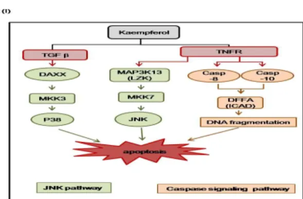
확인하였다(Fig. 4I).
Fig. 4. Genome-Wide RNA Expression Profiling Revealed Kaempferol Induced Apoptosis and G1 Arrest.
The experimental conditions were the same as those described in Figure 3, were used. (A-B) The lists of Up- or Down-regulated genes by kaempferol. (C) Analysis of gene ontology from KEGG. (D) Gene clustering file for apoptosis. Gene clustring files revealed 2-fold up- (red color) or down-regulated genes (green color) in kaempferol-treated Hep3B cells(Hep3B-K) compared with DMSO-treated Hep3B cells (Hep3B). (E-F) Apoptosis (E) and MAPK poptosi(F) from KEGG. Genes up-(red color) or down- regulated (green color) by kaempferol were (E) d on the microarray results. (G-H) Parallel total RNA with microarray was u) d to generate rever) -transcribed and amplified by real-time qPCR u)ing indicated primers (T (le 1). The fold-increases in mRNA expression by real-time qPCR are normalized to the mRNA level of GAPDH. Data shown represent the means±S.D. from three independent experiments(P<0.05). (I) Schematic diagram. Kaempferol leads to apoptosis through MKK3-p38 and MKK7-JNK signaling cascade. Also, kaempferol-induced caspases are trigger to apoptosis.고 찰
간암은 간에 발생하는 악성 종양으로, 原發性과 續發性 (轉 移性)으로 나눌 수 있다. 간암은 문맥을 통한 혈행성 전이가 잘 되므로, 원발성 암보다 전이암의 발생빈도가 더 높다. 간의 원발 성 암은 85% 이상이 간세포암이며, 원인은 B형, C형 간염 바이 러스 감염, 알코올, mycotoxins, 기생충, 남성 호르몬이나 피임약 복용, 비만 및 대사증후군
3,4)등이 추정되고 있으나, 가장 중요한 인자는 B형, C형 간염 바이러스로 보고 있다. 우리나라의 경우 간암환자의 80%에서 만성 바이러스성 간질환에서 이행되었다고 발표하고 있다
4).
우리나라의 간암 발병률은 1999년도에는 전체 암 중 2위였 으나, 점점 발병률이 낮아져 2009년도 기준으로는 전체 암 중 5 위의 빈도로 발생하고, 성별에 따라서는 남성에게서는 4위, 여성 에게는 6위의 빈도로 발병한다고 보고되고 있다. 간암은 다른 암 에 비해 예후가 불량하고 치사율이 높은 편이어서, 주요 암 사망 분율이 전체 암 중 2위를 차지하며, 5년 생존률이 25% 전후로 보 고되고 있다
2,3).
간암의 증상은 매우 다양한데, 주로 호소하는 증상은 복부 팽만감, 체중감소, 미열, 心窩部와 右上腹部 및 背部의 지속적인 鈍痛, 위장관 증후, 식욕부진, 변비 등이다. 증상이 진행되면 腹 水나 黃疸이 발생하며, 간암 말기에는 극렬한 복통, 식도정맥류 출혈 등이 나타날 수 있다
3).
서양의학적 치료는 간절제술, 간이식, 국소치료술, 경동맥화 학색전술, 방사선치료, 항암화학요법 등이 시행되고 있다
4). 최근 들어, 상위의 항암치료법 이외에 한약재들이 항암치료에 사용되 고 있으며, 그 효과가 국내외 연구자들에 의해 보고되고 있다
6-9).
대표적으로, 미국의 Alexander S. Sun 박사는 19종의 식물 및 한 약재로 구성된 SV (Selected Vegetables)가 폐암에 효과적임을 밝 혀 임상실험 중에 있으며, 이 등
6)의 국내 연구진에 의해서 牧丹 皮에서 추출한 메틸 갈레이트 (methyl gallate)가 항암 효과가 있 다고 보고되었으며, 이 등
7), 서 등
8)의 연구에 의해서 當歸, 白芨,
山慈菇 외 10여종의 한약재로 구성된 加味啓膈湯
(Kamikaekyuk-tang)은 암세포의 증식을 억제하는 항암 효과가 있음이 보고되었다. 그 외 야채, 과일, 곡물, 차, 포도주 등에서 함유된 폴리페놀 화합물인 flavonoid는 抗酸化, 抗癌, 抗炎症 작 용에 관여하는 것으로 알려져 있다
25,26).
黃精은 백합과에 속한 다년생 본초인 둥굴레의 根莖으로, 性 은 平하고, 味는 甘하며, 脾, 肺, 腎經으로 歸經하여 補氣養陰, 健 脾, 潤肺益腎, 除風濕, 安五臟 등의 효능을 가지고 있다. 補陰之 劑에 속하여 脾胃虛弱, 虛損寒熱, 肺虛燥咳, 病後體虛食少, 精血 不足, 筋骨軟弱 등의 치료에 활용되고 있다
11-13).
최근의 실험 연구에 따르면, 黃精은 항균, 항진균, 항고혈압 작용이 있음이 보고되어 있고, 당뇨병이 유발된 쥐에게 장기복 용시킨 경우 혈당 감소가 나타났으며, 고지혈증이 유도된 쥐에 게 투여했을 경우 혈액 내 지질수치가 감소되는 것으로 보고되 었다
13-18,25).
黃精의 주요 성분인 kaempferol은 자연에서 발견되는 flavonoid 중 하나로, 채소 및 과일에 많이 함유되어 있으며, 녹 차 및 홍차와 같은 茶類, 한약재 중 黃精, 白果, 松葉, 前胡, 枳實, 枳殼 등에 함유되어 있다
14,17,18). Kaempferol은 고지혈증, 고혈압, 당뇨 등의 증세 완화
25-30)및 암세포의 사멸(apoptosis)을 유도하 여 폐암, 난소암, 자궁암, 골육종, 신경교종 등의 암을 억제하는 작용이 있는 것으로 보고되어 있으며
19-24,31-38), 간암 억제에도 효 과가 있는 것으로 보고되어 있다
39,40).
본 실험에서는, 黃精의 주요 성분인 Kaempferol이 간암 세 포의 성장에 미치는 영향을 알아보고자 하였다. MTT를 이용한 세포 독성 실험에서, Kaempferol은 정상 세포에서는 독성을 일 으키지 않았고, 간암 세포주인 Hep3B와 HepG2 세포에서는 각 각 175 μM과 290 μM의 농도에서 50% 수준으로 세포의 성장을 억제하는 것으로 나타났다 (Fig. 1A). 또한, 黃精 추출물은 정상 세포에서는 독성을 일으키지 않았고, 간암 세포주인 Hep3B 세포 에서는 2.31 mg/ml 농도에서 세포의 성장을 50% 수준으로 억제 하였다(Fig. 1B).
이러한 세포 성장 억제 기전을 좀 더 자세히 알아보기 위하 여 FACS, Western blot, DAPI 염색법, Trypan blue 염색법 및 DNA fragmentation법을 이용하여 분석한 결과, kaempferol은 간암 세포의 사멸을 유도시키는 것으로 확인 간암 세포간암 세 포 중 특히 Hep3B에 대한 세포 사멸 효과가 큰 것으로 확인되었 다(Fig. 2-3).
또한 microarray를 이용한 유전자 발현양상 분석실험을 통
해, kaempferol이 간암세포의 사멸을 유도하는 경로를 분석하고
자 하였다. 그 결과, MAPK pathway에 관여하는 유전자들의 발
현양상이 크게 영향 받는 것을 확인하였다(Fig. 4A-4C). 유전자
발현 분석의 결과, kaempferol에 의해서 TNF receptor가 증가되
었는데, 이는 caspase-10과 caspase-8을 발현을 증가시키고, MKK7-JNK pathway (Fig. 4D-4I)를 유도함과 동시에, TGF beta signal을 통한 MKK3-P38 pathway를 유도함으로써, 궁극적으로 간암 세포의 사멸을 유도하는 것으로 확인되었다(Fig. 4).
이상으로 본 연구에서는, kaempferol은 caspase signaling, MKK7-JNK pathway 및 MKK3-P38 pathway를 유도함으로써 간 암 세포의 사멸을 유도함을 밝혔다. 본 연구를 바탕으로 한 kaempferol의 효과는 간암 억제 기전 확립과 항암 치료 및 치료 제 개발을 위해 기여할 것으로 사료된다.
결 론
黃精의 주요 성분인 kaempferol의 간암 억제 효과를 규명하 기 위하여, kaempferol을 간암 세포주에 처리하여 세포의 생존률 및 성장률을 분석하였으며, microarray를 통하여 유전자 발현 양 상을 분석하였다. Kaempferol은 caspase signaling, MKK7-JNK pathway 및 MKK3-P38 pathway를 통해 간암 세포의 사멸을 유 도하였다. 이로써 kaempferol은 간암 세포 사멸 과정을 촉진함으 로써 간암 억제 효과를 나타내는 것으로 추정된다.
참고문헌