Kor. J. Hort. Sci. Technol. 31(3):299-307, 2013 DOI http://dx.doi.org/10.7235/hort.2013.12211
Characterization of the Gene Encoding Radish (Raphanus sativus L.) PG-inhibiting Protein
Byung-Ho Hwang
1,2†, Hun Kim
1†, Sooyeon Lim
1, NaRae Han
1, and Jongkee Kim
1*1
Department of Integrative Plant Science, Chung-Ang University, Anseong 456-756, Korea
2
Institute of Molecular Plant Sciences, University of Edinburgh, King’s Buildings, Mayfield Road, Edinburgh EH9 3JH, UK
Abstract. A radish (Raphanus sativus L.) polygalacturonase-inhibiting protein (PGIP) gene was cloned and compared to the PGIP gene (BrPGIP2) from Chinese cabbage (Brassica rapa ssp. pekinensis) in order to gain more information on controlling a disease and improving produce quality. To clone the radish PGIP gene, primers were designed based on conserved sequences of two PGIP genes (BnPGIP1 and BnPGIP2) from rape (B. napus L. ssp. oleifera), Chinese cabbage and Arabidopsis thaliana. PCR cloning was performed with cDNA from the stigma of radish ‘Daejinyeoreum’ as a template to confirm DNA fragments which were about 600 base pair in size. Sequence analysis revealed 84.1% homology with BrPGIP2 and 70.1% with BnPGIP1. DNA walking was conducted to confirm the open reading frame of 972 bp, and the gene was named RsPGIP1. RsPGIP1 consisting with 323 amino acids (aa) has a high leucine content (54/323) and contains 10 leucine-rich repeat domains, as do most BrPGIPs of Chinese cabbage. The gene expression of RsPGIP1 was induced by abiotic stresses and methyl jasmonate. It showed enrichment in the stigma and the primary root than a leaf. Cloning RsPGIP1 will aid to further apply practices on postharvest quality maintenance and disease control of the root.
Additional key words: PGIP, polygalacturonase, postharvest disease, produce quality, radish
*Corresponding author: [email protected]
†
These authors are contributed equally to this work.
※ Received 15 November 2012; Revised 24 December 2012; Accepted 29 December 2012. This work was supported by the Young Investigator Research Program of Chung-Ang University 2008 year, the GRRC program of Gyeonggi province [GRRC-CAU-2012-A01, Development of Baemoochae kimchi and postharvest technology] and the Korea Research Foundation Grant (KRF-2007-357-F00006) funded by the Korean Government (MOEHRD), Korea. The authors thank B. Han (Yeungnam University, Gyeongsan) for critical reading of the manuscript.
Introduction
Radish roots are one of the most popular vegetables in Korea, where people consume in a number of different food recipes, including a main ingredient of ‘Kimchi’. Radish roots produced in spring are less susceptible to low temperature, whereas fall season types are very sensitive.
It is widely accepted that the firmness of radish root is one of the most important quality criterion that is used to processing ‘Radish Kimchi’ as well as other food recipes made of radish root (Cheigh and Park, 1994). Several studies suggested that pectins in cell walls play an important role in maintaining tissue firmness during processing or cooking of the root (Fuchigami, 1987; York et al., 1985).
The plant cell wall is an important barrier to pathogen invasion. Most plant pathogens secrete a cocktail of cell wall degrading enzymes capable of depolymerizing the polysaccharides in the plant cell wall. In response, plants have evolved a
diverse battery of defense responses including proteinaceous inhibitors of pectin degrading enzymes such as PG (PGIP, PG inhibiting protein), pectinmethyl esterase (PMEI, PME inhibitor) and pectin lyase (PNLIP, PNL inhibitor protein), as well as hemicellulose degrading enzymes such as endoxylanase (XI, xylanase inhibitor), xylanase (XIP, X inhibitor protein) and xyloglucan endoglucanase (XEGIP, XEG inhibiting protein) (Juge et al., 2006). The majority of plant pathogenic fungi and bacteria secrete PG, which can degrade cell wall pectic components consisting of a rhamnogalacturonan main chain, and this seems to be involved in development of disease symptoms (Lebeda et al., 2001).
PGIPs that are present in plant cell wall have a leucine-
rich repeat (LRR) domain, one of the features of plant
resistance proteins, and directly reduce the aggressiveness
of fungal PG (Liang et al., 2005). The existence of PGIPs
has been confirmed in leek and onion, which are monocots,
and also in a number of dicotyledons including soybean,
apple, pear, orange, and kiwifruit. A variety of PGIP genes have been cloned from 103 plant species (De Lorenzo et al., 2001; NCBI gene bank). Recently, PGIP genes in rape, rice, strawberries and Chinese cabbage have been identified (Ahsan et al., 2005; Mehli et al., 2004). Most PGIP genes are composed of multi-gene families clustered in a particular chromosomal region. In the case of the two Arabidopsis thaliana PGIPs, they are located tandemly on chromosome 5 with a distance of 507 bp (Ferrari et al., 2003).
Numerous studies on PGIPs have investigated their ex- pression in relation to invasion of fungal pathogens, the response to treatment with elicitors such as salicylic acid (SA), jasmonic acid (JA), and oligogalacturonides (OGs), and expression pattern after abiotic stress (De Lorenzo et al., 2001). In addition, the expression pattern of PGIP during developmental processes such as rice floral maturation and organ number has been reported (Jang et al., 2003). The expression of PGIPs of Brassica napus and Arabidopsis is regulated by some abiotic stresses (Ferrari et al., 2003).
The effect of apple PGIP was tested on a well characterized cell wall destructing enzyme, PG from a postharvest soft rot pathogen, Colletotrichum acutatum (Gregori et al., 2008).
Recently, the genes encoding PGIPs were cloned and cha- racterized in Chinese cabbage which is a main crop belonging to Brassicaceae family same as radish (Hwang et al., 2010).
Bacterial soft rot in radish root, caused by Pectobacterium carotovorum ssp. carotovorum (Pcc) is a soil epidemic disease.
Pcc is a facultatively gram-negative bacterium and attacks plant tissue through the secretion of a cocktail of cell wall degrading enzymes mainly including PG during high temperature and high humidity like rainy spell in summer. Therefore, the study on inhibiting PG activity of Pcc is necessary.
In this paper, we cloned cDNA using RT-PCR after confirming the genomic sequence of RsPGIP gene from radish through PCR cloning and DNA walking, and investigated RsPGIP expression pattern in relation to environment stress and signaling molecules to find a useful way of applications for managing radish quality and diseases in postharvest.
Materials and Methods Plant Material and Sample Preparation
Raphanus sativus L. ‘Daejinyeorum’ (Monsanto-Korea, Seoul, Korea) radish seed was immersed in water in dark conditions at 28°C for 36 h, sowed in sterilized soil, and cultivated in a greenhouse for 4 weeks with a day temperature of 18 to 20°C and a night temperature 10 to 12°C. For preparation of genomic DNA, 4-weeks-old radish leaves were extracted and stored at -70°C after being frozen in liquid N
2. We attempted to clone the PGIP gene from
radish stigma through cDNA library screening based on the method used for the isolation of Chinese cabbage PGIP from stigma (Hwang et al., 2010). Ten young plants were placed at 4°C with 16 h day and 8 h night for 30 days to induce flower. After vernalization, plants were transplanted into 5 L pots and cultivated for 6 weeks in the greenhouse.
When plants were in full bloom, stigmas were excised with a scalpel, immediately frozen in liquid N
2,and stored at -70°C for total RNA extraction.
For analysis of the expression patterns of RsPGIP1 in various plant tissues, the 4-week-old radish plants were placed at 4°C for 30 days under 16 h light conditions to induce flowering and then grown in the greenhouse until flowering. Stigma, leaf blade, and primary root tissues from flowering plants were harvested, frozen in liquid N2, and stored at -70°C for later use.
To investigate the expression pattern after treatments with low temperature, salt stress, mechanical wounding, and defense response activators of RsPGIP1, radish seeds were germinated at 28°C for 36 h, and seedlings were grown in the greenhouse for 4 weeks. After selecting young uniform plants, they were conditioned for 5 days at 23°C with 16 h light and 8 h dark.
RT-PCR was performed after those treatments.
PCR Cloning and Sequence Analysis
For PCR cloning, a primer CL (PCR cloning 5' primer)
and CR (PCR cloning 3' primer) were designed based on the
conservative base sequences of PGIP genes from Arabidopsis,
rape and Chinese cabbage. PCR primer sequences for all
experiments are given in Table 1. Total RNA was extracted
with an RNeasy plant mini kit (Qiagen, Valencia, CA, USA) from
the stigma of blooming flowers of radish and quantified with
a spectrophotometer (Beckman Coulter DU 650, Indianapolis,
IN, USA). The synthesis of 1st strand cDNA was completed
using a Superscript 1st-Strand Synthesis Kit (Invitrogen,
Carlsbad, CA, USA) from 5 µg total RNA according to the
manufacturer’s recommendation. Genomic DNA was extracted
from young leaves of radish using a DNeasy Plant Mini
Kit (Qiagen) and quantified with a spectrophotometer. The
PCR template used was a 1:20 dilution of the 1st strand
cDNA product (1 μL). PCR was conducted using primer
5' CL and 3' CR. The same primer set was used for the
PCR with genomic DNA. PCR amplifications were carried
out in a final volume of 20 μL containing 10 mM Tris-HCl
(pH 8.4), 1.5 mM MgCl
2, 50 mM KCl, 0.25 mM each
dNTP, 0.2 μM each primer, 20 ng template DNA and 1
unit of Taq DNA polymerase using the following: 94°C for
5 min, then 35 cycles of (94°C for 1 min, 58°C for 1 min,
72°C for 1 min), with final extension step of 72°C for 10
min. PCR products were analyzed on a 1.2% agarose gel
Table 1. Primer sequences used for PCR and RT-PCR analyses.
Gene Primer Sequence (5' → 3')
PCR cloning CL (PCR cloning 5' primer) CGCCAAGCTCAAGTATCTCC CR (PCR cloning 3' primer) TCGAATTCCTGAAGTGTCCC
DNA walking 5' TSP1 AAGCATCGCCTCCGAGCTT
5' TSP2 GTTTTCGGTATATAACCGGAGAGCTGG
5' TSP3 TGGTTGTGTGATAGGTAAAGGTCAGGTCA
5' TSP4 GGGATCTGACCGGTGAGGTTA
5' TSP5 GTGAGTTTGCGGAAGACAAGAGTCTGTAG
5' TSP6 AAGTCACCGACTTCAGGCGGGAT
3' TSP1 GATCACAGGGAATATCCCGGTT
3' TSP2 TGGACCGAAGCTCCTCTCCAGTT
3' TSP3 TGTGTGGACACATCCCCACAGGA
RsPGIP1 RsPGIP-FW ATAGGGATCCCCATGTGTGGTAAAACAACGACA
RsPGIP-RV AGTCGTCGACTCACTTGCAACTATCAAGAGGTGC
RsACTIN RsACTIN-FW ATGACATGGAGAAGATCTGG
RsACTIN-RV TGAGCTTGTTTTGGAAGTCT
(Agarose M, Biosesang, Seongnam, South Korea) containing EtBr (Sigma, St. Louis, MO, USA) and the corresponding DNA bands were recovered and cloned into pGEM-T Easy Vectors (Promega, Madison, WI, USA) for sequencing.
DNA sequences of the insert (about 600 bp) were deter- mined by Genotech Co. Ltd. (South Korea). Sequence analysis was performed using the software BioEdit ver 7.0.5 (www.
mbio.ncsu.edu/BioEdit/) and Basic Local Alignment Search Tool (BLAST; Altschul et al., 1990) in the GenBank database (www.ncbi.nlm.nih.gov).
DNA Walking Experiments
Two walks were performed in the 5' direction and also in the 3' direction for a total of three DNA walking experiments using a primer designed based on the confirmed base sequence of RsPGIP. The first experiment in the 5' direction was carried out by using a DNA walking SpeedUp
TMPremix Kit (Seegene, Seoul, South Korea) after designing primers 5' target-specific primer (TSP) 1, 5' TSP2, and 5' TSP3.
Since a region that includes the start codon of RsPGIP was not acquired through the first experiment, primers 5' TSP4, 5' TSP5, and 5' TSP6 were designed using the sequence of the acquired 5' region; the second experiment in the 5' direction was then performed. Walking the 3' direction was done using primers 3' TSP1, 3' TSP2, and 3' TSP3, providing sequence data to confirm the gene as RsPGIP.
Sequence Analysis
The NCBI website was used for the database search and
the analysis of the deduced amino acid sequence similarity.
A multiple sequence alignment was done with the ClustalW program (www.ebi.ac.uk/clustalw/; Larkin et al., 2007). A phylogenetic tree was generated using the neighbor-joining method by Mega4 (http://www.megasoftware.net/; Kumar et al., 2008). Signal peptide prediction and motif searches were performed using SignalP ver 3.0 (www.cbs.dtu.dk/
services/SignalP/; Bendtsen et al., 2004) and PROSITE (www.expasy.org/prosite/; Hulo et al., 2006), respectively.
Stress Treatments
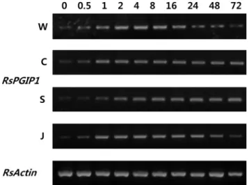
To induce wound, leaves were mechanically damaged by crushing the boundary point at the mid-rib with sterile forceps.
For cold treatment, 4-week old plants were kept at 4°C under 16 h of daylight for 3 days. For salt treatment, plants were watered, and then their leaves were sprayed with 0.5 M NaCl every 12 h for 3 days. For drought treatment, watering was withheld for 4 days, at which time plants began to wilt.
For all the treatments, samples were collected from 4-week-old plants at 0, 0.5, 1, 2, 4, 8, 16, 24, 48, and 72 h after treatment. The same age plants treated with Methyl-jasmonic acid (MeJA) were used as positive control, and those without any treatment were used as negative control. After each treatment, leaves were collected and frozen immediately in liquid N
2from three individual plants and stored at -70°C for RNA extraction.
Treatments of Different Defense Response Activators
The biggest leaves of 4-week-old plants were selected to
A
B C D E
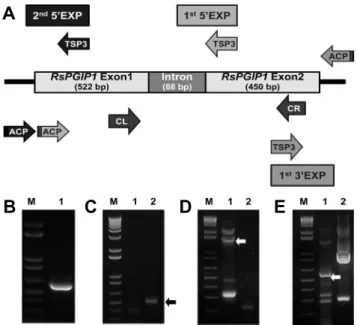
Fig. 1. Gene structure and amplification of the RsPGIP1 gene region. (A) Gene structure of RsPGIP1. CL, PCR cloning 5' primer; CR, PCR cloning 3' primer; TSP, target specific primer;
ACP, annealing controlled primer. (B) PCR cloning product.
(C) PCR products of 1st 5' region DNA walking experiment, Lane 1: ACP1, Lane 2: ACP2. (D) PCR products of 2nd 5' region DNA walking experiment, Lane 1: ACP1, Lane 2: ACP2.
(E) PCR products of 3' region DNA walking experiment, Lane 1: ACP1, Lane 2: ACP2. DNA fragments for sequencing are indicated with an arrow. M, wide range DNA logical marker (100 bp-10 kb).
examine the effects of different defense response activators.
MeJA (100 μM, Sigma, St. Louis, USA) and salicylic acid (50 mM, SA; Sigma, St. Louis, USA) were sprayed on the aerial parts of plants. Plants sprayed with distilled water were used as a control. Leaves were harvested at 0, 0.5, 1, 2, 4, 8, 16, 24, 48, and 72 h after treatment.
RNA Purification and RT-PCR Analysis
For RT-PCR analysis, the total RNA was purified from the frozen sample using the RNeasy Plant Mini Kit (Qiagen) and according to the manufacturer’s instructions. cDNA synthesis was performed with the same method of PCR cloning. RT-PCR was carried out using redesigned RsPGIP-FW and RsPGIP-RV primers.
Results Isolation of the RsPGIP Gene
In this study, PCR cloning primers prepared based on the conserved regions of PGIP genes from Arabidopsis, rape, and Chinese cabbage were used to amplify DNA fragments from radish stigma cDNA and genomic DNA. DNA product with an expected size of about 600 bp was amplified by PCR (Fig. 1B). The DNA fragment of 676 bp amplified from genomic DNA was approximately 100 bp longer than
that amplified from stigma cDNA. Cloning and sequencing of the two products revealed that the 88 bp DNA fragment was an intron of the radish PGIP gene. The intron has good 5' and 3' splice site consensus CAG-GT and ACAG-GT respectively (Fig. 1A). High similarities in amino acid sequences were found between radish PGIP cDNA and PGIP from Chinese cabbage, rape, Arabidopsis. The PGIP gene from radish was named RsPGIP1 (Raphanus sativus L. PGIP gene copy No. 1).
The full length ORF sequence of RsPGIP1 was confirmed by performing 5' DNA walking twice and 3' DNA walking once. The first 5' DNA walking experiment was performed using primers 5' TSP1, 5' TSP2, and 5' TSP3, constructed based on the sequence of partial RsPGIP1 gene and confirmed by PCR cloning. The band with the most distinct 0.5 kilo base (kb) size was confirmed in the lane of DW (DNA Walking) -ACP (Annealing Control Primer) 2. The sequence of this band included the intron (88 bp) area from the location of 5' TSP3 (587) to 165, and was confirmed as being 510 bp in size (Fig. 1C). Since the translation start site in the DNA walk experiment of the first 5' DNA walk was not confirmed, primers 5' TSP4, 5' TSP5, and 5' TSP6 were used in the second 5' DNA walking experiment. They were designed after analysis of the first 5' DNA walk. As a result of performing the second 5' DNA walk, clear bands of 0.3, 0.6, 1.6, and 2.0 kb size were detected in the lane of DW-ACP1. Among them, fragments of 0.3 kb and 1.6 kb size were cloned in the pGEMT-easy vector and their sequence analyzed. These two bands represented the RsPGIP1 gene sequence and the sequence of 1631 bp including the RsPGIP1 promoter region from 5' TSP6 (258) to -1373 (Fig. 1D).
DNA walking from the 3' direction was performed using primers 3' TSP1, 3' TSP2, and 3' TSP3, constructed based on the DNA sequence of partial RsPGIP1 and confirmed with PCR cloning. A variety of clear bands were detected in the DW-ACP1 lane as well as other lanes, but in the DW-ACP1 lane, bands of 5.5 kb and 1.3 kb size were detected. Both bands were from the RsPGIP1 gene sequence and from the location of 3' TSP6 (868) to 2160, the sequence of 1292 bp was confirmed (Fig. 1E).
Primers RsPGIP-FW and RsPGIP-RV were designed based
on the sequence of the full-length ORF of RsPGIP1. cDNA
from the RsPGIP1 gene was cloned by PCR using the
prepared primers and stigma cDNA. The DNA sequence
was analyzed by cloning the cDNA of RsPGIP1 into pGEMT-
easy. The RsPGIP1 gene sequence was confirmed from cDNA
and exactly matched the sequence of RsPGIP1 confirmed
from genomic DNA. The RsPGIP1 gene was deposited in
GenBank database with accession no. EF392661.
Fig. 2. Alignment of predicted amino acid sequences of PGIPs. The signal peptide of RsPGIP1 is underlined, and the cysteines and glycosylation sites are indicated with a star and upward arrow. The amino acid sequences shown are for AtPGIP1, Arabidopsis thaliana AtPGIP1 (accession no. NM120769); AtPGIP2, Arabidopsis thaliana AtPGIP2 (accession no. NM120770); BnPGIP1, Brassica napus BnPGIP1 (accession no. AF529691); BnPGIP2, Brassica napus BnPGIP2 (accession no. AF529693); RsPGIP1, Raphanus sativus RsPGIP1 (accession no. EF392661).
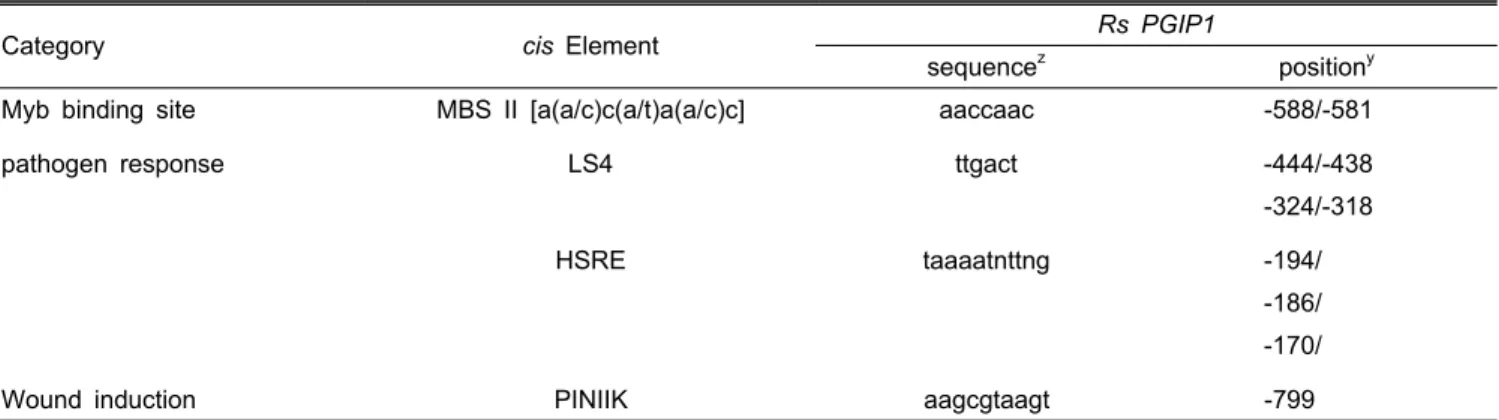
Table 2. Notable putative cis-acting elements in the RsPGIP1 promoter.
Category cis Element Rs PGIP1
sequence
zposition
yMyb binding site MBS II [a(a/c)c(a/t)a(a/c)c] aaccaac -588/-581
pathogen response LS4 ttgact -444/-438
-324/-318
HSRE taaaatnttng -194/
-186/
-170/
Wound induction PINIIK aagcgtaagt -799
z
Sequence is indicated from the 5' to the 3' end.
y