Copyright © 2011 Korean Journal of Bronchoesophagology 19
Korean J Bronchoesophagol 2011;17:19-22 ISSN 1226-0916
REVIEW ARTICLE
서 론
식도암의 치료결과는 식도암의 병기와 연관된다. 내시경초 음파(Endoscopic ultrasonography; 이하 EUS)는 식도암의 T, N 병기의 정확한 설정을 가능하게 함으로써 식도암의 치 료전략을 세우는데 중요한 역할을 한다.1) EUS는 CT, PET 과 비교하여 식도암의 침윤도 진단과 식도주변 림프절, 복강 내 림프절의 진단 정확도가 높으며 전이가 의심되는 림프절 에서 EUS 유도 세침 흡인(EUS-guided fine needle aspira- tion)을 이용하여 림프절 전이 진단율을 높힐 수 있다.2) 표재 성 식도암에서 내시경 절제, 즉 내시경 점막하박리술(Endo- scopic submucosal dissection)같은 최소침습치료가 가능한 환자를 선별하는 데 중요하다.
정상 식도벽의 EUS 소견
식도벽의 층 구조는 초음파 탐촉자의 주파수에 따라 차이 가 있다. 7.5~10 MHz의 저주파의 탐촉자로 5층이나 7층으 로 관찰된다. 제1층은 식도 점막과 주위경계면이 이루는 에코 와 상피층 점막, 저에코의 제2층은 고유판층과 점막근판에 해당된다. 제3층은 고에코로 점막하층, 제4층은 저에코로 고 유근층, 제5층은 외막층이다. 제4층 중간에 고에코층이 보이 는 데 이를 기준으로 내윤상근과 외종주군으로 구별되므로 이 경우 식도벽은 7층으로 관찰된다. 20~30 MHz의 고주파수 탐촉자에서 식도벽은 통상적으로 9층을 묘출되는데 이는 5 층 구조에서 고에코로 묘출되는 제3층(점막하층)이 고-저-고 에코의 3층으로 세분화되어 보이기 때문이다. 9층 중 3층째 (3/9)의 고에코층은 점막근층(muscularis mucosa)이며 9층 중 4층째(4/9)의 저에코층이 점막하층을 반영한다(Fig. 1).
조기 식도암에서 EUS 심달도 진단
EUS의 식도암의 심달도 진단방법은 위암의 심달도 진단
조기 식도암에서 내시경초음파의 역할
가톨릭대학교 서울성모병원 소화기내과
조 유 경
Role of Endoscopic Ultrasound in the Assessment of Superficial Esophageal Cancer
Yu Kyung Cho
Division of Gastroenterology, Department of Internal Medicine, The Catholic University, Seoul, Korea
Endoscopic ultrasound in the diagnosis of esophageal carcinoma is an indispensable procedure, not only to discuss the preop- erative staging of the lesion, but also to evaluate the therapeutic effect of chemo–radiation therapy. The recent increase in the incidence of superficial esophageal cancer and promising developments in potentially curative endoscopic therapies have placed EUS to a central position in decision making. Recent data have called into question the staging accuracy of EUS to dis- tinguish mucosal from submucosal lesions, particularly in patients with early disease. In those cases, diagnostic endoscopic resection may be useful for staging and curative in superficial lesions. Nonetheless, EUS has been regarded as the most accu- rate staging tool and should be performed to identify potential candidates for endoscopic resection.
Korean J Bronchoesophagol 2011;17:19-22 KEY WORDSZZ Endoscopic ultrasound ㆍ Esophageal cancer.
논문접수일: 2011년 5월 13일 / 심사완료일: 2011년 5월 16일 교신저자: 조유경, 110-744 서울 서초구 반포동 505 가톨릭대학교 서울성모병원 소화기내과
전화: 02-2258-2044 ㆍ전송: 02-2258-2055 E-mail: [email protected]
online©MLComm
20
Korean J Bronchoesophagol
█2011;17:19-22
과 동일하다. 식도병변보다 보통 층이 두터운 위 병변에서 EUS를 시행하는 것이 더 쉽지만 오히려 식도암의 EUS 심달 도진단 정확도가 위암보다 높다. 이는 위암에서 궤양에 의한 점막하층의 섬유화를 동반한 경우가 더 흔하고 식도암에서 는 이런 경우가 더 적기 때문이다.
일반적으로 식도암은 식도내강에 원주상 혹은 미만형의 저에코의 벽 비후 소견 또는 벽내 종괴로 관찰된다. 침윤이 진 행되면 식도벽의 층 구조의 구별이 되지 않고 병변 외연이 불 규칙해지며 주위 장기와의 경계가 소실된다. 점막암은 상피내 암(m1), 점막고유층암(m2), 점막근판암(m3)으로 세분하고 점막하층암(sm)은 상부층암을 sm1, 중간층암을 sm2, 하부 층암을 sm3로 분류한다. 점막암의 경우 점막층인 제1층이 부분적으로 소실되고 제2층이 저에코로 비후된 소견을 보이 며 제3층인 점막하층에 변화를 보이지 않는다(T1m, Fig. 2).
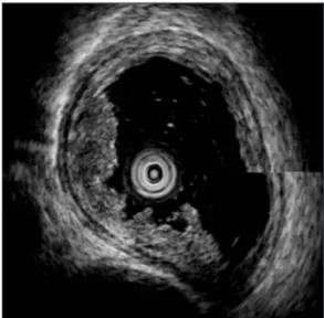
점막하층암의 경우는 점막층의 저에코 비후와 점막하층의 연 속성의 불균질이나 부분적인 단락을 보인다(T1sm, Fig. 3).
점막하층이 완전히 단렬되거나 점막층, 점막하층, 고유근층이 구별되지 않은 저에코 병변으로 외막층의 변연이 규칙적이면 고유근층암(T2), 외막층의 변연의 저에코가 불규칙하거나 식 도벽 구조 주위조직으로 돌출될 경우 외막침윤암(T3), 주위 의 혈관구조나 고에코의 늑막, 기관지와 경계를 보이지 않을 경우 주위 장기 침윤암을 시사한다(T4). T 병기 결정에 있어 서 CT와 EUS의 정확성을 비교한 연구에서 EUS는 CT 45(40~50)%에 비해 85(59~92)%로 우월하였다.3)
조기식도암에서 EUS의 역할
내시경 점막하 절제술, 광역동 치료(photodynamic ther- apy), radiofrequency ablation과 같은 ablative procedure의 발달하며서 표재성 식도암에서 종양 침윤깊이의 정확한 진 단이 필요하게 되었다. 조기식도암에서 EUS의 가장 중요한 역할은 내시경 절제 같은 국소치료가 가능한 표재성 암을 선 별하는 것이다. 림프절전이와 관계없이 점막이나 점막하층에 국한된 식도암을 표재성 식도암이라고 하는데 종양침윤의
심달도는 림프절 전이와 상관이 있다. 내시경 점막하 절제술을 시행하기 전에 정확한 T 병기설정이 중요하다. 암이 상피층 (m1)이나 점막고유층(m2)에 국한되어 있는 경우는 림프절 전 이가 거의 없지만(0~3%), 점막근층(m3)을 침윤하면 12~20%
정도 림프절 전이가 있고 점막하층의 상부 1/3(sm1)을 침윤하 면 26.5%, 점막하층 전체(sm3)를 침윤하면 46%에서 림프절 전이가 동반된다.4) 따라서 식도암의 내시경 점막절제술은 위 나 대장에 비해 더욱 제한적으로 적응증은 점막암이다. 점막 근층 침윤암(m3암) 경우, 고유근판 침윤암과 점막하층의 중 간 정도로 림프절 전이의 위험성이 있고 23% 정도에서 림프 절 전이의 위험인자이 맥관침습 양성이 보고되는 경우가 있으 므로 내시경 절제술의 적응증이 되는지에 대한 아직도 논쟁 이 있다. 최근에 일본에서는 저분화형, 미만성 침윤형 등 림프 절 전이의 위험이 높은 예를 제외하고 그 이외의 m3암에 대 해서도 적극적으로 내시경 절제를 확대 적용하는 추세이다. 이 에 필수적으로 정밀한 심달도, 분화도의 판정이 요구되므로
Mucosa layer
Submucosal layer Muscularis propria
Outer later
(border echo of connective tissue)
Fig. 1. Layered structure of esophageal wall divided into nine
layers.Fig. 2. An examples of esophageal cancer infiltrated into lami-
na propria of mucosa. Hypoechogenic thickening of mucosa is observed in the direction of 9-11 o’clock.Fig. 3. An examples of esophageal cancer infiltrated into sub-
mucosa. Destruction of mucosa and submucosa caused by in- filtration of the cancer is observed in the direction of 6-10 o’clock.http://www.korbes.org 21
EUS in the Superficial Esophageal Cancer
█YK Cho
표재성 식도암에서는 방사형 내시경초음파보다 20~30 MHz 의 고주파 미세 탐촉자를 이용하여 점막암 특히 고유판 이 내의 식도암을 구별하도록 노력하여야 한다. 최근 고주파미 세 탐촉자연구에서 내시경초음파검사의 진단율은 m1과 m2, 81%, m3과 sm1, 60%, sm2과 sm3, 87%이었고 m1, m2와 m3를 구분하는 정확도는 81%에서 100%까지 보고 되었다.5) EUS는 벽 단층상을 얻을 수 있는 유일한 진단 방법으로 암과 비암성 병변의 감별이 어려우며 미소침윤암의 경우 EUS의 진단에 한계가 있다. 하지만 융기나 비후가 눈에 띄는 병변의 평가에는 고주파 초음파 탐침자 EUS가 도움이 된다.
표재성 식도암에서 심달도의 진단정확도를 높이는 최신 내시경 기법
최근 여러 최신내시경 기법이 발달하여 상세한 심달도의 분류가 가능하여졌다. 최근 일본에서 협대역 내시경(narrow band imaging)과 확대내시경을 이용하여, 통상 내시경 관찰 에는 진단할 수 없는 식도상피의 미세 혈관패턴으로 점막암 의 상세분류(m1암, m2암, m3암)를 하려는 노력을 하고 있다.6) 확대내시경에서 심부 침윤에서는 multi-layered vessel, ir- regulary branched vessel, reticular vessel 등의 혈관패턴이 관찰되고 이로 둘러싸인 avasucular area의 크기로 심달도 를 짐작할 수 있으므로 융기나 비후가 눈에 띄지 않는 병변이 나 미소 병변에서는 도움이 된다. 하지만 저분화형이나 미만성 침윤형태를 띄는 암, 융기나 비후가 눈에 띄는 병변, 점막하종 양의 형태를 나타내는 병변에는 역시 EUS가 심달도 진단에 유용하다.
조기식도암의 치료전략 수립(Decision Making)에서 EUS의 역할에 대한 논쟁
최근 일부에서 고주파 초음파탐촉자가 그 동안의 기술적
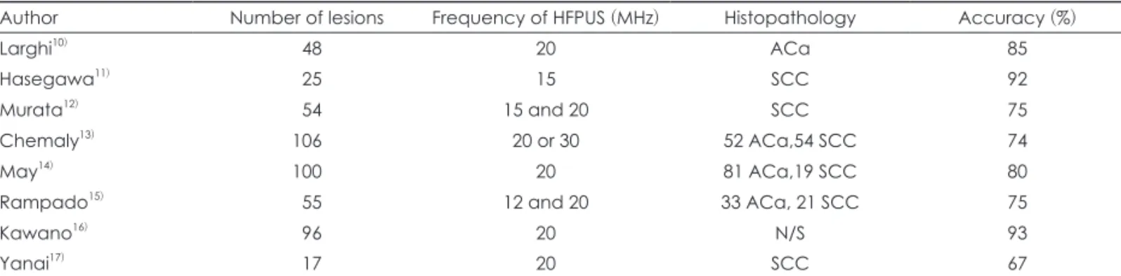
진보에도 불구하고 점막층과 점막하층을 구별하는 정확도가 65~93% 정도로7) 기관마다 차이가 많이 나며, 아주 만족스럽 지 않다는 점을 제기한다(Table 1).8) 그러므로 EUS의 결과와 상관없이, 또는 내시경 육안 소견만으로 내시경절제가 가능 하다고 판단된다면 EUS를 시행하지 않고 바로 진단적+치료 적 목적의 내시경 절제를 우선 시행해 보는 방법이 오히려 정확한 병리 진단을 가능하게 함으로써 overtreatment나 undertreatment를 피할 수 있다는 주장도 제기되고 있다.9)
하지만 그럼에도 불구하고 아직까지는 EUS가 점막층암, 고분화암, 2 cm 미만의 크기로 내시경 절제의 적응이 되는 경 우 시행되어야 하며, 전이가 의심되는 림프절은 EUS 유도하 세 침흡인으로 가능한 샘플을 얻어야 한다는 점에 대부분 동의 하고 있다.7) EUS로 점막암과 점막하층암을 확실히 구별할 수 없다면 앞서 언급한대로 우선 내시경 절제를 시행해 볼 수 있다.
요 약
내시경 초음파검사는 식도벽의 조직학적 층구조를 가장 유사하게 반영하는 검사법으로 식도 병변의 진단과 치료전략 에 필수검사이다. 표재성 식도암에서 고주파 미세탐촉자를 이 용한 EUS는 내시경 절제 같은 국소치료가 가능한 환자들을 선별하는 데 중요한 역할을 한다.
REFERENCES
1) Eloubeidi MA. EUS in esophageal cancer. In: Hawes RH, Fockens P, ed Endosonography, 2nd ed. Philadelphia: Saunders Elsevier, pp59-70, 2010.
2) Rosch T. Endoscopic staging of esophageal cancer: a review of lit- erature results. Gastrointest Endosc Clin North Am 1995;5:537-47.
3) Kelly S, Harris KM, Berry E, Hutton J, Roderick P, Cullingworth J, et al. A systematic review of the staging performance of endoscop- ic ultrasound in gastro-esophageal carcinoma. Gut 2001;49:534-9.
4) Kodama M, Kakegawa T. Treatment of superficial cancer of the esophagus: a summary of responses to a questionnaire on superfi- cial cancer of the esophagus in Japan. Surgery 1998;123:432-9.
Table 1. Accuracy of high frequency probe ultrasonography in the assessment of the depth of invasion of superficial esophageal
carcinoma7)Author Number of lesions Frequency of HFPUS (MHz) Histopathology Accuracy (%)
Larghi10) 48 20 ACa 85
Hasegawa11) 25 15 SCC 92
Murata12) 54 15 and 20 SCC 75
Chemaly13) 106 20 or 30 52 ACa,54 SCC 74
May14) 100 20 81 ACa,19 SCC 80
Rampado15) 55 12 and 20 33 ACa, 21 SCC 75
Kawano16) 96 20 N/S 93
Yanai17) 17 20 SCC 67
ACa: Adenocarcinoma, N/S: not specified, SCC: squamous cell carcinoma, HFPUS: high frequency probe ultrasonography