http://dx.doi.org/10.4174/astr.2016.91.3.118 Annals of Surgical Treatment and Research
Risk factors for lymph node metastasis in mucosal gastric cancer and re-evaluation of endoscopic submucosal
dissection
Si-Hak Lee1,5, Cheol Woong Choi2, Su Jin Kim2, Chang In Choi3, Dae-Hwan Kim3, Tae-Yong Jeon3, Dong-Heon Kim3, Hyun Jung Lee4, Ki-Hyun Kim1, Sun-Hwi Hwang1,5
Departments of 1Surgery and 2Internal Medicine, Pusan National University Yangsan Hospital, Yangsan, 3Department of Surgery, Pusan National University School of Medicine, Yangsan, 4Department of Pathology, Pusan National University Yangsan Hospital, Yangsan, 5Research Institute for Convergence of Biomedical Science and Technology, Pusan National University Yangsan Hospital, Yangsan, Korea
INTRODUCTION
The increased interest in cancer screening—represented by the national cancer screening program in Korea that includes biannual upper endoscopy for all individuals aged >40 years—
and the development of endoscopic equipment and techniques
have facilitated the early detection of gastric cancer. Early gas
tric cancer (EGC) is defined as a cancer confined to the mucosal or submucosal layers of the stomach, regardless of the presence of lymph node (LN) metastasis [1].
The prognosis of patients with EGC has improved with surgi
cal treatment. In fact, the 5year survival rate after curative Purpose: The selection of the appropriate treatment strategy for patients with mucosal gastric cancer (MGC) remains controversial. In the present study, we aimed to determine the risk factors for lymph node (LN) metastasis in MGC and reassess the role of endoscopic submucosal dissection (ESD).
Methods: We examined 1,191 MGC patients who underwent curative gastrectomy between January 2005 and December 2014. We determined the clinicopathologic risk factors for LN metastasis among the MGC patients.
Results: Among 1,191 patients with MGC, 42 patients (3.5%) had LN metastasis. Univariate analysis indicated that age ≤ 50 years (P = 0.045), tumor invasion to the muscularis mucosa (P < 0.001), tumor size > 2 cm (P = 0.014), presence of ulcera- tion (P = 0.01), diffuse type as per Lauren classification (P = 0.005), and undifferentiated-type histology (P = 0.001) were associated with LN metastasis. Moreover, multivariate analysis indicated that tumor invasion to the muscularis mucosa (P
= 0.001; odds ratio [OR], 4.909), presence of ulceration (P = 0.036; OR, 1.982), and undifferentiated-type histology (P = 0.025;
OR, 4.233) were independent risk factors for LN metastasis. In particular, LN metastasis was observed in some MGC cases with indications for ESD, including absolute indications (1 of 179, 0.6%) and expanded indications (9 of 493, 1.8%).
Conclusion: Although MGC patients can be treated via ESD, we recommend that they undergo a more aggressive treatment strategy if they have tumor invasion to the muscularis mucosa, ulceration, or undifferentiated-type histology in the final pathology report.
[Ann Surg Treat Res 2016;91(3):118-126]
Key Words: Lymph nodes, Neoplasm metastasis, Stomach neoplasms, Risk factors
Reviewed January February March April May June July August September October November December
Received March 31, 2016, Reviewed May 19, 2016, Accepted May 19, 2016
Corresponding Author: Sun-Hwi Hwang
Department of Surgery, Pusan National University Yangsan Hospital, Pusan National University School of Medicine, 20 Geumo-ro, Mulgeum-eup, Yangsan 50612, Korea
Tel: +82-55-360-2124, Fax: +82-55-360-2154 E-mail: [email protected]
Copyright ⓒ 2016, the Korean Surgical Society
cc Annals of Surgical Treatment and Research is an Open Access Journal. All articles are distributed under the terms of the Creative Commons Attribution Non- Commercial License (http://creativecommons.org/licenses/by-nc/4.0/) which permits unrestricted non-commercial use, distribution, and reproduction in any medium, provided the original work is properly cited.
resection is >90% in cases of EGC [2,3]. However, in cases with mucosal gastric cancer (MGC), minimally invasive treatments such as endoscopic submucosal dissection (ESD) have been actively substituted for conventional radical gastrectomy with LN dissection, particularly among those with advanced age and comorbidities, and those wishing to maintain their quality of life after treatment, in countries with a high prevalence rate of gastric cancer [46].
Despite the development of novel diagnostic and treatment methods, we often treat the patients diagnosed preoperatively as MGC without LN metastasis, but they are pathologically confirmed as having LN metastasis after the surgery (Fig. 1).
The prognostic factors for EGC include depth of tumor invasion, LN metastasis, grade of histologic differentiation, and curative surgery, and many studies have reported that LN metastasis is the most important risk factor for MGC recurrence [79].
Endoscopic ultrasonography (EUS), computerized tomography, and positron emission tomography are used to predict LN metastasis before surgery, or frozen sections may be used to evaluate LN metastasis intraoperatively. However, the diagnostic accuracy is a limitation of these methods, and hence, the minimally invasive trend in treatment for MGC such as ESD could consequently lead to an increased risk of early recurrence in MGC. Accordingly, the prediction of LN metastasis would
be useful for selecting the appropriate therapeutic strategy in MGC.
In the present study, we examined surgically resected cases of MGC, wherein tumor invasion was confined to the mucosa, in terms of their clinicopathological outcomes, as well as the frequency and risk factors of LN metastasis. Moreover, we evaluated the indications for ESD that were recently established, and reassessed their role in the treatment of MGC.
METHODS
We enrolled a total of 1,191 patients with MGC who under
went curative gastrectomy between January 2005 and December 2014. They were diagnosed with MGC without LN metastasis preoperatively based on the findings of imaging studies such as esophagogastroduodenoscopy, CT, or EUS. Among these patients, 42 (3.5%) were pathologically confirmed as having LN metastasis after resection.
To determine the possible relationship between LN meta
stasis and MGC, we analyzed the demographic and clinico
pathological characteristics of all the patients. These data included sex, age, tumor location, macroscopic type, depth of tumor invasion, tumor size, presence of ulceration, Lauren classification, histologic type (differentiated or undiffer
Fig. 1. A 51yearold woman with a preoperative clinical diag no sis of mucosal gastric can cer with
out lymph node (LN) meta sta sis, who was eventually path olo
gically confirmed as hav ing LN metastasis after sur gery. (A) Endo
scopic image: early gas tric can cer (EGC) gross type IIc with irregular mar gin at the le s ser curvature of the lower body. (B) Endoscopic ultra sound im age: a hypo echoic dis rup tion of the superficial and deep mu co sal layers is noted.
The third (sub mu cosal) layer is in tact. (C) Abdo minal com puted tomo graphy image: no evidence of fo cal wall thickening or a mass in the stomach is observed. (D) Fi nal histological report.
A B
Stomach, laparoscopic (-assisted) distal gastrectomy:
Early gastric carcinoma
(Muc2 ( )/Muc5AC (+)/Muc6 ( )/CD10 ( ), G type) (cERB2-negative) 1. Location: middle third, center at angle, anterior wall
2. Gross type: EGC type 0-IIc
3. Histologic type: adenocarcinoma, tubular moderately differentiated (tub2)
4. Histologic type by Lauren: intestinal 5. Size: 1.7 cm x 0.6 cm
6. Depth of invasion: invades mucosa (muscularis mucosa) (pT1a) 7. Resection margin: free from carcinoma
safety margin: distal 10.0 cm, proximal 4.5 cm
8. Lymph node metastasis: metastasis to 3 out of 63 regional lymph nodes (pN2)
(lesser curvature 1/31, greater curvature 2/32, LN 0/0) 9. Lymphatic invasion: present, mild
10. Venous invasion: present, mild 11. Perineural invasion: not identified 12. Associated findings: pit dysplasia
C D
entiated), lymphatic invasion, vascular invasion, and perineural inva sion. The stomach was anatomically divided into 3 por
tions—the upper, middle body (MB), and lower parts—using lines connecting the trisected points on the lesser and greater curvatures. Tumor location was described based on the parts involved. The primary lesions were macroscopically classified according to the Japanese Classification [10] as follows: type 0I (protruded type), type 0IIa (superficial elevated type), type 0IIb (flat type), type 0IIc (superficial depressed type), and type 0III (excavated type). In the present study, the primary lesions were classified as follows: elevated type (protruded or elevated: I, IIa, IIa + IIb, and IIa + IIc), flat type (IIb, IIb + IIa, and IIb + IIc), and depressed type (depressed or excavated:
IIc, IIc + IIb, IIc + III, IIc + IIa, and III). The depth of tumor invasion of MGC was classified as either invasion of the lamina propria or invasion of the muscularis mucosae without penetration (Fig. 2). The degree of differentiation was classified
into 2 groups: differentiated type, which included papillary, welldifferentiated or moderately differentiated type; and undifferentiated type, which included poorly differentiated or signet ring cell carcinoma and mucinous carcinoma. The indications for ESD were reassessed according to the Japanese gastric cancer treatment guidelines 2010 (ver. 3) outlined by the Japanese Gastric Cancer Association [11].
All statistical analyses were conducted using IBM SPSS Statistics ver. 21.0 (IBM Co., Armonk, NY, USA). The chisquare test or Fisher exact test was used to compare differences in categorical variables, whereas Student ttest was used to com
pare differences in continuous variables. The independent risk factors associated with LN metastasis in MGC were analyzed using logistic regression analysis. Accordingly, the odds ratios (ORs) and 95% confidence intervals (CIs) were estimated. A Pvalue of <0.05 was considered significant for all statistical analyses.
Fig. 2. Mucosal gastric cancer within the lamina propria. (A1) Welldifferentiated adenocarcinoma only invading the lamina propria (H&E, ×40). (A2) Cancer only invading the lamina propria (H&E, ×100) and mucosal gastric cancer with muscularis mucosa invasion. (B1, 2). mucosal gastric cancer with muscularis mucosa invasion; (B1) Welldifferentiated adenocarcinoma invading the lamina propria and muscularis mucosa, in the background of ulcerative inflammation (H&E, ×40). (B2). Tumor invading the muscularis mucosa, in the background of ulcerative inflammation (H&E, ×100) (arrow).
A-1 A-2
B-2 B-1
RESULTS
Comparisons between the LN metastasis-positive group and LN metastasis-negative group
The clinicopathological features of the 2 groups are pre sented in Table 1. Significant differences were observed bet ween the LN metastasispositive group and LN metastasisnegative group.
In particular, the LN metastasispositive group had younger age (P = 0.019), deeper invasion depth (muscularis mucosa invasion, P < 0.001), larger tumor size (P = 0.002), more frequent ulceration on preoperative endoscopy (P = 0.01), more diffuse type as per Lauren classification (P = 0.005), and more undifferentiated type (P = 0.001); however, the other features did not significantly differ between the groups.
Univariate and multivariate analyses of risk factors for LN metastasis in MGC
Patients from both the groups we further classified based on age (≤50 years or >50 years) and tumor size (≤2 cm or >2 cm).
Univariate analysis of the clinicopathological features of MGC indicated that age ≤50 years (P = 0.045), tumor invasion to the muscularis mucosa (P < 0.001), tumor size >2 cm (P = 0.014), presence of ulceration (P = 0.01), diffuse type as per Lauren classification (P = 0.005), and undifferentiatedtype histology
(P = 0.001) were associated with LN metastasis in MGC. These 6 factors were entered into the multivariate analysis, which indicated that tumor invasion to the muscularis mucosa (P = 0.001; OR, 4.909), presence of ulceration (P = 0.036; OR, 1.982), and undifferentiatedtype histology (P = 0.025; OR, 4.233) were independent risk factors for LN metastasis in MGC (Table 2).
Frequency of LN metastasis according to differentiation, ulceration, and tumor size based on the indications for ESD
The patients with MGC were divided into 2 groups based on tumor invasion depth: invasion of the lamina propria and invasion of the muscularis mucosae without penetration.
Moreover, the frequency of LN metastasis in each group was assessed according to differentiation, ulceration, and tumor size, based on the indications for ESD (Fig. 3). Among the cases of MGC invading the lamina propria, 5 of 444 (1.1%) exhibited LN metastasis, whereas only 1 of 186 (0.5%) had extended indications for ESD (Fig. 3, Table 3). Furthermore, among the cases of MGC invading the muscularis mucosae without penetration, 37 of 747 (5.0%) exhibited LN metastasis, whereas only 1 of 107 (0.9%) had absolute indications and 8 of 307 (2.6%) had expanded indication for ESD (Fig. 3).
Table 1. Relationship between clinicopathologic factors and lymph node metastasis in 1,191 cases of mucosal gastric cancer
Variable Lymph node metastasis
Pvalue Positive (n = 42) Negative (n = 1,149)
Sex
Male:female 23:19 (54.8:45.2) 714:435 (62.1:37.9) 0.333
Age (yr) 53.1 ± 12.0 57.2 ± 11.3 0.020
Location
UB:MB:LB 3:12:27 (7.1:28.6:64.3) 80:334:735 (7.0:29.1:64.0) 0.997
Macroscopic type
Elevated:flat:depressed 6:4:32 (14.3:9.5:76.2) 188:262:699 (16.4:22.8:60.8) 0.088 Depth of invasion
Lamina propria:muscularis mucosa 5:37 (11.9:88.1) 439:710 (38.2:61.8) <0.001
Tumor size (cm) 3.78 ± 2.06 2.79 ± 2.07 0.002
Ulcer
Positive:negative 18:24 (42.9:57.1) 290:859 (25.2:74.8) 0.010
Lauren classification
Intestinal:diffuse 12:30 (28.6:71.4) 584:565 (50.8:49.2) 0.005
Differentiation
Differentiated:undifferentiated 9:33 (21.4:78.6) 554:595 (48.2:51.8) 0.001
Lymphatic invasion
Positive:negative 1:41 (2.4:97.6) 8:1141 (0.7:99.3) 0.277
Vascular invasion
Positive:negative 1:41 (2.4:97.6) 3:1146 (0.3:99.7) 0.134
Perineural invasion
Positive:negative 0:42 (0:100) 1:1148 (0.1:99.9) 0.965
Values are presented as number (%) or mean ± standard deviation.
UB, upper body; MB, middle body; LB, lower body.
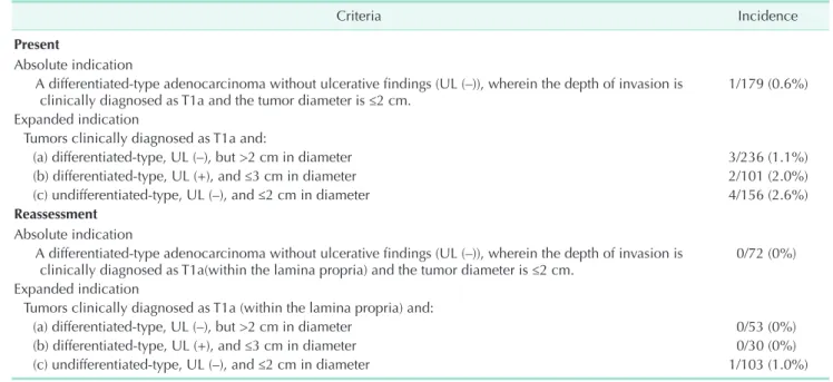
Review of LN metastasis-positive cases with indications for ESD
A total of 10 patients (10 of 672, 1.5%) with indications for ESD, including 1 with absolute indications (1 of 179, 0.6%) and 9 with expanded indications (9 of 493, 1.8%), also had LN metastasis. The clinicopathological outcomes of these 10 patients are described in Table 4. Of these 10 patients, nine had tumor invasion of the muscularis mucosae without penetration and four had undifferentiated histologic features without any ulceration but with tumor size >2 cm. Moreover, 2 patients exhibited ulceration, although they exhibited differentiated
type histology and tumor size <3 cm.
DISCUSSION
The treatment guidelines were recently modified to indicate that therapeutic strategies such as ESD can be applied in pa
tients with MGC who exhibit a low possibility of LN metastasis during preoperative diagnosis. This preference is associated with the increased interest in maintaining the quality of life and the potential complications after conven tional standard gastrectomy [46].
Although LN metastasis is rare in MGC, it should never
theless be considered when selecting the ideal treatment mo
dality; hence, it is important to clarify the clinicopathologic characteristics of MGC patients with LN metastasis. In fact, LN metastasis is often encountered in the clinical setting, with an incidence of approximately 2.6%–4.6% [12,13]. The incidence of LN metastasis in MGC patients was 3.5% (42 of 1,191) in the present study, consistent with that reported previously.
Moreover, studies have indicated that tumor size, depth of tumor invasion, lymphatic invasion, and undifferentiated histology are risk factors for LN metastasis in EGC. However, the definite indications for ESD or the standard treatment of MGC are varied and inconsistent among those studies [7
9]. In the present study, we observed that tumor invasion to the muscularis mucosa, presence of ulceration, and undiffer
entiatedtype histology were independent risk factors for LN metastasis. The number of lymphatic vessels in the mucosal
layer is lower than that in the submucosal layer. Hence, the presence of tumor invasion to the muscularis mucosa and presence of ulceration indicate the destruction of the mus
cularis mucosa, which usually acts as a barrier against lymph
atic vessel invasion (Fig. 2). Hence, these findings can be explained by the breakdown of the muscularis mucosa due to histological ulceration that resulted in interchange between lymph flow in the mucosa and submucosa, and consequently led to an increase in the risk of regional LN metastasis [3].
In fact, MGC patients with the abovementioned risk factors had a significantly higher incidence of LN metastasis in the present study. In particular, LN metastasis was detected in 16 of 216 cases (7.4%) of MGC with invasion to the muscularis mucosa and the presence of ulceration, and in 11 of 105 cases (10.5%) of MGC with all three risk factors (tumor invasion to the muscularis mucosa, presence of ulceration, and undiffer
entiatedtype histology; Fig. 3). Although the mean tumor size was larger in the LN metastasis group, it did not serve as an independent risk factor for LN metastasis in MGC when applied as 2 cm based on the conventional ESD indication (Table 3). In addition, there may be discrepancies in tumor size between surgically resected specimens and endoscopically resected specimens. In the case of surgical resection, the tumor size is measured after fixation with formalin, which could cause shrinkage. In contrast, in endoscopic resection, the specimen is creased and fixed with a pin, which could cause exaggeration of the size. Thus, endoscopically resected cases would be more likely to have expanded indications for tumor size, as compared to surgically resected cases [14]. Hence, this discrepancy in tumor size should be carefully considered when applying the indications for ESD.
Furthermore, lymphatic invasion in cases with MGC is very rare. In the present study, only 9 of 1,149 MGC patients (0.8%) showed lymphatic invasion, and this feature did not have any significant effect on LN metastasis.
In addition, the conventional indications of endoscopic resection for EGC (which are based on the established treatment guidelines for gastric cancer in Japan) are described in Table 3 [11]. However, these indications are primarily based on data Table 2. Univariate and multivariate analyses of risk factors for mucosal gastric cancer (logistic regression analysis; P < 0.10)
Pathologic factor Univariate analysis Multivariate analysis
Pvalue 95% CI Odds ratio Pvalue 95% CI Odds ratio
Age, <50 yr vs. ≥50 yr 0.045 1.005–3.595 1.901 0.175
Lamina propria vs. muscularis mucosa 0.001 1.785–11.73 4.575 0.001 1.877–12.833 4.909
Tumor size, ≤2 cm vs. >2 cm 0.014 1.175–5.583 2.561 0.215
Ulcer vs. no ulcer 0.010 1.189–4.152 2.222 0.036 1.046–3.756 1.982
Intestinal vs. diffuse 0.005 1.310–5.097 2.584 0.723
Differentiated vs. undifferentiated 0.001 1.619–7.199 3.414 0.025 1.196–14.987 4.233 CI, confidence interval.
from Japanese patients, and hence may not be completely applicable in Korea or other countries. In the present study, 672 MGC patients had indications for ESD, including 1 of 179 (0.6%) with absolute indications and 9 of 493 (1.8%) with expanded indications; of these, 10 patients (10 of 672, 1.5%) had LN metastasis. The incidence of LN metastasis among these cases of MGC with indications for ESD exceeds the risk of mortality
of standard surgery for gastric cancer in Korea (0.6%) [15]. Hence, to ensure that the prognosis is better than that of surgical resection, LN metastasis should be thoroughly ruled out before switching conventional radical gastrectomy with ESD. However, as noted in Table 4 in the present study, there are certain limitations to the prediction of LN metastasis in MGC even when considering the indications for ESD or the current risk Fig. 3. (A) The frequency of lym
ph node metastasis according to dif ferentiation, ulceration, and size, based on the indications of endoscopic submucosal dis
sec tion (ESD) in mucosal gastric can cer within lamina propria.
(B) The frequency of lymph node meta stasis according to dif fer
en tia tion, ulceration, and size, based on the indications of ESD in with muscularis mu co sa in
va sion. *Absolute in di ca tions ac cor d ing to the new Japa nese classi fi ca tion and treat ment guidelines for gastric can cer.
**Expanded in di ca tions ac cord
ing to the new Ja pa nese classi fi
ca tions and treat ment guidelines for gastric can cer.
A
Mucosal gastric cancer within lamina propria
(n = 444)
Differentiated (n = 162/444)
Ulceration ( ) (n = 125/162)
Ulceration (+) (n = 37/162)
Ulceration ( ) (n = 227/282)
Ulceration (+) (n = 55/282) Undifferentiated
(n = 282/444)
*Size < 2 cm (n = 72/125) : 0/72 (0%)
**Size > 2 cm (n = 53/125) : 0/53 (0%)
**Size < 3 cm (n = 30/37) : 0/30 (0%)
Size > 3 cm (n = 7/37) : 0/7 (0%)
**Size < 2 cm (n = 103/227) : 1/103 (1.0%)
Size > 2 cm (n = 124/227) : 2/124 (1.6%)
(n = 55) : 2/55 (3.6%)
B
Mucosal gastric cancer with muscularis mucosa invasion
(n = 747)
Differentiated (n = 401/747)
Ulceration ( ) (n = 290/401)
Ulceration (+) (n = 111/401)
Ulceration ( ) (n = 241/346)
Ulceration (+) (n = 105/346) Undifferentiated
(n = 346/747)
*Size < 2 cm (n = 107/290) : 1/107 (0.9%)
**Size > 2 cm (n = 183/290) : 3/183 (1.6%)
**Size < 3 cm (n = 71/111) : 2/71 (2.8%)
Size > 3 cm (n = 40/111) : 3/40 (7.5%)
**Size < 2 cm (n = 53/241) : 3/53 (5.7%)
Size > 2 cm (n = 188/241) : 14/188 (7.4%)
(n = 105) : 11/105 (10.5%)
factors for LN metastasis.
Previous studies have indicated that the recurrence rate of EGC with LN metastasis was relatively higher than that of EGC without LN metastasis due to the pathologic characteristics.
Hence, LN metastasis is the most powerful and important prognostic factor; moreover, the longterm followup results after ESD remain unclear due to insufficient data, whereas the longterm survival rate has improved to 99%–100% in cases Table 3. Incidence of lymph node metastasis in mucosal gastric cancer: the present and reassessment of the criteria for endoscopic submucosal dissection
Criteria Incidence
Present
Absolute indication
A differentiatedtype adenocarcinoma without ulcerative findings (UL (–)), wherein the depth of invasion is
clinically diagnosed as T1a and the tumor diameter is ≤2 cm. 1/179 (0.6%)
Expanded indication
Tumors clinically diagnosed as T1a and:
(a) differentiatedtype, UL (–), but >2 cm in diameter 3/236 (1.1%)
(b) differentiatedtype, UL (+), and ≤3 cm in diameter 2/101 (2.0%)
(c) undifferentiatedtype, UL (–), and ≤2 cm in diameter 4/156 (2.6%)
Reassessment Absolute indication
A differentiatedtype adenocarcinoma without ulcerative findings (UL (–)), wherein the depth of invasion is
clinically diagnosed as T1a(within the lamina propria) and the tumor diameter is ≤2 cm. 0/72 (0%) Expanded indication
Tumors clinically diagnosed as T1a (within the lamina propria) and:
(a) differentiatedtype, UL (–), but >2 cm in diameter 0/53 (0%)
(b) differentiatedtype, UL (+), and ≤3 cm in diameter 0/30 (0%)
(c) undifferentiatedtype, UL (–), and ≤2 cm in diameter 1/103 (1.0%)
UL, ulcer.
Table 4. Lymph nodepositive cases with indications for endoscopic submucosal dissection Patient
No. Age
(yr) Depth WHO
classification Ulcer Size
(cm) Ly Vs Pn Lo LN
(P/T) Lauren
classification Gross type 1 51 Muscularis
mucosa MD – 1.7 + + – MB 3/63 Intestinal Depressed
2 33 Muscularis
mucosa MD – 5 – – – UB 1/97 Diffuse Depressed
3 58 Muscularis
mucosa MD – 4.8 – – – LB 1/23 Intestinal Elevated
4 64 Muscularis
mucosa MD – 5.8 – – – MB 2/59 Intestinal Elevated
5 52 Muscularis
mucosa MD + 2.8 – – – MB 1/30 Intestinal Depressed
6 72 Muscularis
mucosa MD + 1.5 – – – MB 1/21 Intestinal Depressed
7 46 Muscularis
mucosa PD – 1 – – – LB 1/32 Diffuse Depressed
8 62 Muscularis
mucosa SRC – 1.5 – – – LB 5/74 Intestinal Flat
9 43 Muscularis
mucosa SRC – 1.7 – – – MB 2/68 Diffuse Depressed
10 46 Lamina
propria SRC – 1.5 – – – MB 1/21 Diffuse Depressed
WHO, World Health Organization; Ly, lymphatic invasion; Vs, vascular invasion; Pn, perineural invasion; Lo, tumor location; LN (P/T), lymph node (positive lymph node/total harvest lymph node); MD, moderately differentiated; MB, middle body of the stomach; UB, upper bodyof the stomach; LB, lower bodyof the stomach; PD, poorly differentiated; SRC, signet ring cell carcinoma.
treated by conventional radical gastrectomy with LN dissection [1619]. These findings suggest that ESD may represent an incomplete treatment in MGC patients if LN metastasis is present, which would have a negative influence on the recur
rence and prognosis of MGC.
Thus, the selection of the ideal treatment option for MGC depends on the accurate diagnosis of tumor invasion to the muscularis mucosa, presence of ulceration, and undiffer
entiatedtype histology. However, these factors cannot be estimated by forceps biopsy during routine endoscopy, EUS, or abdominal CT before surgery. Instead, these features can be identified during the final histological examination of re
sected specimens. Hence, ESD can yield precise histological information, as the resected ESD specimen (obtained via en bloc resection) includes the full thickness of the submucosal layer, and hence facilitates the evaluation of all 3 factors of LN metastasis. However, if MGC patients have specific conditions
such as old age or significant comorbidities that could result in postoperative complications, or if the MGC patients are surgically inoperable, ESD should be carefully considered as an alternative treatment option based on their life expectancy, and additional therapy such as conventional radical gastrectomy with LN dissection can be scheduled depending on the final pathologic results after ESD. Thus, in addition to assessing whether MGC should be treated by ESD or conventional radical gastrectomy, this study also considers whether ESD can be utilized not only as a diagnostic modality, but also as a therapeutic strategy in cases without these risk factors.
CONFLICTS OF INTEREST
No potential conflict of interest relevant to this article was reported.
1. Katai H, Sano T. Early gastric cancer: con
cepts, diagnosis, and management. Int J Clin Oncol 2005;10:37583.
2. Ahn JS, Bang HY, Lee JI, Noh WC, Hwang DY, Choi DW, et al. Recurrence of early gastric cancer. J Korean Gastric Cancer Assoc 2001;1:1806.
3. Yamao T, Shirao K, Ono H, Kondo H, Saito D, Yamaguchi H, et al. Risk factors for lymph node metastasis from intramucosal gastric carcinoma. Cancer 1996;77:6026.
4. Lee JH, Han HS, Lee JH. A prospective ran domized study comparing open vs lapar oscopyassisted distal gastrectomy in early gastric cancer: early results. Surg Endosc 2005;19:16873.
5. Kitano S, Shiraishi N, Fujii K, Yasuda K, Inomata M, Adachi Y. A randomized con trolled trial comparing open vs lapar
os copyassisted distal gastrectomy for the treatment of early gastric cancer: an interim report. Surgery 2002;131(1 Suppl):
S30611.
6. Kim HH, Hyung WJ, Cho GS, Kim MC, Han SU, Kim W, et al. Morbidity and mor
tality of laparoscopic gastrectomy versus open gastrectomy for gastric cancer: an
inte rim report: a phase III multicenter, pro spective, randomized Trial (KLASS Trial). Ann Surg 2010;251:41720.
7. Lo SS, Wu CW, Chen JH, Li AF, Hsieh MC, Shen KH, et al. Surgical results of early gas tric cancer and proposing a treatment stra tegy. Ann Surg Oncol 2007;14:3407.
8. Shiraishi N, Sato K, Yasuda K, Inomata M, Kitano S. Multivariate prognostic study on large gastric cancer. J Surg Oncol 2007;
96:148.
9. Kim DH, Kim SM, Hyun JK, Choi MG, Noh JH, Sohn TS, et al. Changes in post
operative recurrence and prognostic risk fac tors for patients with gastric cancer who underwent curative gastric resection during different time periods. Ann Surg Oncol 2013;20:231727.
10. Japanese Gastric Cancer Association. Japa
nese classification of gastric carcinoma:
3rd English edition. Gastric Cancer 2011;
14:10112.
11. Sano T, Aiko T. New Japanese classifica
tions and treatment guidelines for gastric can cer: revision concepts and major revi
sed points. Gastric Cancer 2011;14:97100.
12. Gotoda T, Yanagisawa A, Sasako M, Ono H,
Nakanishi Y, Shimoda T, et al. Incidence of lymph node metastasis from early gas
tric cancer: estimation with a large num
ber of cases at two large centers. Gastric Cancer 2000;3:21925.
13. Hirasawa T, Gotoda T, Miyata S, Kato Y, Shimoda T, Taniguchi H, et al. Incidence of lymph node metastasis and the feasi
bility of endoscopic resection for undif fer
entiatedtype early gastric cancer. Gastric Cancer 2009;12:14852.
14. Kim SG. Endoscopic treatment for early gastric cancer. J Gastric Cancer 2011;11:
14654.
15. Park DJ, Lee HJ, Kim HH, Yang HK, Lee KU, Choe KJ. Predictors of operative mor
bi dity and mortality in gastric cancer sur
gery. Br J Surg 2005;92:1099102.
16. Seto Y, Nagawa H, Muto T. Impact of lymph node metastasis on survival with early gastric cancer. World J Surg 1997;21:
1869.
17. Yokota T, Ishiyama S, Saito T, Teshima S, Narushima Y, Murata K, et al. Lymph node metastasis as a significant prognostic fac
tor in gastric cancer: a multiple logistic regres sion analysis. Scand J Gastroenterol
REFERENCES
2004;39:3804.
18. Saka M, Katai H, Fukagawa T, Nijjar R, Sano T. Recurrence in early gastric cancer with lymph node metastasis. Gastric
Cancer 2008;11:2148.
19. Lee JH, Kim HH. The extended indica
tions of endoscopic submucosal dissec
tion (ESD) for early gastric cancer are thus not entirely safe. J Gastric Cancer 2010;
10:8790.