• 검색 결과가 없습니다.

Adhesion of Human Osteoblasts Cell on CrN Thin Film Deposited by Cathodic Arc Plasma Deposition

N/A
N/A
Protected

Academic year: 2021

Share "Adhesion of Human Osteoblasts Cell on CrN Thin Film Deposited by Cathodic Arc Plasma Deposition"

Copied!
5
0
0

로드 중.... (전체 텍스트 보기)

전체 글

(1)

한국표면공학회지 J. Kor. Inst. Surf. Eng.

Vol. 42, No. 5, 2009.

<연구논문>

Adhesion of Human Osteoblasts Cell on CrN Thin Film Deposited by Cathodic Arc Plasma Deposition

Vuong Hung Pham

a

, Sun Kyu Kim

b*

a

Department of Immunology and Biomedicine, University of Ulsan, Ulsan 680-749, Korea

b

School of Materials Science and Engineering, University of Ulsan, Ulsan 680-749, Korea (Received March 12, 2009; revised April 10, 2009; accepted October 30, 2009)

Abstract

Interaction between human osteoblast (hFOB 1.19) and CrN films was conducted in vitro. CrN films were produced by cathodic arc plasma deposition. The surface was characterized by atomic force microscopy (AFM).

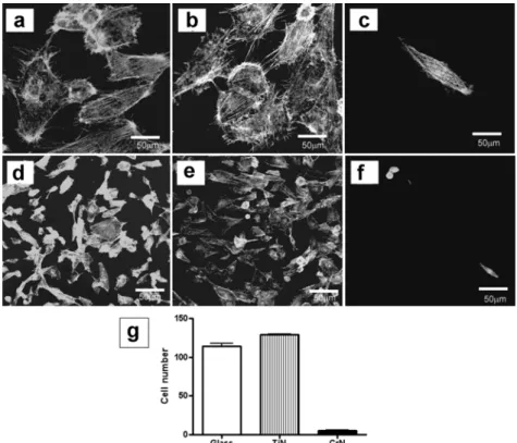
CrN films, glass substrates and TiN films were cultured with human osteoblasts for 48 and 72 hours. Actin stress fiber patterns and cell adhesion of osteoblasts were found less organized and weak on CrN films com- pared to those on the glass substrates and the TiN films. Human osteoblasts also showed less proliferation and less distributed microtubule on CrN films compared to those on glass substrates and TiN films. Focal contact adhesion was not observed in the cells cultured on CrN films, whereas focal contact adhesion was observed well in the cells cultured on glass substrates and TiN films. As a result, the CrN film is a potential candidate as a surface coating to be used for implantable devices which requires minimal cellular adhesion.

Keywords: Human osteoblast, CrN film, Cell adhesion, Surface coating, Cytoskeleton

1. Introduction

Chromium nitride (CrN) films belong to an interesting group of transition metal nitrides films

1)

. Like TiN films, CrN films are considered as hard coating materials. Previous studies have focused on TiN thin film as a hard coating to enhance osteoblast cells adhesion

2,3)

. CrN films have many advantages, such as wear resistance, corrosion resistance, oxidation resistance and low electrical resistivity

4-8)

. The softer and less brittle CrN with a microhardness comparable to TiN has many advantages if one needs to protect relatively soft substrates such as stainless steels, unhardened steels, light metals and light metal alloys

9)

. Numerous studies have been done on the application of CrN films on tools and casting mold dies

10)

, diffusion barriers

7)

and solar selective absorbers

11)

. Concerning cell-based devices, there are two common strategies for designing artificial surfaces in biological application. One involves creating surfaces not allowing the cellular adhesion

12,13)

. The other, a more common strategy, is to create surfaces promoting cell adhesion

14)

.

Williams et al. investigated the effect of CrN and CoCr wear products on the viability of fibroblast and macrophage cells

15)

. Their report indicated the CoCr wear particles reduced cell viability more than CrN wear particles. In this work, we evaluated the osteoblast cell adhesion to CrN films deposited by cathodic arc plasma deposition. The choice of cathodic arc plasma deposition is from the fact that good adhesion between the coated layer and the substrate can be achieved. For comparative purpose, TiN film, which was found as a promoting cellular adhesion surface in the previous work was used as a reference surface

2,3)

.

2. Experimental

Standard round glass coverslips of 12mm (Marienfeld, Germany) were used as substrates. Prior to deposition, the substrates were ultrasonically cleaned with ethanol (95%) for 20 min and then dried by Ar gas.

Finally, they were loaded into the deposition chamber.

The detailed experimental procedures were described elsewhere

16)

. Cr and Ti cathodes were used for deposition of CrN and TiN films, respectively. The

*

Corresponding author. E - mai l : [email protected]

(2)

arc current was kept in constant at 60 A for deposition of CrN films, while it was 45 A for deposition of TiN films. Deposition was done without applying bias potential at 200

o

C. Surface roughness was measured by AFM. The apparatus used was thermomicroscope (CP-research system) with a cantilever (ARROW-CONTRO50, Nanoworld).

Human osteoblasts (hFOB 1.19, ATCC, CRL- 11372) were used as a model for studying the interaction between the cell and the surface. The cells were maintained in Dulbecco’s modified Eagles Medium (DMEM) containing 10% fetal bovine serum and 1% antibiotics at 37

o

C in humidified air and 5% CO

2

.

For actin staining, attached cells were rinsed twice with phosphate buffer saline (PBS), fixed with 4%

paraformaldehyde for 15 min. Then they were permeabilized with 0.1% Triton X-100 in PBS before and after PBS washing. They were stained with Alexa-flour phallodin 488 (Invitrogen) for 30 min.

Adherence of the cells on the thin films was determined by counting in seven randomly chosen fields of view at magnification 100 under an optical microscope (Axiovert 135). For the cell adherence analysis, the t- test was used to assess the statistical significance of results between surfaces. The statistical analysis was performed with the software GraphPad

Prism 4 at the confidence level of 95%. A probability value of p<0.05 was considered significant.

For tubulin staining, anti-tubulin monoclonal antibody (Sigma-Aldrich) was used as the primary antibody.

Alexa 488 goat anti-mouse (Invitrogen) was used as the secondary antibody. Cells were rinsed twice with phosphate buffer saline (PBS), and fixed with 4%

paraformaldehyde for 10 min. Then they were perme- abilized with 0.1% Triton X-100 in PBS after PBS washing, blocked with 1% bovine serum albumin (BSA) and stained with the primary antibody for 30 min. They were rinsed again with PBS and then stained with the secondary antibody for 30 min. Cell proliferation was observed by visualization of the immunofluorescent cells after 48 and 72 h of incubation.

For focal contact adhesion analysis, anti-vinculin (hVIN-1, Sigma-Aldrich) was used as the primary antibody, and Alexa 488 goat anti-mouse (Invitrogen) as the secondary antibody. Cells were rinsed twice with PBS, and fixed with 4% paraformaldehyde for 10 min. Then the cells were permeabilized with 0.1%

Triton X-100 in PBS, blocked with 1% BSA and stained with the primary antibody for 45 min. The cells were then rinsed with PBS and stained with the secondary antibody for 30 min. All visualization of the stained cell was done by a confocal microscope (Olympus 1 × 81).

Fig. 1. Cytoskeleton analysis of osteoblast on tested specimens and its cellular adhesion after 48 h of culturing. (a

and d: glass substrate, b and e: TiN film, c and f: CrN film. (a-c) Actin cytoskeleton, (d-f) visualization of cell

adhesion, (g) quantification of cell adhesion.

(3)

3. Results and Discussion

Using AFM, the average surface roughness of CrN film, TiN film and glass substrate were inspected.

The surface roughness of CrN and TiN film was 0.725 nm and 0.480 nm, respectively while the value on glass substrate was only 0.161 nm.

Examination of actin cytoskeleton organization of the cells focused on the analyzing the cells cultured on the films on which stress fibers were formed (Fig. 1a, b, c). Actin stress fibers were oriented in parallel direction with the main cellular axis on the glass substrates and TiN films, whereas actin stress fibers were almost invisible in the cells cultured on CrN films. Interestingly, filopodia evidences were observed in the cells cultured on glass substrates and TiN films, whereas filopodia evidences were not detected in the cells cultured on CrN films.

Fig. 1d, e, f, and g show the visualization and quantification of cell adhesion on three tested surfaces after 48 h of incubation. The number of attached cells was significantly lower on the CrN films than on the glass substrates (p<0.05) or the TiN films (p<0.05).

The cell proliferation on the films and the glass substrates after 48 and 72 h of incubation is shown in Fig. 2. Cells on the TiN films and glass substrates considerably more proliferated than on the CrN films after 48 h of culturing (Fig. 2a, b, c). It is also clear from the micrographs that the cells nearly reached the confluence after 72 h of incubation on the TiN films (Fig. 2h) and the glass substrates (Fig. 2g), whereas cells were far from obtaining the confluence after 72 h of culturing on the CrN films (Fig. 2i).

Fig. 2(d-f) and (k-m) shows the microtubule organization of cells on glass substrates, TiN films

Fig. 2. Cell proliferation and microtubule cytoskeleton of cells on the tested specimens after 48 and 72 h of culturing

(a, d, g, and k) glass substrate, (b, e, h and l) TiN film, (c, f, i, and m) CrN film, (a-f) 48 h of incubation, (g-

m) 72 h of incubation.

(4)

and CrN films after 48 and 72 h of incubation. As shown in Fig. 2, microtubules of osteoblasts were expanded on the TiN films and the glass substrates, whereas cells on the CrN films were narrower and had a lower expression of tubulin represented by less microtubles in the cells (Fig. 2f, m). We found much difference in the development of microtubules between the TiN films or glass substrates and CrN films after 48 and 72 h of incubation.

Fig. 3 shows the focal contact adhesion of osteoblasts on the glass substrates and the films. Focal contact adhesions were found on the glass substrates and the TiN films. However, focal contact adhesion was not formed in the cells cultured on CrN films. Focal adhesions are signaling pathways resulting in the regulation of cell behavior such as cell adhesion and spreading

17)

. In the cell culture, the medium containing serum proteins does not directly adhere to the substratum surface but to an adsorbed layer of serum components

18)

. CrN surface chemistry might inhibit absorption of extracellular matrix (ECM) protein from cultured medium which made the cells not able to reorganize these molecules to access the ligands for integrin receptors and recruit these receptors into focal adhesion plaques which is prerequisite for delivery of signal ensuring the attachment and growth of anchorage-dependent cells

19,20)

.

Cathodic arc plasma deposition produced many particles thus cause the surface of the TiN and CrN films to be rougher than that of glass substrates. In this paper, we found that cell adhered less and proliferated less on CrN films than the corresponding glass substrates and TiN films. Actin cytoskeleton and microtubule cytoskeleton analysis indicated that cells were not able to develop well organized actin cytoskeleton and well distributed microtubule cyto- skeleton on CrN films, suggesting lack of adhesion

strength and spreading of the cell.

It is interesting to compare our results with the work of Cai et al.

21)

. They found measurably higher osteoblast proliferation on rough (chitosan/gelatin) modified titanium surfaces compared with that of titanium films. However, osteoblasts were found to attach and proliferate at similar rates on CoCrMo and stainless steel substrates, even though these two surfaces had significantly different roughness

22)

. In our study, the CrN films revealed a much lower number of attached osteoblasts compared to the glass substrates and TiN films, even though CrN films had high degree of roughness. The actin cytoskeleton patterns, microtubules organization and focal contact adhesions were also reduced in the cells cultured on CrN films.

This is probably due to the effects of surface charge and surface chemistry of the CrN thin films.

Den Braber et al .

23)

and Chien et al .

24)

have shown that characteristics such surface charge and chemistry have influence on the conformation of the adsorption of proteins on the substrates. In the cell culture, the medium containing serum proteins do not directly adhere to the substratum surface but to the adsorbed layer of the serum components which is crucial to cell adhesion.

4. Conclusions

In this study, osteoblast response to glass substrates, TiN and CrN thin films were evaluated. Actin stress fiber patterns and cell attachments on CrN films were much lower than those on the TiN films and the glass surfaces. Microtubule organizations, proliferation and focal contact adhesions were also much reduced in the cells cultured on the CrN films suggesting that CrN coatings have the potential for designing surfaces for minimizing cells adhesion.

Fig. 3. Fluorescent image focal adhesion of cells on the films after 48 h of incubation. (a) glass substrate, (b) TiN film.

(c) CrN film.

(5)

Acknowledgement

This research was supported by the 2009 University of Ulsan Research Fund.

References

1. H. Y. Chen, C. J. Tsai, F. H. Lu, Surf. Coat. Technol., 184 (2004) 69.

2. H. H. Huang, C. H. Hsu, S. J. Pan, J. L. He, C.

C. Chen, T. L. Lee, Appl. Surf. Sci., 244 (2005) 252.

3. V. H. Pham, S. K. Kim, V. V. Le, B. S. Kwon, J. Kor. Inst. Surf. Eng., 41 (2008) 264.

4. A. Aubert, R. Gillet, A. Gaucher, J. P. Terrait, Thin Solid Films, 108 (1983) 165.

5. O. Knotek, W. Bosch, M. Atzor, W. D. Münz, D.

Hofmann, J. Goebel., High Temp. High Press, 18 (1986) 435.

6. A. Schröer, W. Ensinger, G. K. Wolf, Mater. Sci.

Eng. A., 140 (1991) 625.

7. M. A. Nicolet, Thin Solid Films, 52 (1978) 415.

8. B. Navinšek, P. Panjan, I. Milošev, Surf. Coat.

Technol., 97 (1997) 182.

9. E. J. Bienk, H. Reitz, M. J. Mikkelsen, Surf. Coat.

Technol., 76-77 (1995) 474.

10. F. H. Lu, H. Y. Chen, Thin Silid Films, 398-399 (2001) 368.

11. C. Gantier, J. Machet, Thin Solid Films, 295 (1997) 12. J. Lee, B. S. Kang, B. Hicks, T. F. Chancellor, Jr., 43.

B. H. Chu, H. T. Wang, B. G. Keselowsky, F. Ren,

T. P. Lele, Biomaterials, 29 (2008) 3743.

13. A. D. Cook, J. S. Hrkach, N. N. Gao, I. M. Johnson, U. S. Pajvani, S. M. Cannizzaro, R. Langer, J.

Biomed. Mater. Res., 35 (1997) 513.

14. D. V. Shtansky, N. A. Gloushankova, I. A. Bashkova, M. A. Kharitonova. T. G. Moizhess, A. N. Sheveiko, F. V. Kiryukhantsev-Korneev, M. I. Petrzhik, E. A.

Levashov, Biomaterials, 27 (2006) 3519.

15. S. Wliliams, G. Isaac, P. Hatto. M. H. Stone, E.

Ingham, J. Fisher, J. Arthroplasty, 19 (2004) 112.

16. S. K. Kim, P. V. Vinh, J. H. Kim, T. Ngoc, Surf.

Coat. Technol., 200 (2005) 1391.

17. A. Bershadsky, M. Kozlov, B. Geiger, Curr. Opin.

Cell Biol., 18 (2006) 472.

18. J. A. Jansen, J. P. van der Waerden, K. de Groot, J. Invest. Surg., 2 (1989) 29.

19. S. Huang, C. S. Chen, D. E. Ingber, Mol. Biol.

Cell, 9 (1998) 3179.

20. A. J. Garcia, M. D. Vega, D. Boettiger, Mol. Biol.

Cell, 10 (1999) 785.

21. K. Cai, A. Rechtenbach, J. Hao, J. Bossert, K. D.

Jandt, Biomaterials, 26 (2005) 5960.

22. C. Hendrich, U. Noth, U. Stahl, F. Merklein, C. P.

Rader, N. Schütze, R. Thull, R. S. Tuan, J. Eulert, Clin. Orthopedics Relat. Res., 394 (2002) 278.

23. E. T. den Braber, J. E. de Ruijter, L. A. Ginsel, A. F. von Recum, J. A. Jansen, Biomaterials, 17 (1996) 2037.

24. C. C. Chien, K. T. Liu, J. G. Duh, K. W. Chang,

K. H. Chung, Dental. Mater., 24 (2008) 986.

수치

Fig. 1. Cytoskeleton analysis of osteoblast on tested specimens and its cellular adhesion after 48 h of culturing
Fig. 2. Cell proliferation and microtubule cytoskeleton of cells on the tested specimens after 48 and 72 h of culturing (a, d, g, and k) glass substrate, (b, e, h and l) TiN film, (c, f, i, and m) CrN film, (a-f) 48 h of incubation,  (g-m) 72 h of incubati
Fig. 3 shows the focal contact adhesion of osteoblasts on the glass substrates and the films

참조

관련 문서

Titanium surface modification was carried out by depositing an thin polymeric layer containing carboxyl, amine and hydroxyl groups through plasma

In this study, a titanium surface was modified with acrylic acid (AA) using a plasma treatment and immobilized with bioactive arginine- glycine-aspartic acid (RGD) peptide,

In this study, 3D PCL scaffolds were fabricated by using notch type nozzle via fused deposition modeling (FDM) to improve the cell adhesion and

In this study, we have deposited poly acrylic acid films on three-dimensional (3D) PCL scaffolds using plasma polymerization and then plasma-polymerized 3D

The untreated surfaces and AA plasma treated with 3D-PCL surfaces were also examined for their in vitro pre-osteoblast (MC3T3-E1) cells proliferation and

The surface of untreated Ti, SLA treated Ti, and AA plasma polymerized SLA/Ti were also examined for their proliferation and differentiation of

Determining optimal surface roughness of TiO₂blasted titanium implant material for attachment, proliferation and differentiation of cells derived from

 Often found connected to other molecules on the outsides of cells --- cellular recognition, cell signaling, cell