On the Development of Parthenogenetic Oocytes by Cytochalasin B and Production of Cloned Mice by SCNT
Bo-Woong Sim
1,2and Kwan-Sik Min
1,†1
Animal Biotechnology, Graduate School of Bio. and Information Technology, Institute of Genetic Engineering, Hankyong National University, Ansung 456-749, Korea
2
National Primate Research Center, Korea Research Institute of Bioscience and Biotechnology, Ochang 363-883, Korea
ABSTRACT
This study was conducted to optimize the efficiency of cloning and to produce cloned mice. The majority of cloned mammals derived by nuclear transfer (NT) die during gestation and have enlarged and dysfunctional placentas. In this study, the optimized conditions were established to produce clone mice. The parthenogenetic oocytes were activated after 6 h regardless of cytochalasin B (CB) concentration. CB treatment (2 µg/ml) was found second polar body.
Lower concentration of CB was decreased the activation rate, but the second polar body was the best highly increased during 6 h incubation. The small fragments were exhibited in the 5 µg/ml treatment of CB, but it was not found in lower concentration groups (> 2.5 µg/ml). To examine effects of SrCl
2on the adult cumulus cells, somatic cell NT oocytes were exposed during 0.5, 1 and 6 hrs. The second polar body was significantly greater in 0.5 h exposure group (6.6%) than 1, 6 hrs. Developmental rate from 2-cell to 4-cell was the lowest in 7.5 mM Strontium chloride (SrCl
2) groups (84.1% and 64.3%) than 5, 10 m MSrCl
2. The implantation rate was not significantly difference among 5, 7.5 and 10 m MSrCl
2group. Three live fetuses were produced by SCNT. SCNT placentas were remarkably heavier than IVF group (8 fetuses) (0.34, 0.34, 0.33 vs 0.14 g) compared with the placenta weight of IVF and SCNT clones.
(Key words : parthenogenetic oocytes, cytochalasin B, cloned mice)
*
This work was supported by a grant from the Next-Generation BioGreen 21 Program (PJ0095042014), Rural Development Administration, Republic of Korea.
†
Correspondence : [email protected]
INTRODUCTION
The breakthrough in the study of cloning was the birth of the first clone from a somatic cell (Wilmut et al., 1997). Successful somatic cell cloning has been reported in various animal, such as cows (Kato et al., 1998; Cibelli et al., 1998; Wells et al., 1999), goats (Baguisi et al., 1999), pigs (Onishi et al., 2000;
Polejaeva et al., 2000), cats (Shin et al., 2002) and mice (Wa- kayama et al., 1998). Mice have been cloned using a piezo- actuated nucleus transfer (NT) method with cumulus cells (Wakayama et al., 1998), tail-tip cells (Wakayama and Yanagi- machi, 1999), sertoli cells (Ogura et al., 2000), embryonic stem cells (Wakayama et al., 1999b) and germ cells (Miki et al., 2005) as nucleus donors.
To improve development of SCNT embryos, cytochalasin B should be applied at a concentration that varies with the degree of spindle formation after SCNT. Cytochalasin D, like CB, is a microfilament-disrupting reagent that has been used to induce polyploidy in pre-implantation embryos (Snow, 1973;
Siracusa et al., 1980; Bos-Mikich et al., 1997). Mammalian cells adapted to in vitro culture generally undergo mutations with time, including gross karyotypic alterations, and such genomic changes are unlikely to be compatible with normal embryonic development (Wakayama et al., 1999). Molecular analysis of cloned embryos reveals abnormal epigenetic modi- fication, such as DNA methylation and histone modification (Dean et al., 1998, 2001; Kang et al., 2001; Ohgane et al., 2001; Rybouchkin et al., 2006). Cloning mice from embryonic stem (ES) cells also has practical implications for manipulating the genome. Gene targeting in ES cells has been widely used to create strains of mice with targeted mutations (Capecchi, 1989; Ramirez-Solis et al., 1993).
Here, we report that the parthenogenetic oocytes were acti- vated with varied parameters (CB, SrCl
2, exposure time). We cloned three live-born offspring from the cumulus cell.
MATERIALS AND METHODS
1. Animals
B6D2F1 mice (C57BL/6 × DBA/2) were used to prepare oocytes and cumulus cells. Surrogate females were ICR females mated with vasectomized males of the same strain. All experi- ments were conducted according to the Guidelines for the Care and Use of Animals, Hankyong National University.
The pro- tocol was approved by the Committee on the Ethics of Animal Experiments of the Hankyong National University (Permit Number : 2012. 10).
2. Collection of Oocytes and Preparation of Donor Cells
Briefly, MII oocytes were collected from the oviducts of 6
∼12-week-old females that had been induced to superovulate by injection of pregnant mare serum gonadotropin (PMSG) (7.5 IU) followed by human chorionic gonadotropin (hCG) (7.5 IU) 48 h later. Oocytes were collected from oviducts 13 ∼ 14 h after hCG injection, placed in HEPES-buffered CZB medium, and treated with 300 unit/ml hyaluronidase until the cumulus cells dispersed. The oocytes were transferred to fresh droplets of HEPES-buffered CZB medium and were denuded of almost all cumulus cells by gentle pipetting. The oocytes were then placed in synthetic oviductal medium enriched with potassium medium (KSOM) (Summers et al., 1995), covered with paraffin oil (Nacalai Tesque, Kyoto, Japan), and stored at 37 ℃ in a 5% CO
2at mosphere until use.
Cumulus cells of B6D2F1 mice were removed from oocytes using hyaluronidase as described above. Cumulus cells were transferred and maintained in HEPES-buffered CZB medium at 4 ℃. Immediately prior to use, cumulus cells were resuspended in 10% polyvinylpyrrolidine (PVPD) in HEPES-buffered CZB medium.
3. Production of Parthenogenetic Activation Embryos
MII oocytes were collected from superovulated B6D2F1 female mice 14 h after hCG injection. The oocytes were incuba- ted in KSOM at 37 ℃ under 5% CO
2for 3 ∼4 h prior to PA by SrCl
2in Ca
2+-free CZB supplemented with 2, 3, 4 or 5 µg/ml CB for 6 h. After activation, the PA embryos were cul- tured in KSOM at 37 ℃ under 5% CO
2until the morula and blastocyst stage.
4. Enucleation of MII Oocytes and Activation and Culture of Injected Oocytes
Enucleation was performed in HEPES-buffered CZB medium
supplemented with 3 or 5 µg/ml CB. The zona pellucida was
“drilled” by applying several piezo-pulses at the tip of an enu- cleation pipette (~12 µm outside diameter). The chromosome- meiotic spindle complex was then gently pulled away from the oocyte until the cytoplasmic bridge was severed (Kono et al., 1993). Enucleated oocytes were washed three times in CB-free CZB medium, transferred into CB-free KSOM, and stored for up to 1 h at 37 ℃ under 5% CO
2until nucleus transfer.
The injected oocytes were activated by 5, 7.5 or 10 mM SrCl
2in Ca
2+-free CZB at 37 ℃ under 5% CO
2. Oocytes activated for 0.5 or 1 h was cultured for an additional 5.5 or 5 h in the presence of 2.5 µg/ml CB (for a total of 6 h) to prevent the extrusion of polar bodies that contained donor chromosomes.
Oocytes with at least one pseudo-pronucleus and without a second polar body were considered normally activated and were washed and cultured in KSOM at 37 ℃, 5% CO
2until they reached the 2-cell or morula/blastocyst stage.
5. Nucleus Transfer
SCNT embryos were produced by injecting a cumulus cell nucleus into an enucleated B6D2F mature oocyte. Single cu- mulus cells in 10% PVP medium were drawn in and out of an injection pipette (6 ∼7 µm inner diameter) until the plasma membranes were broke. After a nucleus was drawn deep into the pipette, the pipette was then passed through the zona pellu- cida of an enucleated oocyte by applying piezo pulses. The nucleus was then injected into an enucleated oocyte at room temperature using the piezo-driven pipette, as described pre- viously (Wakayama et al., 1998). Injected oocytes were kept in KSOM for 1 ∼2 h before activation treatment at 37℃ under 5% CO
2in air and transferred.
6. Embryo Transfer and Examination of Placentas
Mice induced to superovulate by injection of PMSG (7.5
IU) followed by hCG (7.5 IU) 48 h later. Oocytes were collected
from oviducts 13 ∼14 h after hCG injection. After maturation,
a detailed procedure has described previously (Fukuda et al.,
1987). Some of two-cell embryos (24 h after the onset of
activation) were transferred to the oviducts of foster mothers
(ICR, albino) that had been mated with vasectomized ICR males
1 day previously. SCNT embryos were transferred to the uteri
of foster mothers (ICR, albino) that had been mated with vasec-
tomized ICR males 3 days previously. Each recipient surrogate
female received 10 ∼20 SCNT embryos. Some of the recipient
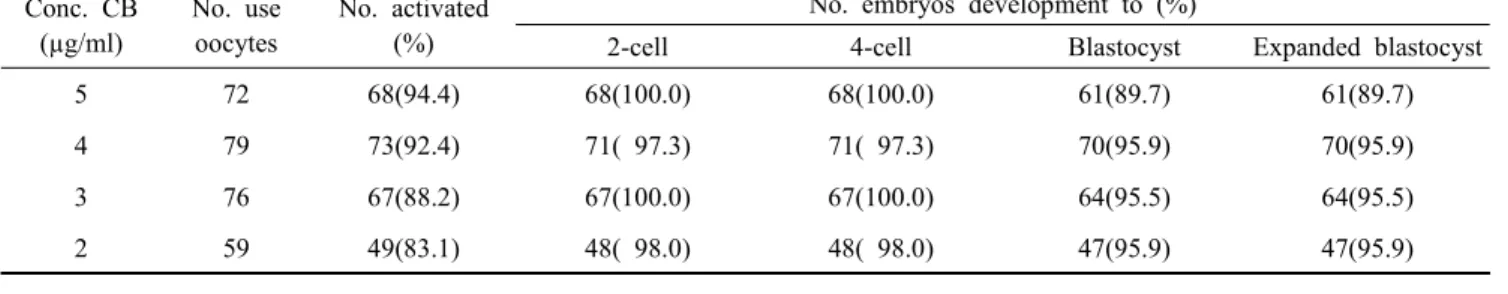
Table 2. Development of PA embryos activated with CB concentration Conc. CB
(µg/ml)
No. use oocytes
No. activated (%)
No. embryos development to (%)
2-cell 4-cell Blastocyst Expanded blastocyst
5 72 68(94.4) 68(100.0) 68(100.0) 61(89.7) 61(89.7)
4 79 73(92.4) 71( 97.3) 71( 97.3) 70(95.9) 70(95.9)
3 76 67(88.2) 67(100.0) 67(100.0) 64(95.5) 64(95.5)
2 59 49(83.1) 48( 98.0) 48( 98.0) 47(95.9) 47(95.9)
There was no significantly different between each value in the same column.
females were euthanized at 9.5 dpc and examined for the pre- sence or absence of fetuses and implantation sites. Live fetuses were raised by lactating foster mothers (ICR). Offspring and placenta obtained by Caesarean section on day 19 of pregnancy were individually weighed.
7. Statistical Analysis
Statistical analyses were performed using Minitab software.
Data were analyzed using one-way ANOVA. Differences were considered significant (P<0.05, P<0.01).
RESULTS
1. Development Rate of PA Oocyte and Embryos by CB Concen- tration
Four different CB concentration (2, 3, 4 and 5 µg/ml) to activate M Ⅱ oocytes was assessed to optimize in activation medium. The majority (83.1 ∼94.4%) of PA oocytes was acti- vated after 6 h regardless of the used CB concentration. The second polar body (10.2%) was found in 2 µg/ml CB treatment group (Table 1). The developmental rate of activated PA embryos was analyzed to the expanded blastocyst. PA embryos were developed to 2-cell (97.3 ∼100%), 4-cell (97.3∼100%) and blastocyst (89.7 ∼95.9%) in different CB concentrations.
Finally, the expended blastocyst rate was not significant diffe- rences as 89.7 ∼95.9% (Table 2).
2. Development Rate of SCNT Embryos by CB Concentration To accurately evaluate the optimal concentration of CB in SCNT embryo’s activation, the activation and development rates of reconstructed embryo were determined in five different CB concentrations (2 ∼5 µg/ml). SCNT oocytes were activated over 84.5% at CB concentration (>2.5 µg/ml). And there was obser- ved two pseudo-pronucleus after 6 h (Fig. 1B). But, activation
Table 1. Activation rate of PA oocytes activated with CB concen- trations for 6 h
Conc.
CB (µg/ml)
No.
used oocyte
No.
>2PP
*(%)
No. not activated
(%)
No. 1pp and 2pb
*(%)
No. lysis (%)
5 72 68(94.4) 0(0.0) 0( 0.0) 4( 5.6)
4 79 73(92.4) 0(0.0) 0( 0.0) 6( 7.6)
3 76 67(88.2) 0(0.0) 0 ( 0.0) 9(11.8)
2 59 49(83.1) 0(0.0) 6(10.2) 4( 6.8)
*
PP: pseudo-pronucleus, 2pb : second polar body.
There was no significantly different between each value in the same column.
rate was decreased to 72.6% in CB 2 µg/ml group. Activation rate in CB 2 µg/ml group was significantly lower than that of CB 5 µg/ml group. Especially, 12.9% of SCNT embryos acti- vated with CB 2 µg/ml was extruded the second polar body (Table 3).
SCNT embryos of all groups were developed to the 2-cell stage (83.3 ∼94.0%) in the developmental rate in different CB concentrations. However, the SCNT embryos activated with
Fig. 1. Reconstruction of the somatic nuclei. (A) The nucleus of
cumulus cell as donor cell were transformated into disarrayed
chromosome at 3 hours after nucleus transfer. (B) Two
pseudo- pronucleus (allow) was formed 6 hours after acti-
vation in SCNT embryos.
Table 4. Development of SCNT embryos activated with CB concentrations Conc.
CB (µg/ml)
No.
injected oocytes
No. activated embryos
(%)
No. embryos development to (%)
2-cell 4-cell Morula Blastocyst Expanded blastocyst
(/activated), (/injected) 5 163 150(92.0)
a138(92.0) 62(41.3)
df47(31.3)
df40(26.7)
df34(22.7)
bdf,(20.9)
d4 105 91(86.7)
a86(94.5) 42(46.2)
bf36(39.5)
bf32(35.2)
bf29(31.9)
d,(27.6)
b3 142 120(84.5)
a100(83.3) 88(73.3)
e70(58.3)
e66(55.0)
ac56(46.7)
e,(39.4)
c2.5 162 140(86.4)
a128(91.4) 102(72.9)
e89(63.6)
e80(57.1)
e74(52.9)
c,(45.7)
ac2 62 45(72.6)
b41(91.9) 33(73.3)
e30(66.7)
ac26(57.8)
c22(48.9)
a,(35.5) Values with different superscripts were significantly different (a vs. b (P<0.05), c vs. d and e vs. f (P<0.01)).
Table 3. Activation rate of SCNT embryos activated with CB con- centration for 6 h
Conc.
CB (µg/ml)
No.
injected oocytes
No.
>2 PP
*(%)
No. not activated
(%)
No. 1pp and 2pb
*(%)
Lysis (%) 5 163 150(92.0)
a6( 3.7) 0 ( 0.0)
b7(4.3) 4 105 91(86.7)
a5( 4.8) 0( 0.0)
b9(8.6) 3 142 120(84.5)
a8( 5.6) 1 ( 0.7)
b14(9.9) 2.5 162 140(86.4)
a20(12.3) 0( 0.0)
b2(1.2) 2 62 45(72.6)
b8(12.9) 8(12.9)
a1(1.6)
*
PP : pseudo-pronucleus, 2pb : second polar body.
Values with different superscripts were significantly different (P<0.05).
CB 4 or 5 µg/ml were resulted in a significant decrease in the development rate from 4 cell to expended blastocyst. Especially, it was greatly decreased in the 4-cell stage of CB 4 or 5 µg/ml groups (P<0.01). The rate was significantly different in the ex- pended blastocyst. It was remarkably increased in CB 2, 3 and 2.5 µg/ml treated groups (48.9%, 52.9% and 46.7%, respectively, P<0.01) (Table 4). The two-cell stage embryos was found small fragments between blastomeres in SCNT embryos activated with CB 5 µg/ml. However, it was not detected in CB 2.5 µg/ml group (Fig. 2).
Thus, results suggest that the highest frequency of pre- implantation development is SCNT oocytes activated with CB 2.5 µg/ml (expended blastocyst rate; 52.9%) (Table 4).
3. Activation Rate of SCNT Embryos by SrCl
2Treatment and Production of Cloned Mice by SCNT
Three different exposure time (0.5, 1 and 6 h) to activate
Fig. 2. Two-cell stage of SCNT embryos. (A) SCNT embryos were activated with 5 µg/ml CB, almost embryos show fragments between blastomeres (arrows). (B) SCNT embryos were activated with 2.5 µg/ml CB.
M Ⅱ oocytes was assessed to optimize in activation time. The majority (86.2 ∼88.0%) of SCNT embryos was activated in exposure time during 1 and 6 h. It was significantly decreased in the 0.5 h exposure group (63.3%) (P<0.01). However, the second polar body was significantly greater in 0.5 h exposure group (6.6%) (Table 5).
To examine effects of SrCl
2concentration on adult cumulus cell clone, SCNT oocytes were activated by SrCl
2(5, 7.5 and 10 mM) treatment for 6 h. Developmental rate from 2-cell to 4-cell was the lowest in 7.5 mM SrCl
2groups (84.1% and 64.3%, respectively). And then, the morula/blastocyst stage was also the lowest (45.2%). The morula/blastocyst stage SCNT embryos were transferred to surrogate mothers. Recipient females were examined the implantation rate at 9.5 day of pregnancy.
There was nosignificantly difference in implantation rate among
5, 7.5 and 10 mM SrCl
2group (data not shown). As shown in
Fig. 3, three live fetuses were produced by SCNT. To compare
the placenta weight of IVF and SCNT clones, SCNT placentas
were remarkably heavier than IVF group (8 fetus) (0.34, 0.34,
0.33 vs 0.14g, respectively).
Table 5. Activation rate of SCNT embryos activated by SrCl
2treatment
Time for exposure No. of treated embryo No. >2PP
*(%) No. not activated(%) No. 1pp and 2pb
*(%) No. lysis(%)
0.5h 316 200(63.3)
c81(25.6)
d21(6.6)
f14(4.4)
1h 181 156(86.2)
a15( 8.3)
e3(1.7)
g7(3.9)
6h 209 184(88.0)
a11( 5.3)
e0(0.0)
g14(6.7)
*