배양된 치유두 유래세포의 조골활성 및 골기질 형성의 평가
박봉욱∙변준호∙최문정∙하영술*∙김덕룡*∙조영철**∙성일용**∙김종렬***
경상대학교 의과대학/의학전문대학원 구강악안면외과학교실, 경상대학교 건강과학연구원, 경상대학교 생명과학연구원
*경상대학교 의과대학/의학전문대학원 생화학교실, 경상대학교 건강과학연구원, 경남지역암센터
**울산대학교 의과대학 구강악안면외과학교실,
***부산대학교 치의학전문대학원 구강악안면외과학교실
EVALUATION OF OSTEOGENIC ACTIVITY AND MINERALIZATION OF CULTURED HUMAN DENTAL PAPILLA-DERIVED CELLS
Bong-Wook Park, June-Ho Byun, Mun-Jeoung Choi, Young-Sool Hah * , Deok-Ryong Kim * , Yeong-Cheol Cho ** , Iel-Yong Sung ** , Jong-Ryoul Kim ***
Department of Oral and Maxillofacial Surgery, College of Medicine and Institute of Health Sciences, Research Institute of Life Science, Gyeongsang National University School of Medicine
*Department of Biochemistry, College of Medicine and Institute of Health Sciences, Gyeongsang National University School of Medicine, Gyeongnam Regional Cancer Center
**Department of Oral and Maxillofacial Surgery, College of Medicine, Ulsan University
***Department of Oral and Maxillofacial Surgery, School of Dentistry, Pusan National University
In the present study, we focused on stem cells in the dental papilla of the tooth germ. The tooth germ, sometimes called the tooth bud, is the primordial structure from which a tooth is formed. The tooth germ consists of the enamel organ, the dental papilla, and the dental follicle. The dental papilla lies below a cellular aggregation of the enamel organ. Mesenchymal cells within the dental papilla are responsible for formation of dentin and pulp of a tooth. Tooth germ disappears as a tooth is formed, but that of a third molar stays in the jawbone of a human until the age of 10 to 16, because third molars grow slowly. Impacted third molar tooth germs from young adults are some- times extracted for orthodontic treatment.
In the present study, we evaluated the osteogenic activity and mineralization of cultured human dental papilla-derived cells. Dental papillas were harvested from mandible during surgical extraction of lower impacted third molar from 3 patients aged 13-15 years. After passage 3, the dental papilla- derived cells were trypsinized and subsequently suspended in the osteogenic induction DMEM medi- um supplemented with 10% fetal bovine serum, 50 g/ml L-ascorbic acid 2-phosphate, 10 nM dex- amethasone and 10 mM -glycerophosphate at a density of 1 × 10
6cells/dish in a 100-mm culture dish. The dental papilla-derived cells were then cultured for 6 weeks and the medium was changes every 3 days during the incubation period. Dental papilla-derived cells showed positive alkaline phosphatase (ALP) staining during 42 days of culture period. The formation of ALP stain showed its maximal manifestation at day 7 of culture period, then decreased in intensity during the culture period. ALP mRNA level was largely elevated at 1 weeks and gradually decreased with culture time.
Osteocalcin mRNA expression appeared at day 14 in culture, after that its expression continuously increased in a time-dependent manner up to day 28. The expression remained constant thereafter.
Abstract
Ⅰ. 서 론
줄기세포 및 전구세포를 이용하는 골조직공학은 현재 구 강악안면영역의 골결손에 대하여 상당히 주목받고 있는 분 야이다. 이와 관련하여 골수에서부터 분리되는 골수기원줄 기세포 (bone marrow-derived mesenchymal stem cells)가 가장 잘 알려져 있지만 골막, 연골, 윤활막 등과 같 은 조직에서도 성체줄기세포를 추출할 수 있다. 골조직공학 을 통한 성공적인 골형성을 위해서 가장 중요한 요소는 자 가 골전구세포의 획득이다. 매복치 발치 등을 포함한 일반 적인 구강내 시술로 쉽게 채취할 수 있는 골막에서 추출한 골막기원세포 (periosteal-derived cells)의 골형성 능력은 이미 본 교실의 이전 연구에 의하여 보고되었다1-5). 이에 본 연구에서는 치배 (tooth germ)의 한 구성요소인 치유두 (dental papilla)에서 추출한 세포들에서 골형성 능력을 연 구하고자 한다.
치배는 법랑기 (enamel organ), 치유두 및 치낭 (dental follicle)으로 구성되어 있으며 정상적으로는 성장하면서 사 라지게 된다. 그러나 일반적으로 제3대구치는 천천히 자라 약 16세 정도까지는 존재하므로 이 시기에는 치유두가 존 재한다. 치유두는 법랑기의 세포밀집층 하방에 존재하는 조 직으로 향후 상아질과 치수 형성을 담당하므로 이와 관련된 간엽조직 세포 (mesenchymal cells)를 함유한다6-9). 임상 적으로는 치배가 존재하는 시기에 교정적 목적으로 매복된 치배를 발치하는 경우가 종종 있다. 이에 본 교실에서는 매 복된 치배의 발치과정에서 추출한 치유두가 골조직공학을 위한 골전구세포의 원천이 될 수 있으리라 생각하여 치유두 유래세포 (dental papilla-derived cells)의 조골활성과 골 기질 형성정도를 평가하고자 한다.
Ⅱ. 연구재료 및 방법 1. 치유두 유래세포의 추출 및 증식
본 병원의 윤리위원회를 따르고 환자 보호자의 동의하에 13에서 15세의 3명의 환자에게서 매복된 하악 제3대구치 치배의 발치과정에서 치유두를 채취하여 몇조각으로 다시 자른다 (Fig. 1).
이를 100-mm culture dish에 넣은 후 넣은 후 10%
fetal bovine serum, 100 IU/mL penicillin, 그리고 100 μg/mL streptomycin이 함유된 Dulbecco’s modified Eagle’s medium (DMEM) 배지에서 37℃, 5% CO2 배 양기 (Model 3546, Forma Scientific Inc, OH, USA)를 통하여 배양한다. 약 90%의 세포군집 (confluence)을 나 타내면 증식된 세포들을 0.02% 트립신과 0.02% EDTA로 5분간 트립신 처리시키고 1,500 rpm에서 원심분리하여 계 대배양을 실시한다.
2. 치유두 유래세포의 표면 표지자 분석 (Cell Sur- face Markers Analysis)
일차배양 후, 치유두 유래세포들을 트립신 처리하고 5 × 10⁵개 정도의 세포를 30분간 얼음에서 일차항체와 배양시 켰다. CD44, CD90, 그리고 CD166에 대한 Fluorescein isothiocyanate (FITC)-conjugated 항체를 사용하였으며 mouse IgG-FITC를 음성 대조군으로 이용하였다. 결과는 fluorescence-activated cell sorter (FACS) calibur flow cytometer를 통하여 히스토그램 점도 (histogram plot)로 평가하였다.
Key words: Dental papilla-derived cell, Osteoblastic differentiation, Mineralization
3. 치유두 유래세포의 조골세포로의 분화
Passage 3을 거친 후, 치유두 유래세포들은 1 × 10⁶ cells/dish의 밀도로 100-mm culture dish에 주입하고 10% fetal calf serum, 50μg/ml L-ascorbic acid 2-phos- phate, 10 nM dexamethasone, 그리고 10 mM β-glyc- erophosphate이 포함된 DMEM로 구성된 골형성 유도 배 지에서 6주동안 배양한다. 50μg/ml L-ascorbic acid 2- phosphate, 100 nmol dexamethasone, 그리고 10 mM β-glycerophosphate이 포함된 골형성 유도 배지를 매 3일 마다 교체해주며 6주동안 배양하였다.
4. 배양된 치유두 유래세포의 조골세포관련 유전인자 의 발현
골형성 유도 배지에서 7, 14, 21, 28, 35, 그리고 42일의 배양기간동안 치유두 유래세포의 조골활성 정도를 평가하 였다.
(1) 알칼리성 인산분해효소에 대한 조직화학적 검사 (Histochemical detection of Alkaline phos- phatase, ALP)
인산염 식염수로 세포층을 세척한 후, 3.7% 포름알데히 드와 90%에탄올로 2분간 고정하고 10분간 TBS (Tris Buffer saline)에 세척하였다. 이후 5-bromo-4-chloro-3- indolyl phosphate와 nitroblue tetrazolium (BCIP/
NBT, Amresco, Ohio, USA) 알칼리성 인산분해효소 기 질로 실온에서 10분간 염색하였다.
(2) 알칼리성 인산분해효소와 osteocalcin에 대한 Reverse Transcription-Polymerase Chain Reaction (RT-PCR) 분석
총 RNA를 각 주의 세포층에서 TRIzol reagent를 처리하 여 추출하였고 oligo (dT) 시발체 (primer)와 Superscript First-Strand Synthesis System (Invitrogen Life Technologies, CA, USA)을 이용한 역전사반응으로 cDNA를 합성하였다. 적절한 시발체를 이용하여 합성된 cDNA로부터 알칼리성 인산분해효소, osteocalcin 및 GAPDH에 대한 PCR 증폭을 실시하였다. PCR을 위하여 사용체 시발체는 다음과 같다 (sense / antisense) : 5’- AATGCATCCTGCACCACCAA-3’, 5’-GTAGC- CATATTCATTGTCAT-3’, 515bp, glyceraldehyde-3- phosphate dehydrogenase (GAPDH), 5’-CCCT- CACACTCCTCGCCCTAT-3’, 5’-TCAGCCAAC- TCGTCACAGTCC-3’, 246bp, osteocalcin, 5‘- CCTCCTCGGAAGACACTCTG-3’, 5’-AGACTGCGC- CTGGTAGTTGT-3’, 238bp, 알칼리성 인산분해효소.
RT-PCR 산물은 1.5% 아가로스겔을 사용하여 전기영동으 로 확인하였다.
5. 배양된 치유두 유래세포의 골기질 형성정도의 평가
골형성 유도 배지에서 7, 14, 21, 28, 35, 그리고 42일의 배양기간동안 치유두 유래세포의 골기질 형성정도를 평가 하였다.
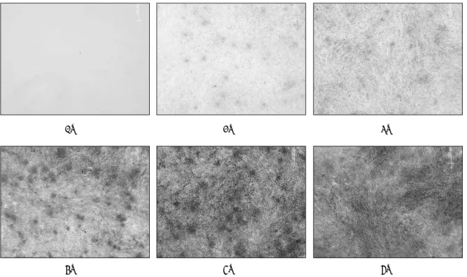
(1) Von Kossa 염색
무기질이 침착된 기질을 갈색으로 표현되게 하는 Von Kossa 염색을 통하여 골기질 형성정도를 평가하였다. 배양
Fig. 1. The photographs showing the dental papilla from extracted tooth germ.
osteocalcin의 정량화적 평가를 위하여 분석하기 48시간 전, 혈청이 배제되고 10 nM dexamethasone, 50μg/ml
L-ascorbic acid 2-phosphate, 그리고 10 mM β-glyc- erophosphate이 포함된 배지로 교체하였다. 48시간 배양 후, 분비되는 osteocalcin의 양은 효소면역분석법 (Enzyme immuno assay (EIA), Intact Osteocalcin EIA kit, Biomedical Technologies Inc, MA, USA)을 통 하여 흡광도 450 nm에서 측정하였으며 측정된 값은 ng/mL로 표시하였다.
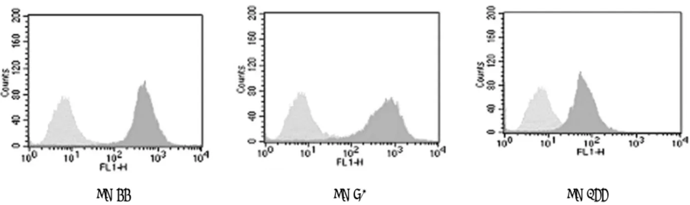
Ⅲ. 연구결과 1. 치유두 유래세포의 표면 표지자 분석
FACS를 이용하여 치유두 유래세포의 표면 표지자를 분 석하였다. 치유두 유래세포는 간엽 줄기세포 (mesenchy- mal stem cells) 표지자로 알려져 있는 CD44, CD90, 그 리고 CD166에 양성을 나타내어 배양된 인간 치유두 유래 세포의 표현형 (phenotype)이 간엽 줄기세포와 유사함을 관찰하였다 (Fig. 2).
(1) 알칼리성 인산분해효소에 대한 조직화학적 검사 알칼리성 인산분해효소의 발현은 배양 7일에 강한 발현을 나타내었으며 이후 배양기간동안 이의 발현은 지속적으로 감소하는 경향을 나타내어 배양 6주째에는 거의 발현되지 않았다 (Fig. 5).
(2) 알칼리성 인산분해효소와 osteocalcin에 대한 RT- PCR 분석
ALP mRNA 발현은 일차배양 및 계대배양을 거치는 비 골형성 유도 배지 (non-osteogenic medium)에서도 강하 게 발현되었고 골형성 유도 배지에서는 배양 7일째 강하게 발현되었으며 이후 이의 발현은 계속 감소하는 경향을 나타 내었다. Osteocalcin mRNA 발현은 일차배양 및 계대배양 을 거치는 비골형성 유도 배지에서는 발현되지 않았고 골형 성 유도 배지에서도 배양 14일째 처음으로 약하게 발현되 어 이후 배양 28일째까지 지속적으로 증가하였으며 이후에 는 일정한 양상을 나타내었다 (Fig. 6).
CD 44 CD 90 CD 166
Fig. 2. FACS analysis of cultured human dental papilla-derived cells. The gray histograms represent the fluorescence intensity of
the cells with negative control mouse IgG-FITC. The green histograms show the fluorecence intensity of the cells with each
antibody. These results indicate that the cultured human dental papilla-derived cells own the phenotypic characterization of
mesenchymal stem cells.
Non-osteogenic
1W 2W 3W
4W 5W 6W
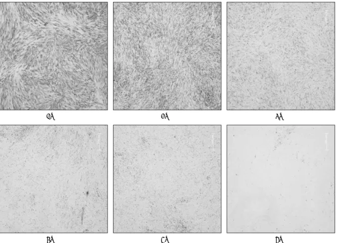
Fig. 3. Confocal laser microscopy photomicrographs of cultured human dental papilla-derived cells. Dental papilla- derived cells presented a fibroblastic morphology in non-osteogenic inductive culture medium for early passages.
Under the osteogenic condition, After 3 passages, periosteal-derived cells were cultured in osteogenic medium as a
monolayer for up to 6 weeks (6W). The cells formed colonies in multiple layers, and the precipitates indicated as
black spots appeared at 2 weeks and gradually increased with respect to culture time. Bar = 200 μ m.
분비는 급격히 증가되었다 (Fig. 8).
Fig. 4. Photograph of 6-week-cultured dental papilla- derived cells. The cells became a membrane-like struc- ture with enough thickness to grasp with forceps.
1W 2W 3W
4W 5W 6W
Fig. 5. Histochemical expression of ALP. ALP-positive cells are indicated by blue/purple staining. ALP was expressed
strongly at 1 week (1W) followed by continuously decreased expression during the culture period. Bar = 500 μ m.
Fig. 6. Expression of ALP and osteocalcin during the osteoblastic differentiation of dental papilla-derived cells. Dental papilla-derived cells were cultured in osteogenic conditions for up to 6 weeks (6W).
OM (-) ; non-osteogenic culture medium, OM (+); osteogenic culture medium.
1W 2W 3W
4W 5W 6W
Fig. 7. Von Kossa staining for mineralized nodule formation in dental papilla-derived cells cultures at sequential time
points. Von Kossa-positive mineralization nodules first appeared after 2 weeks (2W) of culture. Subsequently miner-
alization gradually increased during the entire duration of the culture period.
Ⅳ. 총괄 및 고찰
악안면영역의 골결손에 대하여 최근 줄기세포 혹은 전구 세포를 통한 골조직공학적 해결 방법이 여러 가지 수술적 방법에 대한 대안으로 대두되고 있다. 척추 혹은 장골에서 채취되는 골수는 대표적인 성체줄기세포의 원천이다. 그러 나 골수로부터 관련세포를 추출하는 과정은 악안면 영역의 임상적인 면에서 볼 때 그리 유리한 것은 아니다. 흡입을 통 한 골수의 채취는 그 준비과정의 간단히지 못함과 패혈증 및 시술후 감염과 같은 합병증의 존재가능성으로 임상적인 면에서 유리하지 못한 여건을 제공한다. 골조직공학적 이용 가치가 있고 쉽게 접근하여 채취할 수 있는 원천이 있다면 조직공학적 측면에서 큰 장점을 제공한다고 할 수 있다. 그 리하여 최근 골막기원세포 (periosteal-derived cells) 및 지방기원세포 (adipose-derived cells) 등과 같은 세포들이 골수기원줄기세포 (bone marrow-derived mesenchymal stem cells)와 유사한 조골세포로의 분화능을 포함하여 여 러 가지 간엽조직 세포들로 분화할 수 있는 능력을 가지고 있는 것으로 보고되고 있다2,4,10-13).
본 연구에서는 조직공학적 골형성을 위하여 교정적 목적 등으로 발치하게 되는 치배의 한 구성요소인 치유두에서 유 래하는 세포를 이용하여 조골세포 관련 표현형을 관찰하였 다. 치배는 법랑기 치유두 및 치낭으로 구성되어 있으며 성 숙된 치아로 성장하는 과정에서 치배내의 세포들은 광화과 정에 관여하게 된다. Handa 등14)은 소의 치배에서 추출한 치낭세포들이 백악질과 유사한 광화조직 (cementum-like mineralization tissue)을 형성할 수 있다고 하였다. 그러
telomerase reverse transcriptase) 처리에 의하여 만든 무한증식 치유두 및 치수 세포 (immortalized dental papilla and dental pulp cells)는 ascorbic acid와 β- glycerophosphate의 존재하에서 광화를 형성함을 보고하 였다. 이와 같이 치배의 구성요소 세포들의 광화와 관련된 보고가 일부 있으나 치유두에서 유래하는 세포의 조골세포 분화과정에서 관련된 생화학적 표지자 (biochemical marker)의 순차적 발현 (time-sequential expression)은 연구된 것이 거의 없으므로 본 연구에서는 치유두 유래세 포에서 간엽 줄기세포 표지자의 발현 양상과 조골세포로의 분화과정동안 조골세포 관련 표지자의 순차적 발현을 연구 하였다. 간엽 줄기세포 표지자로 CD44, CD90, 그리고 CD166을 이용하였는데 치유두 유래세포는 CD44, CD90, 그리고 CD166 모두에 양성을 나타내어 배양된 인간 치유 두 유래세포의 표현형은 간엽 줄기세포와 유사함을 관찰하 였다.
일반적으로 미분화 상태에서 기능을 나타내는 활동성 조 골세포로의 분화과정은 알칼리성 인산분해효소와 osteo- calcin의 발현 및 무기질 침착과 같이 특정 시기에서 특정 유전인자들이 관여하는 일련의 과정을 통하여 이루어진다.
알칼리성 인산분해효소의 발현은 분화 초기에 나타나며 osteocalcin의 분비 및 무기질 침착은 조골세포로의 분화 마지막 단계에 나타난다. 알칼리성 인산분해효소는 조골세 포에서 형성되고 무기질 침착을 위한 인산염 혹은 무기성 피로인산염의 국소적 증가를 이루게 하는 역할을 하며 일반 적으로 분화 초기의 조골세포 특이 표지자로 알려져 있다.
반면에 osteocalcin은 거의 독점적으로 조골세포에 의하여 생성되며 분화 후반기인 성숙한 조골세포의 특이 표지자로 알려져 있는 골기질 단백질이다18-21). 본 연구에서 알칼리성 인산분해효소는 배양 7일째 가장 강한 발현을 나타내었으 며 이후 이의 발현은 지속적으로 감소하였다. Osteocalcin 의 발현은 배양 14일째에 처음으로 나타나 이후 배양 28일 째까지 지속적으로 증가하였으며 이후에는 일정한 양상을 나타내었다. 골막기원세포에서 분비되는 osteocalcin은 배 양 1주에서는 관찰되지 않았으나 배양 2주째부터는 그 분 비가 지속적으로 증가됨을 관찰하였다. 효소면역분석법을
Fig. 8. Quantification of osteocalcin secretion from den-
tal papillal-derived cells. Dental papilla-derived cells were cultured in osteogenic conditions for up to 6 weeks (6W). The osteocalcin level was represented as mean and SD (n = 3) of nanogram (ng) of osteocalcin by mL medium at the indicated culture time.
OM(-) OM(+)
통하여 배지로 분비되는 osteocalcin에 대한 정량화적 분석 에서는 osteocalcin의 분비가 배양 7일째부터 나타나 배양 21일째까지 서서히 증가하다가 이후 배양기간 동안에는 이 의 분비가 급격히 증가되었다. Von Kossa 염색을 통한 석 회화된 골기질의 평가에서도 배양 14일째에 von Kossa 양 성의 석회화된 골결절이 나타나 이후 시간의존성으로 계속 적으로 침착되는 양상을 나타냈다. 이러한 결과들은 앞서 언급한 알칼리성 인산분해효소와 osteocalcin의 발현에 대 한 일반적인 사실과도 어느 정도 일치하여 치유두 유래세포 가 성숙한 조골세포로 잘 분화되었다는 것을 알 수 있다. 또 한 저자들의 이전 골막기원세포의 조골세포로의 분화과정 에 대한 연구와 비교하여 볼 때, 치유두 유래세포의 조골세 포로의 분화과정이 좀더 빠르게 진행된다는 것을 알 수 있 었다.
본 연구를 통하여 배양된 인간 치유두 유래세포는 간엽 줄 기세포의 표현형을 나타내고 조골활성을 가지고 있어 향후 골조직공학에서 또 다른 골 전구세포의 원천이 될 수 있으 리라 여겨진다.
Ⅴ. 결 론
경상대학교 병원의 윤리위원회를 따르고 환자 보호자의 동의하에 13에서 15세의 3명의 환자에게서 매복된 하악 제 3대구치 치배의 발치과정에서 치유두를 채취하여 일차배양 및 계대배양을 실시하고 passage 3을 거친 치유두 유래세 포를 50μg/ml L-ascorbic acid 2-phosphate, 10 nM dexamethasone, 그리고 10 mM β-glycerophosphate이 포함된 DMEM 배지에서 6주동안 배양하여 다음과 같은 결론을 얻었다.
1. 치유두 유래세포는 일차배양 후 CD44, CD90, 그리고 CD166에 양성을 나타내어 그 표현형이 간엽 줄기세 포와 유사하였다.
2. 치유두 유래세포는 50μg/ml L-ascorbic acid 2-phos- phate, 10 nM dexamethasone, 그리고 10 mM β- glycerophosphate이 포함된 골형성 유도 배지에서 다 층의 집락을 형성하였고 배양 2주째부터는 무기질 결 절을 형성하기 시작하였다.
3. 치유두 유래세포에서 배양 7일에 알칼리성 인산분해효 소의 발현이 강하게 나타났으며 이후 배양기간동안 이 의 발현은 지속적으로 감소하는 경향을 나타내었다.
4. 치유두 유래세포에서 배양 14일째 처음으로 osteocal- cin의 발현이 나타났고 이후 배양 28일째까지 이의 발 현은 지속적으로 증가하다가 이후에는 일정한 양상을 나타내었다.
5. 치유두 유래세포에서 von Kossa 염색에 양성을 나타 내는 침착된 골기질은 배양 14일째에 처음으로 나타나
이후 배양기간동안 계속적으로 증가하였다.
6. 치유두 유래세포에서 형성되어 배지로 분비되는 osteocalcin은 배양 7일째부터 관찰되어 이후 시간의 존성으로 그 분비가 지속적으로 증가됨을 관찰하였다.
배양 21일째까지는 그 분비양상의 증가가 완만하였으 나 이후, 이의 분비는 급격히 증가되었다.
상기 결론을 통하여 교정적 목적으로 발치되는 치배에서 채취하여 치유두에서 추출한 치유두 유래세포는 간엽 줄기 세포의 표현형을 나타내고 조골활성을 가지고 있어 향후 골 조직공학에서 또 하나의 골전구세포의 원천이 될 수 있으리 라 여겨진다.
참고문헌
1. Takushima A, Kitano Y, Harii K : Osteogenic potential of cultured periosteal cells in a distracted bone gap in rab- bits. J Surg Res 78 : 68, 1998.
2. Meirelles Lda S, Nardi NB : Murine marrow-derived mes- enchymal stem cell : isolation, in vitro expansion, and characterization. Br J Haematol 123 : 702, 2003.
3. Hutmacher DW, Sittinger M : Periosteal cells in bone tis- sue engineering. Tissue Eng 9 Suppl : S45, 2003.
4. Kiramura S, Ohgushi H, Hirose M et al : Osteogenic dif- ferentiation of human bone marrow-derived mesenchymal stem cells cultured on alumina ceramics. Artif Organs 28 : 72, 2004.
5. Park BW, Byun JH, Lee SG et al : Evaluation of osteogenic activity and mineralization of cultured human periosteal-derived cells. J Kor Maxillofac Plast Reconstr Surg 28 : 511, 2006.
6. Kajihara T, Ohnishi T, Arakaki N et al : Expression of hepatocyte growth factor/scatter factor and c-Met in human dental papilla and fibroblasts from dental papilla.
Arch Oral Biol 44 : 135, 1999.
7. Thonemann B, Schmalz G : Immortalization of bovine dental papilla cells with simian virus 40 large t antigen.
Arch Oral Biol 45 : 857, 2000.
8. Kikuchi H, Suzuki K, Sakai N et al : Odontoblasts induced from mesenchymal stem cells of murine dental papillae in three-dimensional cell culture. Cell Tissue Res 317 : 173, 2004.
9. Sonoyama W, Liu T, Seo BM et al : Mesenchymal stem cell-mediated functional tooth regeneration in Swine. PLoS ONE 20 : 1, 2006.
10. Meinel L, Karageorgiou V, Fajardo R et al : Bone tissue engineering using human mesenchymal stem cells: effects of scaffold material and medium flow. Ann Biomed Eng 32 : 112, 2004.
11. Sun JS, Wu SY, Lin FH : The role of muscle-derived stem cells in bone tissue engineering. Biomaterials 26 : 3953, 2005.
12. Laino G, Graziano A, d’Aquino R et al : An approachable human adult stem cell source for hard-tissue engineering.
J Cell Physiol 206 : 693, 2006.
13. Dudas JR, Marra KG, Cooper GM et al : The osteogenic potential of adipose-derived stem cells for the repair of rabbit calvarial defects. Ann Plast Surg 56 : 543, 2006.
14. Handa K, Saito M, Yamauchi M et al : Cementum matrix
formation in vivo by cultured dental follicle cells. Bone 31 :
18. Lawton DM, Andrew JG, Marsh DR et al : Expression of the gene encoding the matrix gla protein by mature
저자 연락처
우편번호 660-702
경상남도 진주시 칠암동 90번지
경상대학교 의과대학/의학전문대학원 구강악안면외과학교실 변 준 호
원고 접수일 2007년 6월 8일 게재 확정일 2007년 7월 20일
Reprint Requests
June-Ho ByunDepartment of Oral & Maxillofacial Surgery, College of Medicine, Gyeongsang National Univ. School of Medicine
90 Chilam-dong, Jinju-city, Gyeongsangnam-do, 660-702, South Korea Tel: 82-55-750-8264 Fax: 82-55-761-7024
E-mail: [email protected] Paper received 8 June 2007 Paper accepted 20 July 2007