Tuberculosis and Respiratory Diseases
결핵 및 호흡기질환, Vol. 54, No. 5, May, 2003□
이달의 X-선□
― 570 ―
우측 폐종괴
을지대학교 의과대학 내과학교실, 병리학교실
*김준형, 한민수, 김동훈*, 고 훈, 이양덕, 조용선
=Abstract=
Right Lung Mass
Junhyoung Kim, M.D., Minsoo Han, M.D., Dong Hoon Kim, M.D.*, Hun Ko, M.D., Yang Deok Lee, M.D., Yongseon Cho, M.D.
Department of Internal Medicine and Pathology
*, Eulji University School of Medicine, Daejeon, Korea
Sarcomatoid carcinomas of the lung are rare malignant biphasic tumors, which contain both a malignant epithelial component and a sarcomatoid component. The majority of patients are men and the mean age of onset is 60 years at the time of diagnosis. A metastasis to the regional lymph nodes and to distant organs is common.
The clinical course of patients with this neoplasm is aggressive, with an overall 5-year survival rate approximating 20%. A sarcomatoid carcinoma of the lung is often observed in the large bronchi and peripheral lung field than in the trachea, and the clinical manifestations are related to their specific location.
We report a case of sarcomatoid carcinoma of the lung in a 79-year-old man who presented with dyspnea on exertion.
(Tuberculosis and Respiratory Diseases 2003, 54:570-573)
Key words : Sarcomatoid carcinoma, Lung malignancy, Biphasic tumor.
Address for correspondence:
Minsoo Han, M.D.
Department of Internal Medicine, Eulji University School of Medicine 24-14, Mok Dong, Jung-Ku, Daejeon, 301-726, Korea
Phone : 042-259-1275 Fax : 042-259-1125 E-mail : [email protected]
증 례환 자 : 이○○, 79세, 남자 주 소 : 운동시 악화되는 호흡곤란
현병력 : 평소 건강히 지내던 환자로 내원 5개월
전부터 운동시 악화되는 호흡곤란 지속되어 입원 하였다.
과거력 : 특이 소견 없음.
흡연력 : 40 갑․년
진찰소견 : 내원 당시 혈압은 160/100 mmHg, 맥
― Right lung mass ―
― 571 ― Fig. 1. Chest PA shows a well-defined mass in
the right lower lung field.
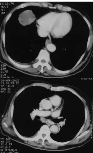
Fig. 2. Chest CT shows a lobulated, heterogene
ous, enhancing mass in the right middle lobe (top), and multiple small mediastinal, both hilar lymph nodes (bottom).
박은 64회/분, 호흡수는 20회/분, 체온은 36.4℃ 이 었으며 청진상 우중폐야에 천명음이 들렸다.
검사실 소견 : 내원시 말초혈액검사상 백혈구 8,200/㎣, 혈색소 12.1 g/dL, 혈소판 289,000/㎣ 이 었다. 대기중에서 시행한 동맥혈 가스분석 검사는 pH 7.50, PaCO2 35.9 mmHg, PaO2 76.9 mmHg, HCO3-
29.3 mmol/L, 동맥혈 산소포화도는 96.4%
이었다. 일반 생화학 검사는 AST 14 IU/L, ALT 5 IU/L, 총단백 7.7 g/dL, 알부민 3.6 g/dL, 총빌리 루빈 0.7 mg/dL, LDH 74 IU/L이었다.
폐기능검사는 FEV1 1.4 L(예측치의 64%), FVC 2.08 L(예측치의 61%), FEV1/FVC 67%로 혼합성 환기장애 소견을 보였다.
방사선 소견 : 단순흉부사진에서 우폐에 경계가 분 명한 종괴가 관찰되었으며(Fig. 1), 흉부전산화단층 촬영에서 우중엽에 5×3.5 ㎝ 크기의 균질성의 조 영증강되는 종괴와, 인접 부위에 흉막비후 및 흉막 삼출이 관찰되었다. 종격동 및 양측 폐문부에 다발 성으로 림프절들이 관찰되었다(Fig. 2). 복부초음파 및 골스캔 검사는 정상 소견이었다.
기관지내시경 소견 : 우중엽기관지와 좌측 주기관 지에 탄분색소(anthracotic pigmentation)가 관찰되 었으며 기관지내 종괴는 관찰되지 않았다.
병리 소견 : 흉부 CT 유도로 종괴부위에서 경피적 세침폐생검을 시행하였다. 광학현미경 소견상 종양 은 방추형세포의 육종성 성분과 부분적으로 분화 가 나쁜 악성상피세포의 두가지 성분으로 구성되 었다. 육종성성분은 적은 양의 호산성 세포질과 둥 글거나 방추형의 핵을 가지고, 핵소체도 자주 관찰 되었다. 비정형적인 세포분열상도 자주 관찰되었으 며, 종종 소용돌이 형태의 소견이 보여 악성섬유조
― J. H. Kim, et al ―
― 572 ― Fig. 3. A biphasic tumor with a spindle cell sar
comatous component and focal poorly differenciated adenocarcinomatous compo
nent (H&E, ×100).
Fig. 4. The sarcomatous component is strongly positive for vimentin (top) and cytokeratin (bottom) immunostaining (avidin-biotin-pe
roxidase complex, ×200).
직구종의 특성이 관찰되었다. 암종성 성분은 둥글 거나 난원형으로 다형성의 핵과 조밀한 핵염색질 을 함유하였고 작은 군집으로 종종 선모양의 형태 를 보이는 구조물도 관찰되었다(Fig. 3). 면역조직 화학 염색상 육종성 성분은 vimentin에 광범위하 게 갈색으로 염색되었으며, cytokeratin에도 진하게 염색되었다(Fig. 4).
임상경과 : 환자는 호흡곤란, 전신쇠약 등의 증상 이 지속되었으며 항암화학요법 및 방사선치료를 거부하고 퇴원하여 외래 추적 관찰 중이다.
고 찰
폐에 발생하는 육종양암종은 악성상피세포와 육종 양 성분으로 구성되며 폐 이외에 식도, 비뇨생식기 관, 피부, 후두, 소화관 등에서도 이와 같은 종양이 발생한다1-3. 이 종양은 대부분 남자에 호발하며, 흡연과의 관련성이 높고 진단시 평균 연령은 60세 로 알려져 있다3,4. 발생빈도는 매우 낮으며 국내에 서는 5편의 보고가 있었다5-9.
육종양암종과 암육종의 조직학적 분류에 대해서 는 논란이 많아서 아직도 결론이 나지 않은 상태 이다. WHO의 분류1에 따르면 폐에 발생하는 육종
양암종은 편평상피세포에서 기원하는 방추형세포의 구성 성분을 가지는 편평상피세포의 변형으로 정의 했지만 편평상피세포암 이외의 폐암에서도 상피세포 기원의 방추세포의 구성성분이 면역조직화학적 염색 과 전자현미경적 소견으로 밝혀졌다10,11. Humphrey 등은 육종성 구성성분에 상피세포의 분화를 보이 는 폐암종은 방추세포성 암종이라 하였고, 악성 골 조직, 연골, 횡문근 조직 등으로 간엽성분화를 보 이거나 광학현미경, 전자현미경, 면역조직화학적 염색에서 상피세포로의 분화를 보이지 않는 것을 암육종이라고 정의 하였다1. 이처럼 조직학적 분류 에 대해서는 아직도 논란이 있으나 이 두가지 종 양이 단지 형태학적으로나 생물학적으로 종양이 진행하는 과정의 연속선상에 있는 시간적으로 다
― Right lung mass ―
― 573 ―
른 시점일뿐이며 임상 경과도 비슷하므로 굳이 분 류 할 필요가 없다는 주장도 있다12.폐육종성암종은 주기관지와 폐의 주변부에 잘 발생하는 것으로 알려져 있으며3,6 임상 증상은 발 생부위와 관련이 있어서 주기관지내에 발생하는 경우 반복적으로 발생하는 세균성 폐렴과 서서히 진행하는 호흡곤란, 기침, 객혈 등의 증상이 나타 나며 천명음이 들리기도 한다. 폐의 주변부를 침범 하는 경우는 증상이 없거나 흉막이나 흉벽의 연조 직이 침범되는 경우 흉통이 주 증상일 수도 있다.
원격전이는 반대측 폐와 간, 뼈, 부신 뇌 등에 발 생할 수 있다2,3,6.
예후는 좋지 않아서 평균 생존기간이 약 15개월2, 5년 생존률은 20%정도이며3, 치료는 수술, 항암화학 요법, 방사선 치료 등이 시행되고 있으나 효과는 그 리 좋지 못한 것으로 알려져있다2,3,6.
요 약
폐에 발생하는 육종양암종은 매우 드문 악성 폐종 양으로 예후가 좋지 않은 것으로 알려져 있다.
저자들은 운동시 호흡곤란을 주소로 내원한 환 자에서 경피적세침폐생검으로 확진한 육종양암종 1예를 경험하였기에 보고하는 바이다.
참 고 문 헌
1. Rainosek DE, Ro JY, Ordonez NG, Kulaga AD, Ayala AG. Sarcomatoid carcinoma of the lung: A case with atypical carcinoid and rhabdomyosarcomatous components. Am J Clin Pathol 1994;102:360-4
2. Ro JY, Chen JL, Lee JS, Sashin AA, Ordo
nez NG, Ayala AG. Sarcomatoid carcinoma of the lung: Immunohistochemical and ultra
structural studies of 14 cases. Cancer 1992;
69:376-86
3. Wick MR, Ritter JH, Humphrey PA. Sarco
matoid carcinoma of the lung: A clinico
pathologic reveiw. Am J Clin Pathol 1997;
108:40-53
4. Kim KI, Flint JD, Muller NL. Pulmonary carcinosarcoma: Radiologic findings in three patients. Am J Roentgenol 1997;169:691-4 5. 정인수, 김영지, 김충현, 김시민, 이상무, 안영
수. 골반강내로 전이되어 발견된 수술적 치료 후 재발한 폐암육종 1예. 결핵 및 호흡기질환 2001;51:453-61
6. 최요안, 김건형, 천석배, 강상우, 류형선, 이영 미 등. 상엽과 하엽에 번갈아 무기폐를 일으킨 폐암육종 1예. 결핵 및 호흡기질환 1995;42:
413-8
7. Kim HM, Shin BS, Song YW, Lee SM, Jung SH, Kim CW, et al. A case of pulmonary carcinosarcoma with persistent mild fever.
Korean J Intern Med 2002;17:78-82
8. 정혜전, 고은주, 이상무, 박만실, 주종은, 윤용 규. 기관지내의 육종양암종 1예 보고. 대한방사 선의학회지 2000;42:633-6
9. 장원기, 조중구. 폐에 발생한 암육종 -치험 2례-.
대한흉부외과학회지 1999;32:1052-6
10. Nakajima M, Kasai T, Hashimoto H, Iwata Y, Manabe H. Sarcomatoid carcinoma of the lung : A clinicopathologic study of 37 cases.
Cancer 1999;86:608-16
11. Humphrey PA, Scroggs MW, Roggli VL, Shelburne JD. Pulmonary carcinomas with a sarcomatoid element: An immunocytochemical and ultrastructural analysis. Hum Pathol 1998;19:155-65
12. Olenick SJ, Fan CC, Ryoo JW. Mixed pul
monary blastoma and carcinosarcoma. Histo
pathology 1994;25:171-4