한수지 51(3), 328-331, 2018
328
Copyright © 2018 The Korean Society of Fisheries and Aquatic Science pISSN:0374-8111, eISSN:2287-8815 Korean J Fish Aquat Sci 51(3),328-331,2018
Note
서 론
바이러스성신경괴사증(viral nervous necrosis, VNN)은 전 세계적으로 50종이상의어류에서발생하는질병으로보고되 어있다(OIE, 2017). VNN은주로자어나치어에발생하지만 능성어(Epinephelus septemfasciatus), 대서양가자미(Hippo- glossus hippoglossus), 유럽농어(Dicentrarchus labrax)에서는 성어에서도발생하여대량폐사를유발한다(OIE, 2017). 병어 는신경세포의이상으로인한이상유영(힘없이유영하거나선 회등의비정상적인유영행동)이나타나며, 병리조직학적으로 척수, 척수신경절, 뇌및망막의신경조직에공포변성이나타난 다(Egusa et al., 2006; OIE, 2017).
VNN의원인병원체인신경괴사증바이러스(nervous necro- sis virus, NNV)는외막이없는정 20면체의 RNA 바이러스로 서(크기, 약 30 nm) 노다바이러스과(Nodaviridae) 베타노다바
이러스속(Betanodavirus)에속한다(Schneemann et al., 2005).
NNV는 5개의유전자형[striped jack NNV (SJNNV) type, tiger puffer NNV (TPNNV) type, barfin flounder NNV (BF- NNV) type, red-spotted grouper NNV (RGNNV) type 및 tur- bot NNV (TNV) type]으로구분되는데(Johansen et al., 2004;
Nishizawa et al., 1997), 국내에서검출되는 NNV는 RGNNV type, SJNNV type 및 BFNNV type에속한다고보고되어있다 (Cha et al., 2007; Gomez et al., 2008; Kim et al., 2012; Kim et al., 2017; Nam et al., 2017; Won et al., 2017). 국내에서보고되 는 NNV의대부분은 RGNNV genotype에속하며, 다양한종 류의양식어류뿐만아니라패류와자연산어류에서검출된다. NNV를 검사하는방법으로는 어류주화세포를이용한분리 배양법, 유전자를이용한분자생물학적방법[reverse transcrip- tase polymerase chain reaction (RT-PCR), real time PCR), 항 체를이용한면역학적인방법(enzyme-linked immunosorbent
신경괴사증바이러스(nervous necrosis virus, RGNNV genotype)에 대한 단클론 항체 생산
김위식·김시우·오명주*
전남대학교 수산생명의학과
Production of Monoclonal Antibodies Against Nervous Necrosis Virus (NNV, RGNNV genotype)
Wi-Sik Kim, Si-Woo Kim and Myung-Joo Oh*
Department of Aqualife Medicine, Chonnam National University, Yeosu 59626, Korea
We developed and subsequently characterized mouse monoclonal antibodies (MAbs) against nervous necro sis virus (NNV, RGNNV genotype). We established six hybridoma clones secreting MAbs against NNV antigen: 2B1, 2B11, 2C12, 13C1-1, 13C1-2 and 14D11. All six MAbs belonged to the IgG2a isotype with a kappa light chain and their reactivity recognized against the 41 kDa coat protein of NNV by Western blot analysis. The affinity constants of the six MAbs were measured by enzyme-linked immunosorbent assay (ELISA). All six MAbs reacted with two NNV isolates (SgNag05 and Gemunodo06), while no reactivity was observed with five know fish viruses, namely marine birnavirus, infectious pancreatic necrosis virus, viral hemorrhagic septicemia virus, hirame rhabdovirus, and infec- tious hematopoietic necrosis virus. Moreover, high ELISA optical density (OD) values (0.87-1.42) were observed in the brain tissues of NNV-infected sevenband grouper, while low OD values (less than 0.12) were recorded in the brain tissues of uninfected fish. These results suggest that these six MAbs are highly competent and useful for the detection of NNV with the RGNNV genotype.
Key words: Nervous necrosis virus (NNV), RGNNV genotype, Monoclonal antibody
This is an Open Access article distributed under the terms of the Creative Commons Attribution Non-Commercial Licens (http://creativecommons.org/licenses/by-nc/3.0/) which permits unrestricted non-commercial use, distribution, and reproduction in any medium, provided the original work is properly cited.
https://doi.org/10.5657/KFAS.2018.0328 Korean J Fish Aquat Sci 51(3) 328-331, June 2018
Received 15 May 2018; Revised 8 June 2018; Accepted 14 June 2018
*Corresponding author: Tel: +82. 61. 659. 3173 $ Fax: +82. 61. 659. 3173 E-mail address: [email protected]
NNV에 대한 MAb 생산 329
assay (ELISA)] 등이사용되고있다(OIE, 2017). 이들검사방 법 중, RT-PCR과분리배양법은 국내에서 NNV를 검출하거 나분리하는데주로사용되고있다. 그러나면역학적인방법은
NNV에대한특이항체가보급되어있지않아사용되지않고
있다. 본연구에서는양식현장에서신속한진단에용이하게적 용될수있는면역학적진단법의개선과보급을위한목적으로 NNV에대한단클론항체(monoclonal antibody, MAb)를제작 한후항체의특성을평가하였다.
재료 및 방법
본 연구에서는 붉바리(Epinephelus akaara)로부터 분리한 SgNag05 분리주(RGNNV genotype)를 실험용 바이러스로 사용하였다(Oh et al., 2012). NNV의배양과정제는 Gye and Nishizawa (2016)와 Gye et al. (2018)의방법에따라실시하였 다. NNV를 SSN-1 세포에접종하여배양한후 4℃에서 12,000 rpm으로 20분간원심분리하여 배양액내의 세포잔유물질을 제거한상층의부유액을 채취하였다. 채취된상층액은 106의 분획분자량의 biotech cellulose ester membrane이들어있는 투석튜브(Spectrum Laboratories, USA)를사용하여 15 mM Tris-HCl (pH 8.0)로 10일간투석한후, Hi-trap Q column을 이용한이온교환크로마토그래피(GE Healthcare, USA)와고 속단백질액체크로마토그래피(ÄKTAprime plus system, GE Healthcare, USA)를사용하여 NNV 정제를실시하였다. NNV 투석액을 column에넣고 15 mM Tris-HCl (pH 8.0)이들어있 는 500, 600, 700, 1000 mM의 NaCl 용액을사용하여 1 mL 씩추출한후, 700 mM의 NaCl 추출물을사용하여 centrifugal ultrafiltration (104분획분자량, Vivaspin, Sartorius, Germany) 으로농축하였다.
NNV에대한 MAb는 Gye et al. (2018)의방법에따라제작 하였다. 정제된 NNV (약 20 μg)와 complete Freund’s adjuvant (Gibco, USA)를동량으로섞어 BALB/c 마우스의복강에 1차 접종한후, 정제된 NNV (약 20 μg)를사용하여 7일간격으로 2 회접종하였다. 최종면역후 3일째, 마우스로부터비장을분리 한후 polyethylene glycol (Roche, Germany)을사용하여 my- eloma 세포(SP2/0-Ag14)와 융합시킨후 fetal bovine serum 이 10% 첨가된 hypoxanthine-aminopterin-thymidine (HAT) 배지로 suspension 시킨후 96 well plate에분주하여 37℃로 설정된 CO2배양기에서배양하였다. 양성 hybridoma는정제 된 NNV를사용하여 ELISA로선별하였고 3회이상제한희석 법으로클로닝하였다. ELISA는정제된바이러스액을 96 well plate에 50 μL (200 ng/well)씩 분주하여 4℃에서 overnight 한후뒷부분에설명된방법에따라실시하였다. 선별된 MAb 의 isotyping은 pierce rapid ELISA mouse MAb isotyping kit (Thermo, USA)를사용하여결정하였다.
본 연구에서 제작된 MAb와 정제된 NNV, NNV (분리주, SgNag05와 Gemunodo06)에감염된 SSN-1 세포와정상 SSN-
1 세포를사용하여 Laemmli (1970)과 Towbin et al. (1979)의 방법에따라 sodium dodecyl sulfate polyacrylamide gel elec- trophoresis (SDS-PAGE)와 Western blot ting을실시하였다. 정 제된 NNV를 12% polyacrylamide gel에 loading 한후 30 mA 에서 전기영동을하였다. 전기영동 종료후 gel을 coomassie brilliant blue를사용하여염색하였다. Western blot은전기영동 한 NNV를 nitrocellulose membrane (Bio-Rad, USA)에옮긴 후, 본연구에서제작한 MAb (1차항체)와 alkaline phospha- tase (AP)가붙어있는 goat anti-mouse IgG (Sigma, USA, 2 차항체)로반응시키고 AP가붙어있는 substrate kit (Bio-Rad, USA)를사용하여발색하였다. 양성대조구로본연구실에서보 유하고있는항 NNV 토끼항체를사용하였다.
제작된 MAb의반응특이성을조사하기위해 RGNNV geno- type에 속하는 2개의 NNV 분리주(SgNag05, 108.55 TCID50/ mL와 Gemunodo06, 108.3 TCID50/mL), 5종류의어류바이러 스[바이러스성출혈성패혈증바이러스(viral hemorrhagic sep- ticemia virus, VHSV: 108.05 TCID50/mL), 넙치랩도바이러스 (hirame rhabdovirus, HIRRV: 108.3 TCID50/mL), 전염성조혈 기괴사증바이러스(infectious hematopoietic necrosis virus, IHNV: 107.3 TCID50/mL), 전염성췌장괴사증바이러스(infec- tious pancreatic necrosis virus, IPNV: 109.55 TCID50/mL), 해양 버나바이러스(marine birnavirus, MABV: 109.3 TCID50/mL)], NNV(분리주: Yeosu08)에감염된능성어의뇌조직마쇄액(3
개체, 108.55-8.8 TCID50/mL) 및정상능성어의뇌조직마쇄액을
사용하여 Jeong et al. (2017)의방법에따라 ELISA를실시하였 다. 바이러스배양액과뇌조직마쇄액을증류수로 320배희석 하여 96 well ELISA microplates (Greiner bioone, Germany) 에각각 50 μL씩분주한후 37℃에서 overnight하여항원을코 팅하였다. T-PBS [0.05% tween-20/PBS (v/v)]로 3회세정한 후, 5% skim milk를 380 μL씩분주하여 25℃에서 1시간동안 blocking하였다. 1차항체로는본연구에서제작한 MAb를 50 μL씩분주하였으며, 2차항체는 horseradish peroxidase (HRP) 가표식되어있는 goat anti-mouse IgG serum (Youngin, Ko- rea)을 5% skim milk로 1,000배희석하여 well 당 50 μL씩분 주였다. ELISA 발색액(100 mM Na2HPO4, 50 mM citric acid, 1 mg/mL o-phenylenediamine, 0.03% H2O2)을각 well에 50 μL씩분주하여발색하였다. 각 well에 2 N H2SO4를 50 μL씩 넣어발색반응을중지시킨후 microplate photometer (Multi- skan, USA)로 492 nm에서흡광도(optical density, OD)값을 측정하였다.
결과 및 고찰
본 연구에서는 RGNNV genotype에 속하는 NNV에 대한 MAb를 생산하고자하였다. 정제한 NNV를 사용하여 SDS- PAGE를실시한결과, 약 41 kDa의분자량이확인되었다(data not shown). 베타노다바이러스의캡시드는 42 kDa의단일구
김위식ㆍ김시우ㆍ오명주 330
조단백질로구성되어있어(Schneemann et al., 2005), 본연구 에서정제된 NNV의 SDS-PAGE의결과는기존에보고된결과 와거의유사하였다.
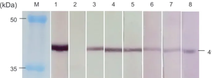
정제된 NNV를마우스에면역시킨후마우스의비장조직과 myeloma 세포를융합시켜 hybridoma를제작하였다. Hybrid- oma로부터생성되는항체를 ELISA법으로선별한후, 제한희 석법으로 3회클로닝하여최종적으로 6개의 MAb를선별하였 다(2B1, 2B11, 2C12, 13C1-1, 13C1-2, 14D11). 6개의 MAb 와정제된 NNV를사용하여 Western blot을실시한결과, 6개 MAb 모두 NNV의 구조단백질을(41 kDa) 인식하였다(Fig.
1). NNV (SgNag05와 Gemunodo06)에감염된 SSN-1 세포와 정상 SSN-1 세포에대한 MAb의반응을조사한결과에서는 6 개 MAb 모두 NNV에감염된 SSN-1 세포에서만 41 kDa에서 밴드가관찰되었다(data not shown). 이상의결과, 제작된 6개 의 MAb는 Western blot에서 NNV에특이적으로반응하는것 이확인되었다.
제작된 MAb의특이도를조사하기위해 6종의어류바이러스,
NNV에감염된능성어의뇌조직마쇄액및정상뇌조직마쇄액
을항원으로사용하여 ELISA를실시하였다(Fig. 2, 3). 6종의 어류바이러스에대해서는 6개의 MAb 모두 NNV (SgNag05와 Gemunodo06)에강하게반응(OD, 0.88-1.42)하였고, 5종의어 류바이러스(MABV, IPNV, VHSV, HIRRV, IHNV)에는반응 하지않았다(OD, 0.06 이하) (Fig. 2). 뇌조직마쇄액의경우, 6 개의 MAb 모두 NNV에감염된뇌조직마쇄액에강하게반응 (OD, 0.87-1.42)하였고, 정상능성어의뇌조직마쇄액에는반 응하지않았다(OD: 0.12 이하) (Fig. 3). 이상의결과, 제작된 6 개의 MAb는 ELISA에서 NNV에특이적으로반응하는것이 확인되었다. 제작된 MAb의 isotyping을분석한결과, 6개항 체모두 H chain은 IgG2a, L chain은 kappa로확인되었다(data not shown).
본연구에서는 RGNNV genotype에속하는 SgNag05 분리주 를사용하여 MAb를제작한결과, 총 6개의 MAb를생산하였 다. 6개의 MAb는 NNV의구조단백질(41 kDa)을인식하였고,
NNV와 NNV에감염된뇌조직마쇄액에특이적으로반응하였 으며, SSN-1 세포, 5종의어류바이러스(MABV, IPNV, VHSV, HIRRV, IHNV) 및정상능성어의뇌조직마쇄액에는반응하 지 않았다. 본연구에서는 붉바리로부터분리한일본분리주 (SgNag05)를 사용하여 MAb를 제작하였으나 제작된 MAb 는 SgNag05뿐만아니라 RGNNV genotype에속하는국내능 성어분리주(Gemunodo06와 Yeosu08; Kim et al., 2012)에도 강하게반응하였다. 이상의결과, 제작된 MAb는 Western blot 과 ELISA에서 RGNNV genotype에속하는 NNV 분리주들을 Fig. 1. Western blot analysis using purified NNV and six mono-
clonal antibodies (MAbs). M, molecular marker; 1, anti-NNV polyclonal antibody (positive control); 2, 2% skim milk (negative control); 3, MAb 2B1; 4, MAb 2B11; 5, MAb 2C12; 6, 13C1-1; 7, 13C1-2; 8, 14D11. NNV, nervous necro sis virus.
M 1 2 3 4 5 6 7 8
41 50
35 (kDa)
0.0 0.2 0.4 0.6 0.8 1.0 1.2 1.4 1.6
NNV(S) NNV(G) MABV IPNV VHSV HIRRV IHNV
ELISA abosrbance (OD 492 nm)
Fish virus
2B1 2B11 2C12 13C1-1 13C1-2 14D11
0.0 0.2 0.4 0.6 0.8 1.0 1.2 1.4 1.6
2B1 2B11 2C12 13C1-1 13C1-2 14D11
ELISA abosrbance (OD 492 nm)
Monoclonal antibody
D1 D2 D3
C1 C2 C3
Fig. 2. ELISA analysis using six fish viruses (NNV, nervous necro- sis virus; MABV, marine birnavirus; IPNV, infectious pancreatic necrosis virus; VHSV, viral hemorrhagic septicemia virus; HIRRV, hirame rhabdovirus; IHNV, infectious hematopoietic necrosis vi- rus) and six monoclonal antibodies (2B1, 2B11, 2C12, 13C1-1, 13C1-2 and 14D11). NNV (S), NNV SgNag05 isolate; NNV (G), NNV Gemunodo06 isolate.
M 1 2 3 4 5 6 7 8
41 50
35 (kDa)
0.0 0.2 0.4 0.6 0.8 1.0 1.2 1.4 1.6
NNV(S) NNV(G) MABV IPNV VHSV HIRRV IHNV
ELISA abosrbance (OD 492 nm)
Fish virus
2B1 2B11 2C12 13C1-1 13C1-2 14D11
0.0 0.2 0.4 0.6 0.8 1.0 1.2 1.4 1.6
2B1 2B11 2C12 13C1-1 13C1-2 14D11
ELISA abosrbance (OD 492 nm)
Monoclonal antibody
D1 D2 D3
C1 C2 C3
Fig. 3. ELISA analysis using brain tissues of NNV-infected sev- enband grouper (D1, 108.55 TCID50/mL; D2, 108.55 TCID50/mL; D3, 108.8 TCID50/mL) and uninfected sevenband grouper (C1, C2 and C3) and six monoclonal antibodies (2B1, 2B11, 2C12, 13C1-1, 13C1-2 and 14D11).
M 1 2 3 4 5 6 7 8
41 50
35 (kDa)
0.0 0.2 0.4 0.6 0.8 1.0 1.2 1.4 1.6
NNV(S) NNV(G) MABV IPNV VHSV HIRRV IHNV
ELISA abosrbance (OD 492 nm)
Fish virus
2B1 2B11 2C12 13C1-1 13C1-2 14D11
0.0 0.2 0.4 0.6 0.8 1.0 1.2 1.4 1.6
2B1 2B11 2C12 13C1-1 13C1-2 14D11
ELISA abosrbance (OD 492 nm)
Monoclonal antibody
D1 D2 D3
C1 C2 C3
NNV에 대한 MAb 생산 331
검출하는데유용하게사용될수있을것으로사료된다. NNV 는 3개의혈청형(serotype)으로구분된다(OIE, 2017). A형에 는 SJNNV genotype의 NNV, B형에는 TPNNV genotype의 NNV, C형에는 BFNNV와 RGNNV genotype의 NNV가속한 다. 따라서향후연구로서본연구에서제작된 MAb를사용하 여 serotype이다른 NNV 분리주에대한반응을조사해야할 것이다.
사 사
본연구는 2017년해양수산부재원으로해양수산과학기술진
흥원의지원을받아수행되었습니다(수산동물바이러스전염 병진단용항체생산).
References
Cha SJ, Do JW, Lee NS, An EJ, Kim YC, Kim JW and Park JW. 2007. Phylogenetic analysis of betanodaviruses isolated from cultured fish in Korea. Dis Aquat Org 77, 181-189.
https://doi.org/10.3354/dao01840.
Egusa S, Wakabayashi H and Muroga K. 2006. Infectious and parasitic diseases of fish and shellfish. Life Science Publish- ing Co., Seoul, Korea. 84-89.
Gomez DK, Baeck GW, Kim JH, Choresca Jr CH and Park SC.
2008. Molecular detection of betanodavirus in wild marine fish populations in South Korea. J Vet Diagn Invest 20, 38- 44. http://dx.doi.org/10.1177/104063870802000107.
Gye HJ and Nishizawa T. 2016. Purification of nervous necrosis virus (NNV) particles by anion-exchange chromatography.
J Virol Method 238, 21-28. https://doi.org/10.1016/j.jvirom- et.2016.10.001.
Gye HJ, Park MJ, Kim WS, Oh MJ and Nishizawa T. 2018.
Heat-denaturation of conformational structures on nervous necrosis virus for generating neutralization antibodies.
Aquaculture 484, 65-70. http://dx.doi.org/10.1016/j.aqua- culture.2017.10.034.
Jeong HN, Jang MS, Oh MJ and Kim WS. 2017. Produc- tion of monoclonal antibodies against viral hemorrhagic septicemia virus (VHSV, genotype IVa) from olive floun- der. J Fish Pathol 30, 149-154. http://dx.doi.org/10.7847/
jfp.2017.30.2.149.
Johansen R, Sommerset I, Tørud B, Korsnes K, Hjortaas MJ, Nilsen F, Nerland AH and Dannevig BH. 2004. Charac- terization of nodavirus and viral encephalophathy and retinopathy in farmed turbot, Scophthalmus maximus (L.).
J Fish Dis 27, 591-601. http://dx.doi.org/10.1111/j.1365- 2761.2004.00581.x.
Kim CS, Kim WS, Nishizawa T and Oh MJ. 2012. Prevalence of viral nervous necrosis (VNN) in sevenband grouper, Epi
nephelus septemfasciatus farms. J Fish Pathol 25, 111-116.
http://dx.doi.org/ 10.7847/jfp.2012.25.2.111.
Kim YC, Kwon WJ, Min JG, Jeong MG and Jeong HD. 2017.
Utilization of two serially connected PCR assay for detec- tion and type-discrimination of betanodaviruses in shellfish.
Korean federation of fisheries science and technology soci- eties international conference 2017. Abstract, Ann Meet. J Fish Pathol, 195.
Laemmli UK. 1970. Cleavage of structural proteins during the assembly of the head of bacteriophage T4. Nature 227, 680- Nam UH, Jeon CH, Seo HJ, Choi DY, Seo JY, Kwon ON, Kim 685.
WS and Kim JH. 2017. Monitoring of viruses in cultured walleye pollock Gadus chalcogrammmus. J Fish Pathol 30, 1-9. http://dx.doi.org/10.7847/jfp.2017.30.1.001.
Nishizawa T, Furuhashi M, Nagai T, Nakai T and Muroga K.
1997. Genomic classification of fish nodaviruses by mo- lecular phylogenetic analysis of the coat protein genes. Appl Environ Microbiol 63, 1633-1636.
Oh MJ, Takami I, Nishizawa T, Kim WS, Kim CS, Kim SR and Park MA. 2012. Field tests of poly(I:C) immunization with nervous necrosis virus (NNV) in sevenband grouper, Epi
nephelus septemfasciatus, (Thunberg). J Fish Dis 35, 187- 191. http://dx.doi.org/10.1111/j.1365-2761.2011.01334.x.
OIE (Office International des Epizooties). 2017. Manual of di- agnostic tests for aquatic animals. Viral encephalopathy and retinopathy. Retrieved from http://www.oie.int/index.php?id
=2439&L=0&htmfile=chapitre_viral_encephalopathy_reti- nopathy.htm. Pdf on May 8, 2017.
Schneemann A, Ball LA, Delsert C, Johnson JE and Nishizawa T. 2005. Family Nodaviridae. Virus taxonomy: Eighth re- port of the international committee on taxonomy of viruses.
CM, Mayo MA, Maniloff J, Desselberger U and Ball LA, eds. Elsevier/Academic Press, London, UK, Fauquet 869- Towbin H, Staehelin T and Gordon J. 1979. Electrophoretic 872.
transfer of proteins from polyacrylamide gels to nitrocellu- lose sheets; procedure and some applications. Proc Natl Acad Sci USA 76, 4350-4354.
Won KM, Lee JT, Cho MY, Kim MS, Kim NY, Jung SH and Lee NS. 2017. A case study of mortality caused by viral en- cephalopathy and retinopathy (VER) in cultured sevenband grouper, Epinephelus septemfasciatus during Winter. K J Ichthyol 29, 157-164.