The medical 3-dimensional image exchange via health level 7 fast healthcare interoperability resource (HL7 FHIR)
Jung Hwan Lee
1, Byung Kwan Choi
2*, In Ho Han
31
Associate Professor, Pusan National University Hospital,
2
Professor, Department of Neurosurgery, School of Medicine, Pusan National University,
3
Associate Professor, Department of Neurosurgery, School of Medicine, Pusan National University
Health level 7 fast healthcare interoperability resource (HL7 FHIR)를 통한 3차원 의료 영상의 교환
이정환
1, 최병관
2*, 한인호
31
부산대학교병원 신경외과 임상부교수,
2부산대학교 의과대학 신경외과학교실 교수,
3
부산대학교 의과대학 신경외과학교실 기금부교수
Abstract For improving interoperability of medical information, health level 7 has initiated the development of a next-generation framework for the exchange of medical information called the Fast health interoperability resources (FHIR). However, there was no attempt to exchange the medical three-dimensional (3D) image with clinical data via FHIR. Thus, we designed a new method. The 3D image to be made from computed tomography was converted to the javascript object notation (JSON) file format, and clinical data was added. We made a test FHIR server, and the client used the postman.
The JSON file was attached to the body, and was then transmitted. The transmitted 3D image could be seen through a web browser, and attached clinical data was identified in the source code. This is the first attempt to exchange the medical 3D image. Additional researches will be needed to develop applications or FHIR resources that apply this method.
Key Words : Health level seven, three-dimensional image, informatics, health information exchange, medical image
요 약 의료 정보의 상호운용성 향상을 위해서 Health level 7은 의료 정보 교환을 위한 차세대 체계인 Fast health interoperability resource (FHIR)를 개발하였다. 그러나, 이를 이용하여 임상 정보를 포함한 3차원 의료 영상을 교환 하려는 시도는 없어 새로운 방법을 제시하고자 한다. CT 영상에서 만들어진 3차원 의료 영상을 javascript object notation (JSON) 형식으로 전환하고, 임상 정보를 추가하였다. 우리는 시험용 FHIR 서버를 만들고, 클라이언트는 postman을 사용하였다. 생성된 JSON 파일은 body에 첨부하여 전송되었다. JSON 형식으로 전송된 3차원 의료 영상 은 웹 브라우저를 통해서 볼 수 있었고, 원시 코드를 확인하여 동봉된 임상 정보를 볼 수 있었다. 우리는 3차원 의료 영상 교환을 최초로 시행하였다. 이 방법을 적용한 앱이나 FHIR 리소스 개발을 위해 추가적인 연구가 필요할 것이다.
주제어 : Health level 7, 3차원 영상, 정보학, 의료 정보 교환, 의료 영상
*This work was supproted by a 2-year Research Grant of Pusan National University.
(이 과제는 부산대학교 기본연구지원사업(2년)에 의하여 연구되었음)
*Corresponding Author : Byung Kwan Choi([email protected])
Received April 3, 2020 Revised June 8, 2020
Accepted June 20, 2020 Published June 28, 2020
1. Introduction
Recently, two-dimensional (2D) images from computed tomography (CT) or magnetic resonance image (MRI) have been reconstructed into three-dimensional (3D) images, which are often useful in the medical field. Medical 3D images can help during preoperative planning[1,2]. And, it can also help patients understand their illness[3]. More importantly, medical 3D images enable the production of customized implants using a 3D printer, resulting in better surgical results[4-6].
Electronic health records (EHRs) are widely used around the world[7]. However, there is no standard system, and these records cannot be exchanged[8]. Thus, the medical costs increase owing to overlapping examinations, and it is difficult to collect big data for research[7]. To this end, health level 7 (HL7) has initiated the development of a next-generation framework for the exchange of medical information called the Fast Health Interoperability Resources (FHIR)[9].
The HL7 FHIR can be used for patient health records (PHRs) as well as exchanging EHRs among hospitals[10]. PHRs are to allow patients to make and manage their health records[10].
HL7 FHIR organizes and exchanges medical information in resource units[11]. These resources also include information related to medical imaging. However, the resource for medical 3D imaging has not yet been configured.
FHIR supports new web standard, such as extensible markup language (XML) or javascript object notation (JSON)[9]. XML and JSON file formats can show not only 2D images but also 3D images in the web browser without any software. Using this, it is expected that medical 3D images can be exchanged through this standard. In addition, if clinical data are added through one of these medical 3D images, it will be easier for healthcare staffs or patients who receive these images to understand the patient’s
illness. Therefore, in this study, we designed a way to exchange medical 3D images through FHIR.
2. Methods
2.1 Preparation of the medical 3D image
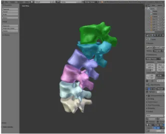
Fig. 1. The medical three-dimensional image reconstructed by Mimics software is converted from the stereolithography file format to the javascript object notation file format through Blender software
To study the exchange of medical 3D images via FHIR, we tested it with a 20-year-old woman.
The diagnosis was fracture-dislocation of thoracolumbar spine. We gained axial CT images of the thoracolumbar spine with the digital imaging and communications in medicine (DICOM) file format. To make the 3D image of the fractured spine, we used the Materialise Mimics version 20 (Materialise Co., Leuven, Belgium) to edit and convert the 3D image to the stereolithography (STL) file format. After opening the Materialise Mimics, we imported the DICOM files of the patient in detail and ran the
“thresholding” function by selecting the “bone”
option. This would automatically select the area
between 226 and 3071 Hounsfield units on all
DICOM files. When the mask corresponding to
the bone was created, we selected the function
“Calculate 3D” to create a 3D model of the thoracolumbar spine from the CT scans. The generated 3D image was then converted into the STL format Fig 1.
2.2 Converting the medical 3D image to JSON format and adding clinical data
Fig. 2. The format of the 3-dimensional image in three.js
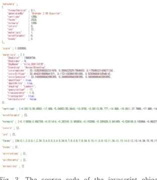
Fig. 3. The source code of the javascript object notation file of the three dimensional image
We used the blender version 2.6.5 (Blender Foundation, Amsterdam, Netherlands) to convert the edited medical 3D images from STL format
into JSON format Fig 1. The JSON file format for the 3D model was used by the format defined by three.js, a kind of the Javascript library (https://threejs.org). The format of 3D data in three.js is shown in Fig 2. An example of the actual data is shown in Fig 3.
We added clinical data to a JSON file converted in Blender using a text editor. The clinical data added include the International Statistical Classification of Diseases and Related Health Problems (ICD) 10 code, Systematized Nomenclature of Medicine (SNOMED) code, model name, age and sex Fig. 4.
Fig. 4. Adding clinical data in the javascript object notation file of the three-dimensional image
2.3 Transmission of data
We made a test FHIR server to confirm the transmission of the medical 3D image with clinical data using FHIR. We used .NET core (Microsoft co., Seattle, USA) for the platform in 2019. The server was programmed to visualize the medical 3D image with clinical data, parse the data, and return some part to the client. The web site was hosted on the AZURE cloud service (Microsoft co., Seattle, USA) for testing. The client used the postman (https://github.com/postmanlabs/
Fig. 4. The function for the data transfer used
“Post”. The data transmission format was set to
“JSON”. The contents of the previously created
JSON file were attached to the body and was
then transmitted. The data returned were the
clinical data and vertices array data Fig. 5.
Fig. 5. The data returned after transmission. Clinical data and vertices array data was returned
3. Results
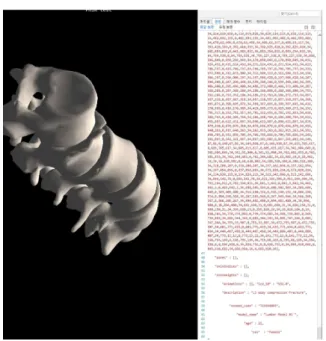
The medical 3D image transmitted under the JSON file could be seen through a web browser Fig. 6. When we viewed the source code of this page, we could see that the attached clinical data was transmitted Fig. 7.
Fig. 6. The javascript object notation file sent via fast health interoperability resources shown in web browser
Fig. 7. The source code of the transferred three-dimensional image file. If the F12 button was pressed, the source code included clinical data was shown
4. Discussion
The HL7 FHIR is an improvement the helps leverage lightweight web services and principles of the newest web and mobile development[10].
The basic organization of HL7 FHIR is a resource, and all the health and related data elements are organized as resources[10]. FHIR resources are classified as follows: clinical, financial, specialized, base, and foundation[10].
Imaging study is included in clinical resources and related with a DICOM imaging study[13]. To the best of our knowledge, this paper is the first study that exchanges medical 3D image via FHIR irrelevantly to DICOM format.
Transmission of healthcare information
through FHIR is possible only with 2D data such
as text, numbers and graphs. This study shows
that transmission of healthcare information via
FHIR is also possible in 3D images. In particular,
the advantage of this method is that it can be
implemented in a general web browser
environment without any other dedicated
application. Medical 3D images from MR or CT scanners can also be viewed in the DICOM file format. Scanned Images can be reconstructed in 3D with the software provided by CT or MR scanners, and reconstructed 3D images can be saved as the DICOM file format[14]. Thus, we can see medical 3D images in the DICOM format without converting them to 3D related formats such as STL files. However, DICOM files require a separate application, which slows down performance and takes longer time.
In this study, we added additional clinical data when exchanging the medical 3D image, which is a general scenario in the real clinical setting. In the DICOM file format used to view medical images, various information related to imaging and clinical information were included in the header[15]. Since this information is included, it can be helpful not only to display medical images but also to interpret the images to medical staffs who view such images. With help of FHIR, we were able to send clinical data while simultaneously exchanging the medical 3D image. Including important information in the exchanged image will be more helpful for the medical staff who interpret the image. In addition, through the FHIR, healthcare information can be exchanged to the patient.
Including clinical data can help the patient better understand their own medical 3D image.
Applying this method will help to improve the function of exchanging EHRs and PHRs.
However, this study has some limitations. First, medical 3D images need to be converted. 3D images have several formats depending on their purpose. A software could be provided to successfully extract a 3D image and convert data to the JSON format, and all these processes can be implemented through a picture archiving and communicating system software. However, in the process of converting these files to the JSON format, loss of information can occur. Our method was to try to transfer, not to create a
perfect application or a FHIR resource.
Therefore, additional development is required to actually apply it.
5. Conclusion
We have presented a new method of transmitting the medical 3D image with clinical data using FHIR, and the 3D image was transmitted successfully. This is the first attempt as far as we know. This process will increase staff and patients’ understanding of the disease when transmitting healthcare information. Additional researches will be needed to develop applications or FHIR resources that apply this method.
REFERENCES
[1] J. H. Lee, I. H. Han, B. K. Choi, K. H. Nam, D. H. Kim, C. S. Lee. (2017). Virtual preoperative simulation for excision of spinal tumors: surgeon processing of medical computer-assisted design software. Korean Journal of Spine, 14(4), 170-174
https://doi.org/10.14245/kjs.2017.14.4.170
[2] M. Jonathan, A. Feridun, H. Bronwyn, B. Kim. (2008).
Preoperative visualization of neurovascular anatomy in trigeminal neuralgia. Journal of Neurosurgery, 108(3), 477-482
https://doi.org/10.3171/JNS/2008/108/3/0477
[3] P. S. Kim, C. H. Choi, I. H. Han, J. H. Lee, H. J. Choi, J.
I. Lee. (2019). Obtaining informed consent using patient specific 3d printing cerebral aneurysm model. Journal of Korean Neurosurgical Society, 62(4), 398-404 https://doi.org/10.3340/jkns.2019.0092
[4] E. K. Park et al. (2016). Cranioplasty enhanced by three-dimensional printing: custom-made three-dimensional-printed titanium implants for skull defects. Journal of Craniofacial Surgery, 27(4), 943-949 https://doi.org/10.1097/SCS.0000000000002656 [5] K. C. Wong, S. M. Kumta, N. V. Geel, J. Demol. (2015).
One-step reconstruction with a 3d-printed, biomechanically evaluated custom implant after complex pelvic tumor resection. Computer Aided Surgery, 20(1), 14-23
https://doi.org/10.3109/10929088.2015.1076039
[6] B. J. Kim, K. S. Hong, K. J. Park, D. H. Park, Y. G.
Chung, S. H. Kang. (2012). Customized cranioplasty implants using three-dimensional printers and polymethyl-methacrylate casting. Journal of Korean Neurosurgical Society, 52(6), 541-546
https://doi.org/10.3340/jkns.2012.52.6.541
[7] J. H. Lee, J. H. Lee, W. Ryu, B. K. Choi, I. H. Han, C.
M. Lee. (2019). Computer-based clinical coding activity analysis for neurosurgical terms. Yeungnam University Journal of Medicine, 36(3), 225-230.
https://doi.org/10.12701/yujm.2019.00220
[8] D. Yoon, B. C. Chang, S. W. Kang, H. Bae, R. W. Park.
(2012). Adoption of electronic health records in Korean tertiary teaching and general hospitals. International Journal of Medical Informatics, 81(3), 196-203.
https://doi.org/10.1016/j.ijmedinf.2011.12.002
[9] M. V. Anderson, I. H. Kristensen, M. M. Larsen, C. H.
Pederson, K. R. Goeg, L. B. Pape-Haugaard. (2017.
May). Feasibility of representing a danish microbiology model using FHIR. Studies in Health Technology and Informatics, 235, 13-17.
https://doi.org/10.3233/978-1-61499-753-5-13 [10] R. Saripalle, C. Runyan, M. Russell. (2019). Using HL7
FHIR to achieve interoperability in patient health record. Journal of Biomedical Informatics, 94, 103188.
https://doi.org/10.1016/j.jbi.2019.103188
[11] C. Chroaki, F. Ploeg. (2016). Towards mhealth assessment guidelines for interoperability: HL7 FHIR.
Studies in Health Technology and Informatics, 224, 164-169.
https://doi.org/10.3233/978-1-61499-653-8-164 [12] M. Khalilia, M. Choi, A. Henderson, S. Iyengar, M.
Braustein, J. Sun. (2015, November). Clinical predictive modeling development and deployment through FHIR web services. American Medical Informatics Association Annual Symposium Process 2015, pp.
717-726. San Francisco : Curran Associates, Inc.
[13] HL7 (2019). Resource index. HL7 FHIR Release 4.
http:s//www.hl7.org/fhir/resourcelist.html
[14] K. J. Mortele, J. McTavish, P. R. Ros. (2002). Current techniques of computed tomography. helical CT, multidetector CT, and 3d reconstruction. Clinical Liver Disease, 6(1), 29-52.
https://doi.org/10.1016/s1089-3261(03)00065-5 [15] C. Prieto et al. (2009). Image retake analysis in digital
radiography using DICOM header information.
Journal of Digital Imaging, 22(4), 393-399.
https://doi.org/10.1007/s10278-008-9135-y
이 정 환(Jung Hwan Lee) [정회원]
․ 2009년 2월 : 부산대학교 의학대학원 신경외과(의학석사)
․ 2020년 3월 ~ 현재 : 부산대학교병원 신경외과 임상부교수
․ 관심분야 : 신경손상, 의료용 3D 영상
․ E-Mail : [email protected]
최 병 관(Byung Kwan Choi) [정회원]
․ 2004년 2월 : 부산대학교 의학대학원 신경외과(의학박사)
․ 2013년 9월 ~ 현재 : 부산대학교 의과 대학 신경외과학교실 교수
․ 관심분야 : 의료용 인공지능, 진료정보 학, 학제간융합연구
․ E-Mail : [email protected]
한 인 호(In Ho Han) [정회원]