약학희지 제42권 제 5 호 487~~493(1998) Yakhak Hoeji Vol. 42. No. 5
영지버섯 생장점 단백다당체 GLB-A, GLB-B의 항암효과 및 면역 활성에 관한 유세포 분석학적 연구
오 정 연 • 정 경수*
충 남 대학 교 약 학 대학 미생 물 약 품 화 학 교 실
(R e c e iv e d A p r il 27. 1998)
Flow Cytometrical Analysis of the Antitumor and Immunomodulatory Activities of GLB-A and GLB-B, the Protein-polysaccharide Fractions
of the Growing Tips of Ganoderma Lucidum
Jung-Yeon Oh and Kyeong-Soo Chung*
Laboratory of Microbial Chemistry, College of Pharmacy, Chung-Nam National University, Taejon 305-764, Korea
Abstract—— In t h e p r e v io u s s t u d y w e d e s c r i b e d t h e a n t i t u m o r e ffe c t o f G L B . a p r o t e i n - p o l y s a c c h a r i d e fr a c t io n s e p a r a t e d fr o m th e g r o w i n g ti p s o f Ganoderma lucidum, a g a i n s t s a r c o m a 180 s o lid t u m o r in iC R m ic e . In t h i s s t u d y , w e s e p a r a t e d a n a c id ic p r o t e i n - p o ly s a c c h a r i d e fr a c t io n , G L B -A . a n d a b a s i c p r o t e i n - p o ly s a c c h a r i d e fr a c t io n . G L B - B , fro m G L B b y d iffe r e n t ia l p r e c ip it a ti o n , a n d e lu c i d a t e d t h e i r a n ti tu m o r a n d im m u n o m o d u l a t o r y a c tiv it ie s . W h e n ip in je c t e d a t t h e d o s e o f 50 m g / k g / d a y in t o t h e IC R m ic e , G L B - A a n d G L B - B in h ib it e d t h e g r o w t h o f ip im p l a n t a t e d s a r c o m a 180 c e l ls b y 3 2 .4 % a n d 2 1 .0 % . r e s p e c t i v e l y . O f t h e s e . G L B - A in c r e a s e d t h e % l y m p h o b l a s t in t h e s p l e e n o f t h e t u m o r - b e a r i n g a n d th e n o r m a l m i c e b y 2 0 .9 % a n d 12 3 .0 % . a n d t h e C D 4 / C D 8 r a tio b y 7 3 .3 % a n d 2 2 .4 % , r e s p e c t i v e l y . G L B - A a l s o i n c r e a s e d th e e x p r e s s i o n o f C D 2 5 (IL-2 r e c e p t o r ot c h a i n ) i n n o r m a l m ic e b y 8 2 .0 % . T h e s e r e s u l t s s t r o n g ly s u g g e s t t h a t G L B - A i s a p r o m is in g c a n d i d a t e fo r a n t i t u m o r i m m u n o m o d u l a t o r y m e d ic in e .
Keywords □ Ganoderma lucidum. a n t it u m o r a c t i v i t y , i m m u n o m o d u l a t io n . ly m p h o b l a s t - f o r m a t i o n . IL-2 r e c e p to r . C D 4 / C D 8 ra tio .
면역을활성화시켜 암을치료하는면역요법에는여러 방법이사용되고 있으나, 그 중BCG(BflC!7/MS Calmet - te-Guerin)Streptococcus 속 세균, 담자균류 등외 미 생물및그성분을이용하는방범은메우활발히 연구되 고 있는 분야 중의 하나이다. 한편 담자균류 중에서는 영지버섯(Gfl«odenjw lu c id u m ) ,표고버섯(Lenh'nus e d o d e s ),구름버섯(Con'o/«s v e r s ic o lo r )치마버섯 (Schizophyllum com m une),잎새버섯(Gn/oZa/romto- s a f 등의 항암작용이 잘알려져 있다, 이중 영지버섯
* 본 논문에 관한 문의는 이 저자에게로 ( 전화) 042-821-5927 ( 팩스) 042-823-9630
은 항 암 작 용 ^ 외 에 도 항 allergy 및 항염증작용,® 던 역 증 강 작 용 에 관한 연구가 활발히 이루어지고 있 다. 그러나그동안의 연구는주로성숙한자실체를대상 으로 이루어졌으며 본 연구에서 사용한 영지버섯 생장 점에 대한연구는보고된 바가없다. 영지버섯 생장점은 자실체 생성 초기 과정에서만 볼 수 있는황백 내지 황 갈색의 점성을지닌선단부를 말하며. 본 연구자등*®은 생장점으로부터 분리한 단백다당체 분획 GLB가 일반 영지버섯의 단백다당체 보다 sarcoma 180에 대한 항 암효과 및 cyclophosphamide-유도 백혈구 감소증에 대하여 억제효과가 더욱 우수함을 보고한 바 있다. 본 연구에서는 GLB로부터 산성 단백다당체 분획 GLB-
488 오정연■정경수
A 및 염기성 단백다당체 분획 GLB-B를 분리 • 정제하 여 그의 항암 ■ 면억활성을 유세포분석기(flow cyto- meter)를 이용하여 비교 ■ 연구하였다.
실험 재료 및 방법
실험동물 및 세포주 - 대한실험동물센터로부터 약 4 주령의 SPF(specific pathogen free) IC R 계 생쥐 암
• 수컷을 구입하여 층남대학교 약학대학 동물실험실에 서 안정화시킨 후 사용하였다. sarcoma 180 암세포는 ICR 생쥐의 복강 내에 1 주일 간격으로 계대 보존 중인 것을 사용하였다.
배앙 - RPMI 1640(Sigma, Missouri, USA) 배지 에 1 M HEPES buffer(Sigma) 10 ml. penicillin- streptomycin solution(Sigma) 10 ml, sodium bi- carbonate(Sigma) 2 g 및 56°C에서 30 분간 불활성화 시킨 fetal bovine serum(Hyclon)을 10% 첨가하여 사 용하였다. 배양은 모든 경우에 있어서 Forma사외 CO2
배양기를 이용하여 37°C. 5% CO2 상태에서 행하였다.
영지버섯 생장점 다당체의 분리정제 - 이미 보고한 방법*®에 따라 영지버섯 생장점으로부터 단백다당체 GLB(6 g)를 분리하여 온수 240 m /에 용해시킨 후 95% 에탄올 70 m /을 첨가하고 20% 초산으로 pH를 3.0 으로 조절하여 산성 분획을 침전시켰다, 이 침전을 원심분리하여 온수에 재용해시킨 후 IN-NaOH로 중 화시키고 투석 , 동결건조하여 건조분말 2.2 금을 얻었고 이틀 "acidic fraction of GLB(GLB-A)" 라 하였다. 한 편 원심분리 상등액은 IN-NaOH로 중화시키고 95%
에탄올 700 m /을 가해 빙욕상에서 첨전을 생성시켰다.
이률 원심분리하여 온수에 재용헤시킨 후 투석, 동결건 조하여 건조분말 3.1 담을 얻었고 이를 "basic fraction of GLB (GLB-B)" 라 하였다.
다당류 및 단백질 함량 측정 - 총 다당류의 분석은 이미 보고된 방법^Ml 따라 anthrone시약과 반응시켜 625 nm 에서 발색도를 측정하여 정량하였으며 포도당 을 표준당으로 사용하였다. 총 단백질의 함량은 Sed- mak 등 의 방법 에 따라 Coomassie brilliant blue를 이용한 발색법으로 즉정하였으며 bovine serum al- bum in을 표준 단백으로 사용하였다.
항암설험 일정 - 실험군마다 IC R 계 생쥐 5~6 마리 를 사용하였고 제 1, 2, 3, 6, 7일에 시료(50 mg/kg) 또 는 생리식염수를 1 일 1 회 복강주사하고 제 5 일에
sarcoma 180 암세포(1x10® cells/mouse)률 복강내 이식하였다. 제 8 일에 실험동물들을 처사시키고 50 IU heparine-saline 5 ml을 복강주사한 후 2—3 분 동 안 마사지하였다. 복막을 절개한 후 복강세척액을 회수 하고 비 장을 적출하여 아래 항에 기술한 방법에 따라 복 강유입세포(peritoneal exudate cell: PEC) 및 sar- coma 180 세포를 계수하고 비장 림프구의 표면항원 등 을 분 석 하 였 다.
정상생쥐에 대한실험 일정- 정상 IC R계 생쥐의 복강내에 생리식염수 또는 시료(50 mg/kg)률 1 일 1 회, 3 일간 연속투여하고 투여 종료 48 시간 만에 동물 을 처사시키고 비장을 적출하였다.
비장백혈구분리*^ - 이미 보고한 바와 같이 적출한 비장을 100-mesh stainless steel screen 위에서 부드 럽 게 문질러 단세포로 분리시킨 후 PBS로 2회 세척하고 red cell lysis buffer(Sigma) 1 m /에 부유시 켜 37°C 수 욕상에서 3 분간 반응시킴으로써 적혈구를 용혈시켜 제 거하였다. 이를 다시 PBS로 3 회 세척하였다.
면역형광염색(ImnHinofluorescence staining) - 비장 백혈구는 Phycoerythrin(PE)-conjugated anti-mou
se CD25 mAb, PE-conjugated anti-mouse CD4 mAb, fluoreseninisothiocyanate (FITC)-conjuga- fed anti-mouse CDS mAb 등을 이용하여 직접 면역형 광 염색을 하였다. 회수된 복강액 50(4를 취해 2 회 세 척한 루 생쥐 백혈구와 반응하는 PE-conjugated an
ti-mouse CD45 mAb를 이용하여 면역형광 염색하였 다, 본 실험에 사용한 항체둘은 Sigma사로부터 구입하 였다.
유세포분석 (flow cytometric analysis) - Becton Dic
kinson 사의 유세포분석기인 FACScalibur와 Cell Quest™ 프로그램을 이용하여 다옴과 같이 분석하였다.
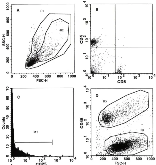
자료취합영역 설정- FSC(forward scatter), SSC (side scatter) 외 dual parameter dot plot 상에서 FSC threshold를 설정하여 적혈구나 세포 debris 등을 제외한 영역(R l)을 설정하고 이 영역내에 포함되는 5,000-10,000 개의 세포에 대한 자료를 취합하였다 (Fig. 1A). Two color 분석의 경우 FL1 로 인한 FL2로 외 bleeding을 보정한 후 자료를 취함하였다.
Lymphoblast 분석 - FSC/SSC dual parameter dot plot 상에서 크기 및 과립도가 큰 세포집단을 lym- phoblast로 간주하되 granulocyte 영역을 피하여 영 억 (R2)을 설정하고 R2/R1 외 백분율을 % lympho-
M l
^*•"1 A ■ 10^
CD25
200 400 600 800 1000 ^
FSC-H
Fig. 1 — Flow cytometrical analysis. Panel A: A FSC/SSC dual parameter dot plot showing the gates R I and R2 which encompass the total lymphocytes and lymphoblasts, respectively. Panel B: A FL1/FL2 dual parameter dot plot for analysis of CD4/CD8 ratio. The splenocytes were stained with PE-conjugated anti-mouse CD4 mAb and FITC-conjugated anti-mouse CDS mAb. Panel C; A FL2 histogram for analysis of expression of CD25 molecules. The splenocytes were stained with PE-conjugated anti-mouse CD25 mAb. Panel D- A FSC/FL2 dual parameter dot plot for differential analysis of the peritoneal exudate cells(PECs) and the sarcoma 180 cells. The ascitics cells were stained with PE-conjugated anti-mouse CD45 mAb.
blast로 분석하였다(Fig. lA).
CD4/CD8 버율 분석 - CD4 또는 CD8 분자를 인식 하는 mAb를 동시에 가하여 2중 형광염색된 세포들을 FL1/FL2 dual parameter dot plot 상에 나타내어 quardrant 분석을 시행하였다(Fig. IB).
CD25(IL-2 receptor a chain) 분석 - FL2 histx)- gram상에서 marker를 설정하여 CD25우 세포집단의
% 및 전체세포 집단외 FL2 평균형광강도(mean flu- orescence)를 분석하였다(Fig. 1C).
Sarcoma 180 세포와 PEC의 분별계수 一 일정 유속
(80 ^Li/sec)-^ 30 초동안 자료를 취합한 후 이를 FSC/FL2 dual parameter dot plot 또는 contour plot으 로 나타내어 CD45우 PEC(R3)와 CD45 sarco
ma 180 세포(R4)를 구분하여 분석하였다(Fig. ID).
결과 및 고찰
다당체 및 단백 함량 - anthrone과 Coomassie brilliant blue를 이용하여 총 다당체 및 총 단백 함량 을 측정한 걸과. GLB-A는 11.4%외 다당체 및 45.9%
영지버섯 생장점 항암 면역활성
B
m -
: r-S-: *■ • , • . •
: ;. ;;■••:.:■ ■■
■y:::가.'r./
... ...
CD8
A R1
1001.008009
0 0^ X . O S
i
o
oT -
Z
0 9
OS
0^
0€
WHJrIOO
490 오정연• 정경수
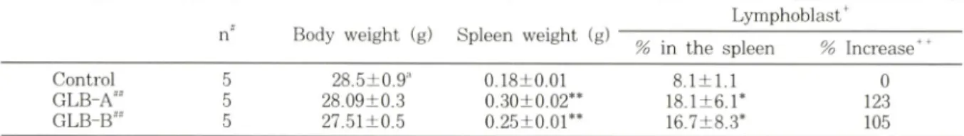
Table I ~ Lymphoblast-formation stimulatory effect of GLB-A and GLB-B on the splenic leukocytes of ICR mouse
n" Body weight (g) Spleen weight (g)
Lymphoblast^
% in the spleen % Increase"*나
Control 5 28.5±0.9^' 0.18±0.01 8.1 + 1.1 0
GLB-A^^ 5 28.09±0.3 0.30+0.02** 18.1±6.r 123
5 27.51±0.5 o.25±o.or* 16.7±8.3* 105
■ The lymphoblasts were distinguished from the small lymphocytes on the FSC/SSC dual parameter dot plot as described in materials and methods.
lOOx (T-C)/C. where T and C is % lymphoblasts of the treated group and the control group, respectively.
° Number of mice used.
테 GLB-A or GLB-B was ip injected at a dose of 50 mg/kg once daily for 3 consecutive days.
mean±S.E.
* significant at p<0.05, ** significant at p<0.01
Table II — Antitumor effect of GLB-A or GLB-B against ip-implanted sarcoma 180 cells and their induction of per
itoneal exudate cells (PEC) in ICR mice
No. of mice
Sarcoma 180 cells PEC
Number""
(x lO ' cells) % Inhibition 우우 Number'
( x l t f cells) % Increase"**
(- ) 6 127.2±5.r 0 28.5±2.1 0
GLB-A** 5 86.0±13.6* 32.4 46.7±8.6* 64.1
GLB-B= 5 100.6 + 25.7 21.0 85.1±12.r* 198.7
*The mice were ip injected with GLB-A (50 mg/kg) or GLB-B (50 mg/kg) before and afler ip implantation of sarcoma 180 (1 x l( f cells/mouse) cells.
The sarcoma 180 cells and PECs were immunofluorescence-stained with PE-conjugated anti-mouse pan- leukocyte mAb CD45. and then counted for 30 sec using a flow cytometer at the flow rate of 80 |i//sec after '" 우 100 X (Cn-Tn)/Cn. where Cn and Tn stands for the number of sarcoma 180 cells of the control group and the
treated group, respectively.
100 X (Tn-Cn)/Cn. where Cn and Tn stands for the number of the peritoneal exudate cells of the control group and the treated group, respectively.
® m e a n iS .E .
' significant at p<0.05. ' significant at p<0.01.
의 단백질을. GLB-B는 55.8%의 다당체 및 6.7%외 단백질을 함유하는 것으로 확인되었다.
정상동물의 체중 및 버장 중량에 미치는 영향 - IC R 계 생쥐의 복강에 GLB-A, GLB-K를 50 mg/kg 농도로 1 일 1 회. 3 일간 연속 투여한 후 제 5 일에 분 석한 결과 이둘 시료가 설험동물의 체중에는 유외성었 는 영향을 미치지 않았으나. 비장 중량은 두 시료 처리 군에서 모두 유외성었게 증가시켰다(Table I). 이러한 현상은 단순한 항원성 단백질의 투여에서도 관찰될 수 있다. 그러 나 GLB-A 및 GLB-B가 sarcoma 180에 대 해 항암효과를 발휘할 뿐만 아니라 CD4/CD8 비율 등 면역 활성 지표를 상승시 킨다는 점을 감안할 때 . 비 장중 량의 증가는 단순한 항원성보다는 면역활성화에 외한 결과라고 사료된다.
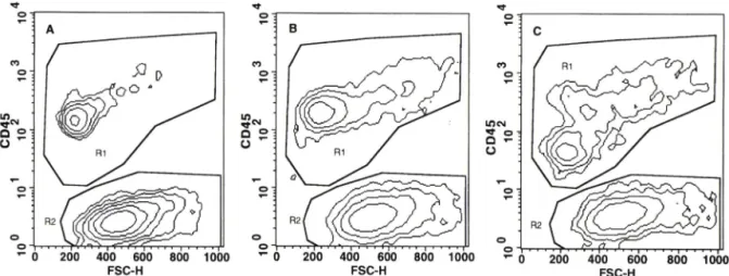
Sarcoma 180 복수암에 대한 항암 효과 - 복강내에 sarcoma 180 세포를 이식한 후 복강내 세포률 회수하 여 유세포분석 기로 분석 한 결과, GLB-A가 GLB-B보
다 우수한 항암효과를 발휘하여 sarcoma 180 복수 암 세포의 중식을 32.4% 유의성있게(p<0.05) 억제하였다 (Table II) . 이는 sarcoma 180 고형 암에 대한 GLB외 항암효과(종양저지율 81.8 % )■"•보다 낮은 수치지만, 급속히 중식하는 복수암에 대해서도 GLB-A/} 효과적 인 억제작용을 발휘할 수 있옴을 암시하는 새로운 결과 이다(Fig. 2). 한편 본 연구자*"*등은 GLB가 직접세포 득성 (cytotoxicity)을 나타내지 않음을 보고한 바 있으 며. 본 연구에서도 GLB-A 및 GLB-B가 sarcoma 180에 직접세포득성을 나타내지 않음을 확인하였다(자 료제시 생략). 이러한 결과들은 GLB-A가 숙주메개성 항암효과를 발휘하는 것을 의미한다.
Lymphoblast 생성자국 효과 - Table I에 나타낸 바 와 같이 정상동물에서 대조군의 lymphoblast 비율이 8.1% 인데 비하여 GLB-A, GLB-B 투여군에서는 각 각 18.1%, 16.7%로서 대조군에 비하여 2.23배 및 2.05배 유의성있게(p<0.05) 증가하였다. 한편 담암동물
0.72±0.16 0 4.08±0.29 0 6.87+1.24 0 1.34±0.87 86.1 5.09±0.36** 11.1 12.50±1.94** 82.0 1.02±0.32 41.7 4.48±0.35 -2.2 8.01±0.97 16.6 Control 5 3.16±0.42
GLB-A"" 5 3.87±0.43 5 3.26±0.19
^ 0 200 400 600 800 1000 FSC-H
Fig. 2 — A FSC/FL2 dual parameter dot with PE-conjugated anti-mouse
1000
plot showing sarcoma 180 cells and peritoneal exudate cells(PECs) stained panleukocyte mAb CD45. Panel A, Panel B and Panel C. respectively, shows sarcoma 180 cells and PECs of the control, the GLB-A-treated and the GLB-B-treated mice. R I and R2.
respectively, contains PECs and sarcoma 180 cells.
Table III — Effect of GLB-A and GLB-B on the CD4/CD8 ratio and the expression level of CD25 (IL-2 receptor a ___________ chain) of the splenic lymphocytes of the normal ICR mice_________________________________________
CD4/CD8 ratio** IL-2 receptor
Group Small
lymphocyte
% Increase^
Lympho- blast***
% Increase
Mean fluorescence
% Increase
% positive cells
% Increase
Number of mice used.
나 GLB-A (50 mg/kg) or GLA-B (50 mg/kg) was ip injected once daily for 3 consecutive days into IC R mice.
* The splenocytes were IF-stained with PE-conjugated anti-mouse CD4 mAb and FITC-conjugated anti-mouse CDS mAb.
The cells with higher FSC and SSC values were counted as lymphoblasts.
^ The splenic leukocytes were IF-stained with PE-conjugated anti-mouse CD25 mAb and the mean flu
orescence intensities of all the leukocytes and the percent of the CD25-positive cells were analyzed.
^ % Increase^ lOOx (T-O/C, where T and C is the values of the treated and the control group, respectively.
* significant at p<0.05. * significant at p<0.01.
Table IV — Effect of GLB-A and GLB-B on the lymphoblast formation and the CD4/CD8 ratio in the spleen of ICR mice which were ip-implanted with sarcoma 180 cells________ _____________
Lymphoblast CD4/CD8
% formation^ % Increase** ratio ^ % Increase
Control GLB-A^"
GLB-B
41.20±1.92 49.80±1.9r 50.44±3.16*
94
o
2
- 2 2
6.49±0.69 11.24+1.54*
9.41+1.27
0 73.3 45.1
^ The lymphoblast was distinguished from the small lymphcytes on the FSC/SSC dot plot.
% Increase= 100 x (T-C)/C, where T and C stands for the value of the treated and the control group, respectively.
* The splenocytes were IF-stained with PE-conjugated anti-mouse CD4 mAb and FITC-conjugated anti-mouse CD8 mAb.
' Number of mice used.
사 GLB-A (50 mg/kg) or GLB-B (50 mg/kg) was ip injected once daily for five days into ICR mice.
흡 Mean+S.E.
* significant at p<0.05. ** significant at p<0.01.
에 대해서는 GLB-A. GLB-B가 lymphoblast 비율을 각각 20.9%, 22.4% 증 가 시 켰 다(Table IV). 일반적으
로 small lymphocyte가 lymphoblast/구^로 전환되면 곧 effect cell 및 memory cell로 분화되어 체내 면역능
영지버섯 생장점 항 암 • 면역활성 491
22.
C
P
혁^ 0
C00
492 오 정 연 ■정경수
을 강화시키기 때문에 이들 시료의 lymphoblast 생성 자극 효과는 곧 면역활성의 증거로 받아들일 수 있다.
이로써 정상동물 뿐만 아니라 담암동물에서도 이돌 두 시료 모두 뚜렷한 면역활성 효과가 있음을 알 수 있다.
CD4/CD8 증가 효과 - CD다인 T„ cell은 다양한 cytokine을 분비하여 B cell, Tc cell, monocyte, m a
crophage 등을 활성화시키기 때문에 Th cell의 증가는 전반적인 면역활성화로 이어질 가능성이 높다. 정상동 물외 비장 백혈구를 small lymphocyte 영역과 lym- phoblast 영역으로 구별하여 분석한 결과, Table III에 나타낸 바와 같이 small lymphocyte 영역에서 GLB- A는 CD4/CD8 비율을 22.4% 증가시켰으며, GLB- B는 3.2% 증가시켰다. 또한 lymphoblast 영역에서 GLB-A는 CD4/CD8 비율을 86.1% 증가시켰으며 GLB-B는 41,7% 증가시켰다. lymphoblast 영역에서 CD4/CD8 비율의 증가가 더욱 두드러진 것은 lym- phoblast로 전환된 세포가 주로 CD4우 T cell이기 때 문으로 풀이 된다. 한편 담암동물에서는 GLB-A 투여로 CD4/CD8 비율이 73.3% 중가되었으며 GLB-B 투여 로 45.1% 중가되었다(Table IV). 즉 이들 시료가 정상 동물은 물론 담암동물에서도 CD4" T cell을 보다 선텍 적으로 활성화시킴을 알 수 있다. 그 중 GLB-A가 GLB-B보다 CD4/CD8 비율을 더 현저히 증가시키는 것으로 확인되었다. 그러나 담암동물의 경우, 생리식염 수 투여 대조군도 CD4/CD8 비율이 정상동물의 수치 보다 높았는데, 이는 복강내에 이식한 sarcoma 180 암 세포에 대한 면역반응으로 CD4^ T cell(TVcell)이 증 가되었기 때문이라 고찰된다.
CD25(IL-2 receptor a chain)의 발현증가 효과 - IL-2는 다양한 면역세포둘을 자극하여 활성화시키고 분화를 촉진하는 cytokine으로 T cell 등의 활성화에 중요한 역할을 한다. 한편 IL-2 receptor를 가진 세포 들은 IL-2에 의해 자극되어질 경우, CD25 분자(IL-2 receptor a chain) 외 발현을 중가시켜 더욱 활성화된 다. 따라서 CD25 분자의 발현정도를 측정함으로써 면 역활성 정도를 추정할 수 있다, 본 연구에서는 정상동물 의 비장 세포 중 CD25 분자의 발현정도를 분석한 결 과, GLB-A가 CD25^ 세포외 비율을 82.0% 증가시켰 으며, GLB-B는 16.6% 증가시켰다(Table III). 한편 GLB-A는 전체 비장 백혈구의 CD25 분자에 의한 평균 형광강도를 유의성있게 11.1% 증가시켰다.
담암동물에서의 PEC 유입 촉전 효과 - GLB-B는
PEC를 198.7%나 증가시켰으며 이는 항암효과가 보다 우수한 GLB-A의 64,1%보다 3 배 이상 높은 수치였다 (Table II). 따라서 복강유입세포 증가와 sarcoma 180 증식 억제효과는 상관관계가 인정되지 않았다.
이상의 실험 결과를 종합하여 볼 때. GLB-A와 GLB-B 모두 뚜렷한 면역 활성이 입증되었으나 이 중 GLB-A가 더욱 현저한 효과를 발휘하였다. 즉 항암효 과가 뛰어난 GLB-A가 PEG 증가 효과만을 제외한 모 든 경우 즉 lymphoblast 형성. CD4/CD8 비율 증가 효과 및 CD25 분자 발현증가 효과면에서 모두 월등히 우수하였던 사실에 비추어 주로 대식세포로 이루어진 복강세포보다는 T cell 등외 면역활성이 GLB-A의 항 암효과에 크게 기여했을 것으로 관단된다.
결 론
영지버섯 생장점으로부터 분리 • 정제한 산성 단백다 당체 분획 GLB-A 및 염기성 단백다당체 분획 GLB- B는 복강에 이식한 sarcoma 180 세포외 증식을 효과 적으로 억제하였으며 담암동물 및 정상동물에서 lym- phoblast 형성, CD4/CD8외 비율 및 CD25(IL-2 re
ceptor a chain) 의 발현을 증가시 킴으로서 강력 한 면 역증강 작용을 나타내었다. 이 중 GLB-A는 항암효과 및 면역활성화 효과가 우수하여 항암성 던역요법제로 의 개발이 기대된다.
문 헌
1) Nikonenko, B. V.. Apt, A. S., Mezhlumova, M.
B., Avdienko, V. G.. Yereneev, V. V. and Mo
roz, A. M. : Influence of the mouse Bcq, Tkxrl and xid genes on resistance and immune res
ponses to tuberculosis infection and efficacy of Bacille Calmette-Guerin (BCG) vaccination. Clin.
Exp. Im munol., 104, 37 (1996).
2) Mizuno, T., Suzuki, E., Maki, K. and Tamaki.
H, : Fraction, chemical modification and an
titumor activity of water-soluble polysaccharide of the fruiting body of Ganoderma lucidum. Nip
pon Nokeikagaku Kaishi,59, 1143 (1985).
3) Chihara, G., Hamura, J., Maeda, Y. Y., Arai, Y.
and Fukuoka, F. '■ Fraction of the polysacchari
de with antitumor activity, especially lentinan
영지버섯 생장점 항암 • 면역활성 493
from the Lentinus edodes (an edible mushroom).
Cancer Res., 30 2776 (1970).
4) Tsukagoshi, S. and Ohasi, F. : Protein bound po- lysacchride preparation. PS-K, effective against mouse sarcoma 180 and rat ascites hepatoma AH-13 by oral use. Gann. 65, 557 (1974).
5) Komatsu. N.. Okubo. S.. Likumoto. S.. Kimura, K.. Saito. G. and Sasaki. S. ; Host-mediated an
titumor action of schizophyllan. a glucan pro
duced by Schizophyllum cotiimiine. Gann. 60. 137 (1969).
6) Adachi. K. : Potentiation of host-^mediated an
titumor activity in mice by p-glucan obtained from Griofola frondosa. Chem. Pharmaceu. Bull., 35, 262 (1987).
7) Maruyama, H., Yamazaki. K.. Murofushi. S..
Konda. C. and Ikekawa, T. : Antitumor activity of Sarcodon asparatus (Berk.) S. Ito. and Canod- erma lucidum (Fr.) Karst. /. Phamacohiodyn., 12,
118 (1989).
8) Wang. G., Zhang, J., Mizuno. T.. Zhuang, C..
Hitx)shi. L. Mayuzumi. H.. Okamoto. H. and Li.
J. : Antitumor active polysaccharides from the Chinese mushroom shongshan Ling Zhi, the fruiting body of Ganoderma tsugae, Biochem.
Biotech. Biochem., 57. 894 (1993).
9) Stavinoha. W. B.. Satsangi. N. and Weintraub.
S. T. ■ Study of the anti inflammatory effect of Ganoderma lucidum. Recent advances in Ganod
erma lucidum research. The Phamaceutical So
ciety of Korea, Seoul, p. 3 (1995).
10) Zhang. L. X.. Mong. H. and Zhou, X. B. ■ Effect of Japanese Ganoderma lucidum on production of IL-2 from murine splenocytes. Chung-Kuo-Chung- Hsi-I-Chieh-Ho-Tsa-Chih. 13. 613 (1993).
11) Lieu. C. W., Lee. S. S. and Wang, S. Y. '■ The effect of Ganoderma lucidumon induction of dif
ferentiation in leukemic U937 cells. Anticancer Res., 12. 1211 (1992).
12) Kino. K.. Hem, L. G.. Vliet, J. A.. Bocken. C. F..
Hoitsma, A. J. and Tax, W. J. : Ling Zhi-8(LZ- 8) ; Studies of a new immunonodulating agent.
Transplantation, 60. 438 (1995).
13) Chung. K. S.. Kim, S. B. and Chung, S. H. ; Im- munoactivities of the protein-polysacchaiides of the tips of the carpophores of Ganoderma lucidum.
YakJmk Hoeji. 41. 105 (1997).
14) Norris. J. R. and Robbons, D. W .(Eds.) : Methods in Microbiology (Vol. 5B). Academic Press. New York. p. 209 (1971).
15) Sedmak. J. J. and Grossberg. S. E. ■ A rapid, sensitive and versatile assay for protein using Coomassie brilliant blue G250. Ami. Biochem., 79.544 (1977).
16)Reeves, J. P. and Reeves P. A. ;Current Pro
tocols in Immunology:Survival surgery, removal of the spleen and thymus. Wiley Interscience.
New York, p. 1.10.1 (1991)
17) Kuby, J. '■ Immunology. W. H. Freeman and Co..
New York. p. 57 (1994).
Vol. 42, No. 5, 1998