Available at http://www.formulastudy.com
HFS
Original Article / 원저
HepG2 세포에서 tert-butyl hydroperoxide로 유도된 간독성에 대한 kurarinone의 세포 보호 효과
김상찬
1, 이종록
2, 박숙자
2✽1
대구한의대학교 한의과대학,
2대구한의대학교 제약공학과
Cytoprotective effects of kurarinone against tert-butyl hydroperoxide-induced hepatotoxicity in HepG2 Cells
Sang Chan Kim
1, Jong Rok Lee
2, Sook Jahr Park
2✽1
College of Korean medicine, Daegu Haany University
2
Department of Pharmaceutical Engineering, Daegu Haany University
ABSTRACT
Objective : Kurarinone is one of the flavonoids isolated from Sophorae Radix with various biological activities including anti-microbial effect. In this study, we investigated the effects of Kurarinone on tert-butyl hydroperoxide (tBHP)-induced oxidative stress finally leading to apoptosis in human hepatoma cell line HepG2.
Methods : To determine the effects on cell viability, the cells were exposed to tBHP (100 μmol/l) after pretreatment with kurarinone (0.5 and 1 ㎍/㎖). Cell viability was measured by MTT assay. To reveal the possible mechanism of cytoprotectivity of kurarinone, levels of reactive oxygen species, intracellular glutathione, mitochondrial membrane potential, and expression of caspase were examined.
Results : tBHP-induced cell death was due to oxidative stress and the resulting apoptosis. Kurarinone dose-dependently protected cells from apoptosis when determined by MTT and TUNEL assay. Consistent with this observation, decreased expression of pro-caspase 3/9 protein by tBHP was restored by kurarinone. Kurarinone also showed anti-oxidative effects by inhibiting generation of ROS and depletion of GSH in tBHP-stimulated HepG2 cells. In addition, kurarinone significantly recovered disruption of mitochondrial membrane potential (MMP) as a start sign of hepatic apoptosis induced by oxidative stress.
ⓒ 2018 The Korean Medicine Society For The Herbal Formula Study
This paper is available at http://www.formulastudy.com which permits unrestricted non-commercial use, distribution, and reproduction in any medium, provided the original work is properly cited.
Conclusion : From these results, it was concluded that kurarinone protected tBHP-induced hepatotoxicity with anti-oxidative and anti-apoptotic activities. Our results suggest that kurarinone might be beneficial to hepatic disorders caused by oxidative stress.
Key words : Kurarinone, Cytoprotectivity, Oxidative stress, Apoptosis.
Ⅰ. 서론
1)인체에서 간은 물질 대사와 해독 작용에 주요한 역 할을 담당하는 장기로서 대사과정을 얻어진 대사산물 들을 필요로 하는 다른 장기로 보내는 역할을 하거나 우리 몸에 해로운 영향을 미치는 여러 물질의 해독작 용에 관여한다1). 따라서 간의 손상 및 기능 장애는 생명을 위협하는 상황을 만들 수 있다. 산화 스트레 스는 간독성과 관련된 일반적인 메커니즘 중 하나로 산화 물질과 항산화 방어 시스템 사이의 불균형으로 정의된다. 유리 라디칼 (free radical)과 같은 산화 물질은 세포막의 다중 불포화 지방산의 과산화를 유 발하여 막 파괴, 반응성 알데히드의 형성 및 환원된 글루타티온 (GSH)의 고갈을 유도해 세포를 손상시 키게 된다2). 산화 스트레스에서 과도한 활성산소종 (reactive oxygen species, ROS)의 발생은 세포 손 상 및 세포 사멸에 기여하는 것으로 알려져 있으며, 이는 괴사 (necrosis) 또는 세포 자멸사 (apoptosis) 일 수 있다3,4). 지속적인 산화 스트레스를 통한 세포 사멸의 유도는 당뇨, 심혈관 질환 및 암을 포함한 만 성적인 건강 문제의 병인을 유발한다5,6). 만성 당뇨병
7)과 심장 기능 장애8)에 산화 스트레스 신호 전달과 세포 사멸의 활성화가 관여한다는 연구 결과가 보고 되어 있다.
Tert-butyl hydroperoxide (tBHP)는 다양한 세포 에서 산화 스트레스를 유도하는 데 널리 사용되는 유 기 과산화물이다9). tBHP는 산화 스트레스를 유발하 는 두 가지 경로로 대사되는데 cytochrome P450에 의해 제공되는 첫 번째 경로에서 peroxyl 및 alkoxyl radical로 대사되어 membrane 인지질의 지방 과산화를 유발하여 membrane의 유동성과 투과성을 변화시키게 된 다10). 두 번째 경로에서 tBHP는 tert-butanol로 무해화
되고 GSH는 산화에 의해 이황화 형태 (GSSG)로 고 갈된다11). GSH의 고갈 및 mitochondrial membrane potential (MMP)의 변화는 산화 스트레스로 인한 세 포 손상을 일으키게 된다12). HepG2 cell line과 간세 포의 일차 배양에서 tBHP가 간세포 및 HepG2 세포 의 일차 배양에서 apoptosis를 통해 세포 사멸을 유 도할 수 있다는 보고가 있다13,14). 간세포의 apoptosis 는 간 섬유증의 주요한 메커니즘으로 알려져 있으며, 간 섬유화가 만성 간 기능 부전으로 진행되기 때문에 비정상적인 간세포의 apoptosis는 인간의 건강과 수 명에 큰 문제가 될 수 있다15).
Kurarinone은 고삼의 주요한 flavonoid 성분으로 HL-60 백혈병 세포에서 세포 독성 활성이 확인되었 으며16), 구강 미생물인 S. mutans 균에 대한 항균활 성이 보고되었다17). 고삼은 Sophora flavescens Aiton의 뿌리를 건조한 것으로 한의학에서는 성질이 차고 강 한 쓴맛을 지닌 약재로서 세균성 이질, 급성 위장염, 급성 전염성 감염의 치료에 쓰이고 피부 소양, 습진, 농포창, 마풍병 등을 치료하여 피부질환에 중요한 약 으로 처방되고 있다18,19). 이러한 고삼의 약효에 성분 화합물들이 관여할 것으로 기대되며, 잘 알려진 고삼 의 성분화합물인 kurarinone이 S. mutans에 대한 항균 작용에 기여함이 보고되었다17). 하지만 아직까 지 산화 스트레스에 의해 유도되는 간세포 손상에 대 한 보호 효과는 보고되지 않았다.
그러므로 본 연구에서는 kurarinone이 tBHP에 의 해 유도된 산화스트레스에 대해 간 보호 효과를 가지 는지 확인하기 위해 human hepatic cell line인 HepG2 세포에 tBHP를 처리하여 kurarinone이 세포 자멸사를 통한 세포 사멸에 미치는 영향을 조사하였 고 항산화 지표물질인 ROS와 GSH의 변화를 조사하 였다.
*Corresponding author : Sook Jahr Park. Department of Pharmaceutical Engineering, Daegu Haany University. 1, Hanuidae-ro, Gyeongsan-si, Gyeongsangbuk-do, 38610, Republic of Korea.
Tel : +82-53-819-1298, E-mail : [email protected]
∙Received : August 13, 2018 / Revised : August 28, 2018 / Accepted : August 30, 2018
Ⅱ. 재료 및 방법
1. Kurarinone의 분리 및 정제
Kurarinone의 분리 및 정제는 이18)의 방법을 활용 하여 실시하였다. 고삼 2 ㎏을 에탄올 15 ℓ에 침지 하여 상온에서 72시간 동안 추출하여 고삼 에탄올 추 출물 180 g을 얻었다. 고삼 에탄올 추출물은 silica gel column (70-230 mesh, 12 × 26 ㎝)에 loading한 후 클로로포름/메탄올의 혼합용매에서 메탄올의 비율 을 순차적으로 높여가며 (1/0→4/1) 분리를 실시하였 다. 250 ㎖씩 회수하여 얻어진 용출액은 TLC pattern에 따라 10개의 fraction으로 분획하였다. 이들 중 10%
황산에 의해 노란색으로 발색되고, UV 램프 (254 ㎚) 에서 비교적 강한 형광을 나타내는 4번 분획물을 Recycling HPLC (LC-9104, JAI, Japan)에 loading 하고 GS-310 column과 메탄올 단일 용매를 사용하 여 순수한 kurarinone으로 분리정제 한 다음 UPLC (AcQUITY UPLCTM System, Waters, UK)로 확인하 였다.
2. 세포배양
Human hepatocyte에서 유래된 HepG2 cell은 ATCC (Rockville, MD, USA)에서 구입하였으며, Dulbecco’s modified Eagle’s medium (DMEM)에 10% fetal bovine serum (FBS), 100 U/ml penicillin 및 100 μg/ml streptomycin을 혼합한 배지를 사용하여 37℃, 5%
CO2 incubator에서 배양하였다. 실험과정의 모든 cells은 80~90%의 confluence에서 실험하였고, 20 passages를 넘기지 않은 것만 사용하였다.
3. 세포 생존율 분석 (MTT assay)
HepG2 cell을 12 well plate에 1×10⁵ cells/well 로 분주한 다음 kurarinone을 농도별 (0.5, 1 ㎍/㎖) 로 처치하여 24 시간 배양한 다음에 100 μM tBHP 를 처치하여 3 시간 더 배양하였다. 배양배지를 제거 하고 MTT (0.1 ㎎/㎖)를 500 ㎕ 넣고 4시간 배양하 여 생성된 formazan crystals을 DMSO에 녹여 ELISA microplate reader (Model MCC/340, Huntsville, AL, USA)를 사용하여 570 ㎚에서 흡광도를 측정하 였다. 세포생존율은 control cell에 대한 백분율로 나 타내었다. [viability (% control) = 100 × (absorbance of treated sample)/(absorbance of control)].
4. TUNEL assay
4 well chamber slide의 well당 1×10⁵개의 농도 가 되게 세포를 배양하고 10 ㎍/㎖의 kurarinone을 12시간 처리한 후에 tBHP (100 μM)에 3시간 동안 노출시켰다. 배양세포는 PBS로 2회 washing한 후에 4% paraformaldehyde로 고정하여 In Situ cell death detection kit-POD (Roche, Mannheim, Germany)를 사용하여 kit에 포함된 protocol에 맞춰 분석하였다. 즉, paraformaldehyde로 고정된 cell을 0.3% H2O2를 사용하여 blocking시키고 0.1% triton X-100/0.1% sodium citrate로 permeabilisation 상 태로 만든 후에 TdT-enzyme이 처리된 용액으로 labeling하였다. PBS로 2회 washing한 후에 Converter- peroxisase (POD)로 37℃에서 30분간 처리하고 DAB로 발색하여 light microscope로 관찰하였다.
5. 미토콘드리아 막투과성 (mitochondrial membrane permeability, MMP) 측정
막 투과성 형광 염료인 Rh123을 사용하여 MMP를 측정하였다. 세포는 0.05 ㎎/㎖의 Rh123로 1 시간 동안 염색한 후에 트립신화하여 회수하였다. 회수된 세포는 생존율을 확인하기 위해 0.25 μg의 propidium iodide (PI)를 5분 동안 염색한 후에 flow cytometer (PAS system, Partec, Münster, Germany)로 MMP의 변화를 측정하였다.
6. ROS 생성 측정
세포 내 ROS는 DCFH-DA를 사용하여 측정하였 다. 배양 세포에 10 μM의 DCFH-DA를 37℃에서 30분 동안 처리한 후에 ELISA reader를 이용해 excitation 494 nm, emission 525 nm에서 형광강 도를 측정하였다. DCF 형광강도의 증가율은 대조군 과 비교하여 퍼센트(%)로 계산하였다.
7. GSH 측정
배양 세포를 5% metaphosphoric acid으로 용해시 킨 후에 4℃, 3,000×g에서 10분간 원심분리 하여 맑은 상등액을 회수하였다. 이 상등액은 시판 assay kit (GSH-400, OXIS International Inc., Portland, OR, USA)를 이용하여 400 ㎚에서 GSH 분석에 사용하였 다. GSH의 변화는 대조군과 비교하여 퍼센트(%)로 계산하였다.
8. Immunoblot analysis
RIPA lysis buffer를 사용하여 배양 세포로부터 total cell lysate를 준비하였고 단백질 농도는 bovine serum albumin (BSA)를 표준물질로 하여 Micro BCA protein assay reagent kit (Pierce, Rockford, IL, USA)로 결정하였다. 30 μg의 단백질 양으로 취 하여 10% gel 전기영동을 실시한 후에 nitrocellulose membrane으로 이전하였다. Nitrocellulose membrane 은 procaspase procaspase 3/9, actin 등의 1차 antibody를 가하여 반응시킨 후에 2차 antibody를 처리 하였다. 면역반응성 단백질은 ECL chemiluminescence detection kit (Amersham Biosciences, Buckinghamshire, UK)를 사용하여 확인하였다.
9. 통계적 검정
모든 실험은 3회 이상 반복 실시하였으며 실험 결
과는 mean ± S.D.로 나타내었다. 유의성 검정은 윈 도우용 SPSS 17.0을 사용하여 one way analysis of varience (ANOVA), Turkey test (multiple comparison) 방식으로 분석하였으며, p값이 0.05 미만일 때 통계 적으로 유의하다고 판정하였다.
Ⅲ. 실험결과
1. Kurarinone의 UPLC diagram
고삼 에탄올 추출물로부터 분리 정제된 kurarinone을 C18 column을 사용하여 용매A (0.1% formic acid in water)와 용매B (0.1% formic acid in acetonitrile) 를 기울기 변화로 흘려보내면서 254 ㎚에서 UPLC로 분석한 결과 단일 peak의 kurarinone을 확인할 수 있었다 (Fig. 1).
Fig. 1. UPLC chromatograms of standard (A) and purified kurarinone from Sophorae Radix (B).
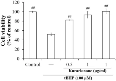
2. tBHP로 유도된 간세포 독성에 대한 kurarinone 의 세포 보호 효과
tBHP에 의한 간세포 독성에 대한 kurarinone의 보호효과를 확인하기 위해 세포에 kurarinone을 12 시간 처리한 다음 100 μM의 tBHP에 3시간 동안 노 출시킨 후에 MTT assay를 실시하였다.
그 결과 tBHP만을 처리한 세포의 생존률이 크게 감소한 반면, kurarinone 처리에 의해 생존율이 회 복되었다. Kurarinone은 단독 MTT assay에서 세포 독성을 나타내지 않은 0.5, 1 ㎍/㎖의 범위에서 실험 을 진행하였으며, tBHP로 유도된 HepG2 세포에서 농도의존적인 세포 보호 효과를 나타내었다 (Fig. 2).
Fig. 2. Hepatoprotective effect of kurarinone against tBHP-induced cell death.
HepG2 cells were pre-treated with kurarinone (0.5∼1 μg/ml) for 12 h, and then the cells were exposed to tBHP (100 μM) for another 3 h. Data represent the mean ± S.D. of three separate experiments (#, significant as compared to tBHP control; ##p<0.01).
3. tBHP로 유도된 세포자멸사 (apoptosis)에 대한 kurarinone의 세포 보호 효과
tBHP에 의해 유도된 세포 손상이 apoptosis에 기 인하는지를 확인하고 apoptosis에 대한 kurarinone 의 효과를 살펴보기 위하여 TUNEL assay를 실시하 였다. Fig. 3에서 보는 바와 같이 control에서는 세 포의 밀도가 높고 TUNEL stain된 세포 (화살표로 표시된 dark brown spot)가 거의 관찰되지 않았으나 tBHP에 의하여 현저하게 증가됨을 확인하였다. 따라 서 tBHP에 의한 HepG2 세포의 사멸이 apoptosis에 기인함을 알 수 있었다. Kurarinone (1 μg/ml)은 세포 밀도의 감소를 억제하였고 TUNEL stain에 양
성인 세포 수를 감소시켜 anti-apoptotic activity를 나타내었다 (Fig. 3A).
Apoptosis에서 주요한 역할을 담당하는 caspase는 proenzyme 형태로 존재하다 세포 신호에 의해 proteolysis되고 cleaved form으로 activation된다.
Western blot을 실시하여 tBHP에 의한 procaspase 3/9의 발현에 어떤 변화가 일어나는지 살펴보고 이러 한 변화를 kurarinone이 회복시킬 수 있는지 조사하 였다. 그 결과, tBHP는 control과 비교하여 procaspase 3/9 단백질의 발현을 감소시켰으며 kurarinone은 농 도의존적으로 단백질 발현을 증가시켰다 (Fig. 3B).
Fig. 3. Protective effect of kurarinone against tBHP-induced apoptotic cell death.
(A) Apoptotic cells were detected by TUNEL staining. Arrows indicate TUNEL positive cells.
(B) Expressions of procaspase 3/9 proteins were determined by Western blotting analysis.
Actin protein was used as internal control.
4. tBHP로 유도된 산화 스트레스에 대한 kurarinone의 항산화 효과
Kurarinone의 항산화 효과를 확인하기 위해 활성 산소종 (ROS)의 생성과 감소된 글루타티온 (GSH)의 농도를 biomarker로 사용하였다. 세포가 kurarinone 으로 12시간 동안 전처리되었을 때 tBHP에 의해 유
도된 ROS 생성이 유의하게 감소되었다 (Fig. 4A).
또한 kurarinone은tBHP에 의한 GSH의 감소도 막을 수 있었다 (Fig. 4B). 이러한 결과는 kurarinone이 HepG2 세포에서 tBHP에 의한 산화 스트레스에 중요 한 항산화 효과를 가질 수 있음을 보여준다.
Fig. 4. Anti-oxidative effects of kurarinone on ROS generation and intracellular GSH levels.
(A) ROS were detected using DCFH-DA, which emits green fluorescence in the presence of ROS. (B) The amount of GSH was calculated as nmol GSH per mg protein and is presented as a percentage of the control value. Data represent the mean ± S.D. of three separate experiments(#, significant as compared to tBHP control; ##p<0.01).
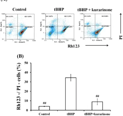
5. tBHP로 유도된 MMP 감소에 대한 kurarinone의 효과
세포에서 에너지 생산에 역할을 담당하는 미토콘트 리아의 막 손상, 즉 MMP의 감소는 세포 사멸을 유 도하는 중요한 매개 변수 중 하나이다. tBHP도 HepG2 세포13)와 쥐의 간세포14)에서 MMP를 감소시킴으로써 세포 사멸을 유도한다고 보고되어져 있다. 따라서 본
실험에서는 MMP에 직접적으로 비례하여 세포에 축 적되는 형광 염료인 Rh123과 죽은 세포를 감시하는 데 사용되는 PI 염료를 이용하여 MMP의 변화에 대 한 kurarinone의 효과를 조사하였다. 그 결과, control (Rh123+/PI-)과 비교해 tBHP에 의해 현저하게 감소 된 MMP (Rh123-/PI-)가 kurarinone에 의해 회복 됨을 확인할 수 있었다 (Fig. 5).
Fig. 5. Flow cytometric analysis of MMP using Rh123/PI staining.
(A) The lower right quadrant (Rh123+/PI-) of each graph represents normal cells with proper mitochondrial function, while the lower left quadrant (Rh123-/PI-) indicates injured cells with decresed MMP. (B) Percent change in loss of MMP (Rh123-/PI-). Data represent the mean ± S.D. of three separate experiments(#, significant as compared to tBHP control;
##p<0.01).
Ⅳ. 고찰
본 연구는 tBHP로 유발된 산화 스트레스와 이로 인한 세포자멸사에 대한 kurarinone의 세포 보호 효 과를 확인하기 위해 수행되었다. 산화 스트레스는 염 증성 질환, 암, 당뇨병과 같은 다양한 질환의 발병에 관여되어 있으며 신장, 간과 같은 주요 기관의 손상
에도 영향을 준다5-8). 특히, 생체 내에서 해독작용에 관여하는 간은 산화 스트레스로 인한 손상이 간 섬유 화와 간 기능 부전으로 진행될 수 있어 생명에 큰 지 장을 초래할 수 있다15). 이 연구에서는 human hepatic cell line인 HepG2 세포 시스템을 활용하여 ROS 증 가가 잘 나타나고 GSH를 고갈시켜 세포 손상과 세포 사멸을 일으키는 tBHP를 산화 스트레스 유도제로 사 용하였다. Kurarinone은 tBHP에 의해 감소된 HepG2
세포의 생존율을 통계적으로 유의하게 증가시켰고, 0.5와 1 μg/ml의 농도에서 농도의존적인 세포 보호 효과를 나타내었다.
자연적으로 발생하는 세포자멸사는 손상된 세포의 제거에 중요하지만19), 산화 스트레스에 의한 세포 사 멸은 해롭고 생물학적으로 부정적인 결과를 초래한다
20). tBHP에 의한 세포의 사멸은 산화적 DNA 손상 및 미토콘드리아 막 잠재력의 파괴에 의해 일어나기 도 하며21), 과도하게 생성된 ROS가 항산화 효소에 의해 제거되지 못할 경우에 세포자멸사를 통한 세포 사멸을 유도한다고 보고되었다9). 본 연구에서도 TUNEL assay를 실시한 결과, tBHP에 의한 세포자멸사를 관 찰하였으며 kurarinone이 세포자멸사에 대한 억제 효과를 나타냄을 확인하였다. Caspase는 세포자멸사 과정에서 핵심적인 역할을 담당하는 단백질로, 처음 에는 비활성 상태의 procaspase로 존재하다가 죽음 수용체 또는 세포자멸사 신호에 의해 급속히 분해되고 활 성화된다22). Kurarinone은 tBHP에 감소된 procaspase 3와 9 단백질의 발현을 증가시켰다. tBHP에 의한 procaspase의 발현 감소는 caspase 3와 9의 활성화 를 의미하기 때문에 kurarinone이 caspase의 활성화 과정을 방해함으로써 세포자멸사를 감소시킨 것으로 사료된다. 하지만 tBHP에 의한 apoptosis에서 좀 더 명확한 caspase의 역할을 설명하기 위해서는 향후의 실험에서 procaspase 뿐만 아니라 cleaved된 active form에 대한 Western blot도 실시할 필요가 있다.
Kurarinone의 tBHP에 의한 세포자멸사 억제효과와 산화 스트레스에 대한 항산화 활성 사이의 연관성을 살펴보기 위해 세포내 ROS의 생성과 GSH의 변화를 조사하였다. 간의 미토콘드리아는 catalase가 부족하 기 때문에 GSH가 산화 스트레스로부터 미토콘드리아 를 보호하는 데 중요한 역할을 한다23). 본 연구에서 tBHP는 과도한 ROS의 생성과 GSH의 고갈을 일으킴 으로써 산화 스트레스를 유도하였으나 kurarinone에 의해 ROS 생성은 감소하고 GSH 수준은 증가되어 세 포내 산화 방어 시스템이 회복되었다. 이러한 결과는 kurarinone의 세포자멸사 억제 효과와 항산화 효과 사이에 큰 연관성이 있음을 나타낸다.
HepG2 세포에서 tBHP에 의한 산화 스트레스는 mitochondrial membrane potential (MMP)의 변화 를 초래할 수 있다. MMP의 변화는 caspase 활성화 보다 먼저 일어나는 것으로 알려져 있기 때문에13) 산화
스트레스와 연관된 세포자멸사에서 미토콘드리아가 중심적인 역할을 수행하고 있다고 할 수 있을 것이 다. 본 연구에서 MMP의 변화를 측정해 본 결과, tBHP 에 의한 MMP의 감소를 관찰할 수 있었으며 kurarinone 이 미토콘드리아 막 전위를 유지함으로써 세포자멸사 를 억제함을 확인하였다.
Ⅴ. 결론
본 연구에서는 산화 스트레스로 인해 유도되는 세 포자멸사에 대하여 kurarinone에 세포보호 효과를 나타내는지 살펴보았다. 그 결과, kurarinone은 tBHP에 의해 유도되는 세포자멸사를 억제하여 세포 생존율을 증가시켰다. 이러한 효과는 caspase의 활성 화 과정을 방해함으로써 나타나는 것으로 사료된다.
또한 kurarinone은 ROS의 생성과 GSH의 고갈을 억 제함으로써 항산화 활성을 나타내었으며 tBHP에 의 해 현저하게 감소된 MMP를 회복시켰다. 이상의 결 과는 kurarinone이 산화 스트레스로 유발되는 간 손 상에 활용될 수 있는 가능성을 보여주고 있다.
Acknowledgments
This work was supported by the National Research Foundation of Korea (NRF) grant funded by the Korea government (MSIP) (Grant No. 2012R1A5A2A42671316).
References
1. Ramadori G, Moriconi F, Malik I, Dudas J.
Physiology and pathophysiology of liver inflammation, damage and repair. J Physiol Pharmacol. 2008;59 Suppl 1:107-117.
2. Wu G, Fang YZ, Yang S, Lupton JR, Turner ND. Glutathione metabolism and its implications for health. J Nutr. 2004;134:489-492.
3. Castro L, Freeman BA. Reactive oxygen species in human health and disease. Nutrition 2001;17:161-165.
4. Al-Gubory KH. Mitochondria: omega-3 in the route of mitochondrial reactive oxygen
species. Int J Biochem Cell Biol. 2012;44:
1569-1573.
5. Finkel T, Holbrook NJ. Oxidant, oxidative stress and the biology of ageing. Nature.
2000;408:239-247.
6. Ames BN, Shigenaga MK, Hagen TM. Oxidants, antioxidants, and the degenerative diseases of aging. Proc Natl Acad Sci USA. 1993;
90:7915-7922.
7. Bitar MS, Al-Saleh E, Al-Mulla F. Oxidative stress-mediated alterations in glucose dynamics in a genetic animal model of type II diabetes. Life Sci. 2005;77:2552-2573.
8. Tsutsui H, Kinugawa S, Matsushima S.
Oxidative stress and heart failure. Am J Physiol Heart Circ Physiol. 2011;301(6):
H2181-90.
9. Martín C, Martínez R, Navarro R, Ruiz-Sanz JI, Lacort M, Ruiz-Larrea MB. Tert-Butyl hydroperoxide-induced lipid signaling in hepatocytes: involvement of glutathione and free radicals. Biochem Pharmacol. 2001;62:
705-712.
10. Davies MJ. Detection of peroxyl and alkoxyl radicals produced by reaction of hydroperoxides with rat liver microsomal fractions. Biochem J. 1989;257(2):603-606.
11. Nishida K, Ohta Y, Ishiguro I. Modulating role of endogenous reduced glutathione in tert-butyl hydroperoxide-induced cell injury in isolated rat hepatocytes. Free Radic Biol Med. 1997;23:453-462.
12. Cao L, Quan XB, Zeng WJ, Yang XO, Wang MJ. Mechanism of hepatocyte apoptosis. J Cell Death. 2016;9:19-29.
13. Piret JP, Arnould T, Fuks B, Chatelain P, Remacle J, Michiels C. Mitochondria permeability transition-dependent tert-butyl hydroperoxide- induced apoptosis in hepatoma HepG2 cells.
Biochem Pharmacol. 2004;67:611-620.
14. Tripathi M, Singh BK, Kakkar P. Glycyrrhizic acid modulates t-BHP induced apoptosis in
primary rat hepatocytes. Food Chem Toxicol.
2009;47:339-347.
15. Wang YP, Cheng ML, Zhang BF, Mu M, Wu J. Effects of blueberry on hepatic fibrosis and transcription factor Nrf2 in rats. World J Gastroenterol. 2010;16:2657-2663.
16. Seo OW, Kim JH, Lee KS, Lee KS, Kim JH, Won MH, Ha KS, Kwon YG, Kim YM.
Kurarinone promotes TRAIL-induced apoptosis by inhibiting NF-κB-dependent cFLIP expression in HeLa cells. Exp Mol Med. 2012;44(11):
653-664.
17. Park SJ, Lee JR, Kim SC. Antimicrobial effects of Sophorae radix extracts against oral microorganisms. Kor J Herbology. 2010;
25(2):81-88.
18. Lee HS. Isolation of antibacterial substances from sophora flavescens ait. Gyeongsang National University Master’s Thesis. 1998:20-31.
19. Kannan K, Jain SK. Oxidative stress and apoptosis. Pathophysiology. 2000;7(3):153-163.
20. Nagata S. Oxidative stress and apoptosis.
Adv Exp Med Biol. 1996;406:119-124.
21. Estaquier J, Vallette F, Vayssiere JL, Mignotte B. The mitochondrial pathways of apoptosis.
Adv Exp Med Biol. 2012;942:157-183.
22. Wang Q, Huang Y, Ni Y, Wang H, Hou Y.
siRNA targeting midkine inhibits gastric cancer cells growth and induces apoptosis involved caspase-3,8,9 activation and mitochondrial depolarization. J Biomed Sci.
2007;14(6):783-95.
23. Marí M, Morales A, Colell A, García-Ruiz C, Fernández-Checa JC. Mitochondrial glutathione, a key survival antioxidant. Antioxid Redox Signal. 2009;11:2685-2700.