ⓒ 2013 The Korean Medicine Society For The Herbal Formula Study
This paper is available at http//:www.ompak.okdanche.com which permits unrestricted non-commercial use, distribution, and reproduction in any medium, provided the original work is properly cited.
Original Article / 원저
Oryeong-san Ameliorates High Glucose-induced Mesangial Cell Proliferation
Jung Joo Yoon
1,2,3· Yun Jung Lee
1,2,3· So Min Lee
1,2,3· Dae Hwan Kim
1,2,3· Ho Sub Lee
1,2,3· Dae Gill Kang
1,2,3*1
College of Oriental Medicine ·
2
Professional Graduate School of Oriental Medicine ·
3
Hanbang Body-fluid research center, Wonkwang University
오령산에 의한 고포도당 유도 사구체간질세포 이상증식 개선효과
윤정주
1,2,3․ 이윤정
1,2,3․ 이소민
1,2,3․ 김대환
1,2,3․ 이호섭
1,2,3․ 강대길
1,2,3*1
한의과대학 생리학교실 ․
2원광대학교 한의학전문대학원 ․
3
한방체액조절연구센터
ABSTRACT
Objectives : Diabetic nephropathy is associated with morbidity and mortality of diabetes mellitus patients.
Mesangial cell proliferation is known as the major pathologic features such as glomerulosclerosis. Oryeong-san, Korean formula, is widely used for the treatment of nephrosis, edema, and uremia. Oryeong-san is composed of five herbs: Alismatis Rhizoma, Polyporus, Atractylodis Rhizoma Alba, Hoelen, and Cinnamomi Cortex.
Methods : The present study was performed to investigate potent inhibitory effect of Oryeong-san on high
glucose (HG)-induced rat mesangial cells (RMC) proliferation.
Results : RMC proliferation under 25 mM glucose was significantly accelerated compared with 5.5 mM glucose, which was inhibited by Oryeong-san in dose dependent manner. Pre-treatment of Oryeong-san induced down-regulation of cyclins/CDKs and up-regulation of CDK inhibitor, p21waf1/cip1 and p27kip1 expression. In addition, Oryeong-san reduced HG-induced RMC proliferation by suppressed the mitogen-activated protein kinase (MAPK) phospholyration such as extracellular signal regulated kinase (ERK), Jun N-terminal Kinase (JNK), and p38. Oryeong-san significantly suppressed HG-induced ROS production.
Conclusions : Oryeong-san consequently inhibited HG-induced mesangial cell proliferation through the inhibition of MAPK and ROS signaling pathway. These results suggest that Oryeong-san may be effective in the treatment of renal dysfunction leading to diabetic nephropathy.
Keyword : Oryeong-san; Mesangial cell; Proliferation; Diabetic nephropathy
Ⅰ. Introduction
1)
Diabetes is the leading cause of end-stage renal failure, and much of the morbidity and mortality of diabetes can be attributed to nephropathy. The pathological changes of diabetic nephropathy include kidney hypertrophy, glomerulus and tubular basement membrane thickening, tubular interstitial fibrosis and arteriosclerosis. Increased mesangial cell pro- liferation is one of the major pathologic features in the early stage of diabetic nephropathy
1). Knowledge of the role of rat mesangial cell (RMC) in normal glomeruli and of their response to pathological stimuli is crucial to the understanding of these disease processes
2). Mesangial cells, which are contractile, smooth muscle-like cells located in the intercapillary space of the glomerular tufts, are thought to be the primary producers of mesangial extracellular matrix (ECM) constituents. The mesangial matrix is normally composed of various macromolecules, including fibronectin, laminin, collagen, and throm- bospondin, as well as various proteoglycans
3).
In many cells, transit through the G1 phase of the cell cycle and entry into the S-phase require
*Corresponding author : D. G. Kang, College of Oriental Medicine, Wonkwang University. Iksan.
․ Tel : 063-850-6933 ․ Fax : 063-850-7260
․ Email : [email protected]
∙ 접수 2013/10/16 ∙ 수정 2013/11/9 ∙ 채택 2013/11/15
the binding and activation of cyclin/CDK complexes, predominantly cyclin D1/CDK4 and cyclin E/CDK2
4). Some studies have shown a role for CDKI p21waf1/
cip1 and p27kip1 in the proliferation of renal mesangial cell
5-7). Mitogen-activated protein kinases (MAPK) including the extracellular signal- regulated protein kinase-1/2 (ERK 1/2), stress- activated c-Jun N-amino terminal kinase (JNK), and p38 MAPK play a key role in the intracellular signal transduction cascade to integrate the transcription of genes responsible for a variety of cellular responses relevant to diabetic nephropathy such as cell growth, differentiation, and ECM synthesis
8). There are few data on the effects of pharmacological intervention on mesangial cell proliferation in diabetic nephropathy.
Oryeong-san (Wulingsan), composed of five herbs:
Alismatis Rhizoma, Polyporus, Atractylodis Rhizoma Alba, Hoelen, and Cinnamomi Cortex, originally recorded in“Treatise on Cold Damage Diseases”
(Shanghanlun). Oryeong-san has been reported to possess renal protective effects from renal diseases such as diabetes induced renal damage
9), nephrocalcinosis
10), and adriamycin-induced nephrotic syndrome in experimental models
11). Recently, it was reported that Oryeong-san induced natriuresis and diuresis along with an inhibition of renin–
angiotensin–aldosterone system
12,13).
Therefore, the present study was performed to determine the possible effects of Oryeong-san decoction on the proliferation of primary RMC in response to high glucose (HG).
Ⅱ. Material and Method
1. Materials
D-glucose, collagenase (type IV), and all other reagents were purchased from Sigma Chemical Company (St. Louis, MO, USA). CM-H2DCFDA was purchased from Invitrogen, Inc (Eugene, OR).
Mouse monoclonal antibodies to p21
waf1/cip1, p27
kip1, JNK, ERK and p38 MAPK were obtained from Santa Cruz Biotechnology (Santa Cruz, CA, USA).
Rabbit polyclonal antibodies to CDKs, Cyclins, β -actin were purchased from Santa Cruz Biotech- nology. Goat anti-rabbit IgG and goat anti- mouse IgG were purchased from Jackson Immu- noresearch (West Grove, PA, USA).
2. Preparation of Oryeong-san
The formula of Oryeong-san consists of five herbs including Alismatis Rhizoma, Polyporus, Atractylodis Rhizoma Alba, Hoelen, and Cinnamomi Cortex were mixed according to the ratio of 5:3:3:3:1 in weight and ground into a crude powder. The Oryeong-san (281 g) was boiled with 2 L of distilled water at 100°C for 2 h. The extract was centrifuged at 990 ×g for 20 min at 4°C and resulting supernatant was lyophilized to produce a powder (65.67 g), which was then kept at -70℃
until using this experiment.
3. Cell cultures
Rat mesangial cell were isolated and cultured by employing a standard collagenase digestion method
14). Briefly, male Sprague-Dawley rats weighing 150-
175 g were anesthetized and their kidneys were removed. Renal cortical tissues were separated from the medulla and minced in D-Hank's balanced buffer using sterile conditions. Minced renal cortical tissues were filtered through 220, 100, and then 76 mm stainless steel mesh filters and subsequently digested in 0.1% collagenase (type IV) solution at 37℃ for 30 min. After centrifuging at 1,000 rpm/
min for 10 min at room temperature, pellets were re-suspended with 5.5 mM glucose DMEM supple- mented with 15% FBS, 100 U/mL penicillin, 100 mg/ml streptomycin, and 5 mg/ml bovine insulin.
The dispersed glomeruli were placed in 100 mm plastic dishes with the same culture medium and incubated in a humidified incubator at 37℃ under 95% air and 5% CO
2. The culture medium was changed every 3 days. Cell outgrowth from glomeruli was observed every 2-3 days after seeding, which would reach confluence after 30 days. The cells from passages 5-10 were employed in the current study.
4. Cell viability and proliferation
The cells were plated in culture flasks and incubated approximately 80% confluent. Cells were stimulated with HG in the presence or absence of indicated concentration of Oryeong-san (1-50 µ g/ml) for 24 h. The assay was dependent on the reduction of tetrazolium salt (MTT) by mitochondrial dehydrogenase of viable cells into dark violet formazan product which was then solubilized in 100 µl of Dimethyl sulphoxide (DMSO). Viability of the cells was determined using a colorimetric assay at 540 nm (Bio-Tek Instrument Inc., Vermont, USA).
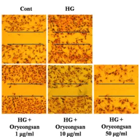
5. Migration assay
The effect of Oryeong-san on the migration of RMC was evaluated using a wound healing assay.
.Briefly, RMC were grown to confluence 6-well
culture plates. A scratch was made with a yellow
tip and then rat mesangial cell were incubated for 24 h with or without HG (25 mM) and various Oryeong-san in serum free media. After incubation, cells were washed 3 times with ice-cold PBS. The cells fixed by methyl alcohol for 5 min and cells were stained with hematoxylin and eosin. The microscopic photographs of migrated cells were kindly producted by fluorescence microscope (Axiovision 4, Zeiss, Germany). In each group, three duplicate wells were assayed, and each assay was conducted at least three times.
6. Western blot analysis
Cell homogenates (40 g of protein) were separated on 10% SDS-polyacrylamide gel electrophoresis and transferred to nitrocellulose paper. Blots were then washed with H
2O, blocked with 5%
skimmed milk powder in TBST [10 mM Tris-HCl (pH 7.6), 150 mM NaCl, 0.05% Tween-20] for 1 h and incubated with the appropriate primary antibody at dilutions recommended by the supplier. Then the membrane was washed, and primary antibodies were detected with goat anti-rabbit-IgG conjugated to horseradish peroxidase, and the bands were visualized with enhanced chemiluminescence (Amer- sham, Buckinghamshire, UK). Protein expression levels were determined by analyzing the signals captured on the nitrocellulose membranes using the Chemi-doc image analyzer (Bio-Rad, Hercules, CA, USA).
7. Determination of hydrogen peroxide
The dye CM-H2DCFDA (2´,7´-dichlorodihydro- fluorescein diacetate) has been used to measure intracellular generation of ROS. Rat mesangial cell were washed twice with phosphate buffered saline (PBS) and incubated 30 min at 37℃ with 10 µM DCF-DA (Molecular Probes, Eugene, OR, USA), a nonfluorescent compound that freely permeates cells. After being washed with cold PBS
twice, the cells were trypsinized from the culture flask. And then centrifugation at 800×g for 2 min at room temperature, the supernatant was removed. Collected cells were suspended in PBS of 500 µl. The cells were analyzed by flow cytometry (FACScalibur, BD, San Diego, CA, USA).
8. Intracellular ROS production assay
The fluorescent probe CM-H2DCFDA was used to determine the intracellular generation of ROS by HG. Briefly, the confluent RMC in the 24-well culture plates were pretreated with Oryeong-san for 24 h. After removing the Oryeong-san from the wells, RMC were incubated with 10 µM CM- H2DCFDA for 30 min. The RMC were then stimulated with HG, and the fluorescence intensity (relative fluorescence units) was measured at an excitation and emission wavelength of 485 nm and 530 nm, respectively, using a spectrofluorometer (F-2500, Hitachi, Tokyo, Japan).
9. Statistical analysis
Values were expressed as a mean ± S.E., the data were assessed by Sigma 10.0 software. Unpaired Student's t test was used for comparison between two groups. For multiple comparisons, data were analyzed by one-way ANOVA with Post Hoc Multiple Comparisons. The independent experiments were performed at least 3 times with similar results.
P<0.05 was considered as statistically significance.
III. Results
1. Effect of Oryeong-san on HG-induced mesangial cell Proliferation and migration
In the experiment to determine the effect of high
glucose (25 mM) on RMC proliferation, this study
examined cell proliferation using MTT. Exposure of
Figure 1. Effect of Oryeong-san on HG-induced cultured RMC proliferation.
RMC were incubated for 24 h with or without HG and Oryeong-san, and then cell viability and proliferation were assessed using a MTT assay. Results are expressed as the mean±S.E. from four independent experiments. *p<0.05, vs. control; #p <0.05, vs. HG alone.
RMC to 25 mM D-glucose for 24 h induced cell proliferation (Fig. 1). HG-induced increase of cell proliferation was significantly reduced by pret- reatment of Oryeong-san. To rule out the involvement of hyperosmolarity in 25 mM glucose-induced increase of cell proliferation, L-glucose and D-mannitol with an equal concentration of glucose were added to RMC. D-mannitol (25 mM) and L-glucose (25 mM) did not affect cell proliferation (data not shown).
To investigate the effect of Oryeong-san on RMC migration, a wound-healing assay was performed in which the rate of wound closure (initial width 1 mm) was measured in confluent cell monolayers.
The wounds healed linearly over 24 h in the HG- treated cells. However, the speed of the wound edge was decreased significantly in a dose-dependent manner in the Oryeong-san pretreatment cells (Fig.
2). These results demonstrate that Oryeong-san has a protective effect on cell growth and renal proliferation in diabetes.
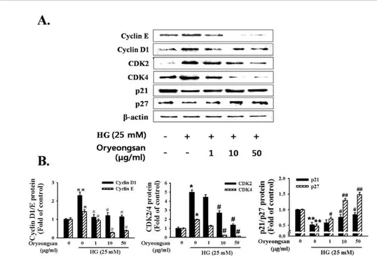
2. Effect of Oryeong-san on G1 cell-cycle related protein expression
Cell proliferation requires the coordinated inter-
Figure 2. Effect of Oryeong-san on HG-induced RMC migration.
Representative photographs of migrating cells that received either control or Oryeong-san treatment. (a) control; (b) HG alone; (c) HG with Oryeong-san (1 μg/ml); (d) HG with Oryeong-san (10 μg/ml); (e) HG with Oryeong-san (50 μg/ml).
Each assay was conducted at least three times.
action of cyclins and cyclin-dependent kinases to drive cells through the cell cycle. Cyclin-dependent kinase inhibitors can bind cyclin-CDK complexes and cause G1 arrest. Using Western blotting, we analyzed the protein expressions of the cyclins and CDKs, which are known to regulate by p21
waf1/cip1and p27
kip1, following treatment with Oryeong-san.
Incubation of RMC with Oryeong-san led to slightly decrease the levels of cyclin D1/E and CDK2/4 expression in HG status. In contrast, HG condition resulted in a reduction in the levels of p21waf1/
cip1 and p27kip1, CDK inhibitory protein. Pretreatment with Oryeong-san attenuated the HG-induced increases in cyclin D1/E, CDK2/4 and decreases in p21
waf1/cip1and p27
kip1expression levels (Fig. 3A, B).
3. Effect of Oryeong-san on HG-induced MAPKs phosphorylation
Since the activation of JNK, ERK and p38 MAPK
signaling is a key step in the proliferation process
of a variety of cell types
15). Western blot analysis
Figure 3. Effect of Oryeong-san on cell-cycle related protein expression.
(A) Cells ere stimulated with HG in the presence or absence of indicated concentration of Oryeong-san at 24 h and Western blot analysis was performed with antibodies specific for CyclinD1/E, CDK2/4, p21waf1/cip1, and p27kip1, respectively. Each electrophoretogram is representative of the results from five independent experiments. (B) Each value represents the means±S.E. of independent experiments. *p<0.05, **p<0.01 vs. control; #p <0.05, ##p <0.01 vs. HG alone.
Figure 4. Effect of Oryeong-san on HG-induced the phosphorylation of MAPK.
Cells were treated with the indicated concentrations of Oryeong-san for 24 h. And then cells were analyzed Western blot analysis for the p-ERK1/2, p-p38, p-JNK protein expressions. Each electrophoretogram is representative of the results from five independent experiments
was performed to evaluate the role of the active form (phosphorylated) of these signaling proteins in HG- nduced proliferation. As shown in Figure 4, pretreatment of Oryeong-san decreased the phosphorylation
level of JNK, ERK and p38 MAPK significantly under HG condition. These results reveal that MAPK
family take part in the regulation of RMC proliferation.
Thus, Oryeong-san improves HG-induced mesangial
cell proliferation through inhibited MAPK signaling.
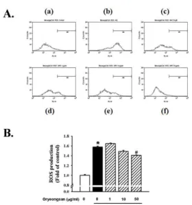
Figure 5. Effect of Oryeong-san on HG-induced ROS production.
(A) DCF-sensitive ROS assay were measured by FACS analysis. (a) control; (b) HG alone; (c) HG with NAC (50 μ M); (d) HG with Oryeong-san (1 μg/ml); (e) HG with Oryeong- san (10 μg/ml); (f) HG with Oryeong-san (50 μg/ml). (B) Alternatively, the relative ROS levels were quantified by fluorescence microplate. Each value represents the means±S.E. of three independent experiments.
*p<0.05 vs. control; #p <0.05 vs. HG alone.
4. Effect of Oryeong-san on HG-induced ROS pro- duction
To investigate the effect of Oryeong-san on HG-induced oxidative stress, cells were collected and treated under 25 mM glucose. Cells were treated with HG in the absence or presence of Oryeong-san (1-50 µg/ml). Cells were labeled with a cell-permeable fluorescent dye, CM-H2DCFDA and analyzed by flow cytometry or spectrofluorometer.
Coincident with the Fluorescence-Activated Cell Sorting Analysis, Oryeong-san was compared with N-acetyl-cysteine (NAC) (50 µM), as an anti- oxidant (Fig. 5A). The data suggest that Oryeong-san inhibited HG-induced ROS production in a concentration-dependent manner. As shown in Figure 5B, intracellular ROS levels after HG treatment were higher than untreated cells (control). However,
pretreatment with Oryeong-san significantly decreased HG-induced ROS levels.
IV. Discussion
Herb, Acupuncture and Natural Medicine (HAN), one of the most ancient and revered forms of healing, has been used to diagnose, treat, and prevent disease for over 3,000 years. HAN is now used worldwide as an effective means of overcoming disease. Oryeong-san is a well-known blended traditional herbal medicine specifically for the treatment of body-fluid disorders.
In the current study, we provided the evidence for the inhibitory effect of Oryeong-san on HG- induced RMC proliferation. Diabetic nephropathy, a progressive kidney disease caused by angiopathy of capillaries in the kidney glomeruli, is one of the most severe complications of type I and type II diabetes, and has become a major cause of end- stage renal disease
16). The earliest detectable change in the course of diabetic nephropathy is an expansion of the glomerular mesangium, which is caused by mesangial cell proliferation and excessive accumulation of ECM proteins
17). Glomerular mesangial cells, an important member of renal glomerulus, play a major role not only in physiological functions but also in pathogenesis of kidney diseases
18).
In the present study, mesangial cell numbers
were reduced by Oryeong-san treatment. In addition,
migration of rat mesangial cell was also reduced
by Oryeong-san in an in vitro mechanical injury
assay. Cell cycle is an elementary process in vital
movements of cells, and it has a close connection
with the cell proliferation, differentiation and
apoptosis. Progression of the cell cycle is regulated
by the balance between the levels and activities
of cyclin/CDK complexes and CDKIs. Some studies have shown a role for the CDKIs p21waf1/cip1 and p27kip1 in the proliferation of rat mesangial cell
5-7)