Application of Magnetic Resonance Imaging and Magnetic Resonance Angiography as Diagnostic Measures for the First
Attack of Suspected Cerebrovascular Diseases in Korea
Kunsei Lee,
1Hyeongsu Kim,
1Jae-Hyeok Heo,
2Hee-Joon Bae,
3Im-Seok Koh,
4and Sounghoon Chang
11Department of Preventive Medicine, School of Medicine, Konkuk University, Seoul;
2Department of Neurology, Seoul Medical Center, Seoul;
3Department of Neurology, Seoul National University Bundang Hospital, College of Medicine, Seoul National University, Seongnam;
4Department of Neurology, National Medical Center, Seoul, Korea.
Received: September 17, 2010 Revised: November 4, 2010 Accepted: December 6, 2010
Corresponding author: Dr. Hyeongsu Kim, Department of Preventive Medicine, School of Medicine, Konkuk University, 1 Hwayang-dong, Gwangjin-gu, Seoul 143-701, Korea.
Tel: 82-2-2030-7942, Fax: 82-2-2049-6192 E-mail: [email protected]
∙ The authors have no financial conflicts of interest.
© Copyright:
Yonsei University College of Medicine 2011 This is an Open Access article distributed under the terms of the Creative Commons Attribution Non- Commercial License (http://creativecommons.org/
licenses/by-nc/3.0) which permits unrestricted non- commercial use, distribution, and reproduction in any medium, provided the original work is properly cited.
Purpose: No precise data are available showing how magnetic resonance imaging (MRI) and magnetic resonance angiography (MRA) can be applied to diagnosis for the first attack of a suspected cerebrovascular disease in Korea. The purpose of this study was to evaluate the application level of MRI and MRA as diagnostic tools and the related factors to the use of these techniques. Materials and Meth- ods: This study used the health benefit claim data of 89,890 patients who were hospitalized for the first time due to suspected cerebrovascular disease in 2007 without having visited medical institutions as an outpatient or inpatient from 2003 to 2006. Results: Of the 89,890 cases, 28.4% took both MRI and MRA, 10.7%
took only MRI and 6.9% took only MRA. The related factors identified in the multivariate logistic regression analysis were gender, type of insurance, type of medical institution, type of department, duration of hospitalization, and type of disease. Conclusion: This study showed that the application level of MRI and MRA as diagnostic measures for the first attack of a suspected cerebrovascular diseases varied depending on several factors. It is necessary to study more accurate levels of computerized tomography (CT), computerized tomography angiography (CTA), MRI or MRA as measures to diagnose a first attack of suspected cerebro- vascular disease.
Key Words: Cerebrovascular disease, MRA, MRI
INTRODUCTION
In Korea, the aging of the population will lead to an increase in cerebrovascular disease including cerebral infarction and cerebral hemorrhage. Korea is an aging society in which the proportion of people aged 65 years or older passed 7.2% in 2000 and accounted for 10.3% of the total population in 2008. Moreover, Korea is expected to be an aged society by 2018 and a post-aged society by 2026.1 The in- cidence and prevalence of cerebrovascular disease were about 100,000 and
applied to the diagnosis of cerebrovascular disease.
This study aimed to investigate the application level of MRI and MRA as diagnostic tools for patients who are sus- pected to have a first attack of cerebrovascular disease, to assess factors related to the use of these techniques, and to provide objective evidence about the current application lev- els of MRI and MRA.
MATERIALS AND METHODS
Study subjects
The process of selecting study subjects is shown in Fig. 1.
Among 168,979 patients hospitalized in Korea with cerebro- vascular disease in 2007, 71,079 cerebrovascular disease pa- tients who had visited medical institutions as an outpatient or an inpatient from 2003 to 2006 were excluded. Moreover, 8,010 patients who were admitted to medical institutes for two days or less were also excluded because it was believed that they did not receive a routine diagnostic examination for suspected cerebrovascular disease including brain imaging, as they were transferred to other medical institutions or died.
Finally, 89,890 patients who were hospitalized for the first time due to suspected cerebrovascular disease for three days or longer in 2007 were chosen for analysis.
Methods
All health care institutions must submit health insurance claim to the Health Insurance Review and Assessment Ser- vice (HIRA) in Korea. The information on the use of MRI or MRA and other variables are included in the claim data.
This study used benefit claim data from the HIRA.
400,000, respectively, in 2004. The incidence of stroke is expected to increase to around 350,000 by 2030.2,3
The first step in treating cerebrovascular disease is an ac- curate diagnosis based on clinical symptoms, a neurologi- cal examination, and brain imaging. Brain magnetic reso- nance imaging (MRI) and computerized tomography (CT) are the most frequently used modalities for diagnosis of cerebrovascular disease.
MRI is superior to CT for detecting acute cerebral infarc- tion within several hours, including lesions in the posterior fossa, small lesions, vascular obstruction, and cerebral ede- ma.4,5 Although cerebral hemorrhage can be easily observed within six hours by both CT and MRI, MRI reveals not only clinically silent parenchymal microbleeds through gradient echo images, but also more helpful than CT in determining the causes of secondary cerebral hemorrhage such as brain tumor, vascular malformation, and aneurysm.6-9 Additional- ly, both magnetic resonance angiography (MRA) and com- puterized tomography angiography (CTA) are useful for ex- amining intracranial vessels and are helpful for diagnosing intracranial stenosis and cerebral aneurysms of over 5 mm.
However, CTA has limitations for diagnosing arterial steno- sis when wall calcification is present around the cavernous portion of the internal carotid arteries, and MRA is relatively more sensitive and specific than is CTA.10-12 Therefore, it has been suggested that both MRI and MRA should be per- formed to maximize the clinical diagnostic accuracy of cere- brovascular disease and to minimize cost.13
Although cases diagnosed by brain MRI or MRA and brain CT have been reported annually in the National Health Insurance Statistical Yearbook,14 there is no precise data showing how many brain images (MRI or MRA) can be
Patients who had visited a medical institute as an outpatient or an inpatient due
to strork between 2003 and 2006: 71,079 (total incidence
of admission: 263,317)
Patients who had stayed one or two days at first admission in 2007: 8,010 (total incidence of admission: 15,191) Patients hospitalized due
to stroke in 2007: 168, 979 (total incidence of admission:
437,727)
Patients who had not visited a medical institute between 2003 and 2006 due to stroke hospitalized in 2007: 97,900 (total incidence of admission:
174,410)
Suspected first-attack stroke patients who had stayed three
days or longer in 2007: 89,890 (total incidence of admission:
159,219)
ing in cerebral infarction), I67 (other crebrovascular dis- ease), I68 (cerebrovascular disorders in diseases classified elsewhere), and I69 (sequelae of cerebrovascular disease) were classified as cerebral infarction.
Statistical analysis
Statistical analysis was conducted using the SAS statistical program version 8.12.
A frequency analysis assessing the application of MRI and MRA in cases of stroke and its relationship to other factors was performed. Multivariate logistic regression analysis was conducted using the significant variables in the univariate analysis, and odds ratio (OR) and 95% confidence interval (CI) were calculated. A significance level of 0.05 was used for all statistical analyses.
RESULTS
MRI and MRA application
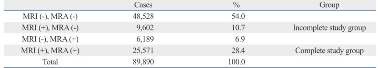
Of the 89,890 cases investigated in this study, 48,528 (54.0%) cases did not employ both MRI and MRA, whereas 25,571 (28.4%) cases involved both MRI and MRA. Only MRI was applied in 9,602 (10.7%) cases and only MRA was applied in 6,189 (6.9%) (Table 1). Therefore, the complete study group included 28.4% of the total cases, whereas the incomplete study group included 71.6%.
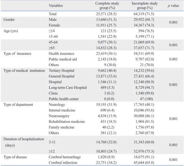
Factors related to application of both MRI and MRA The proportion of males who received a complete study (31.3%) was significantly higher than that of females (25.7%) (p<0.001). The 45-65 year group showed the high- est frequency of complete study (30.1%), whereas the levels for those 65 years or older, 14 years or younger, and 15-44 years were 28.3%, 23.5%, and 22.9%, respectively (p<0.001). The proportion of the complete study group cov- ered by health insurance was significantly higher (30.1%) than that of subjects covered by public medical aid (18.0%) (p<0.001). When level of application was compared accord- The variables presented in the data were classified as de-
pendent or independent variables based on the study pur- pose. The dependent variable was whether MRI and MRA were applied, and the independent variables were patient- related parameters (gender, age, insurance type, disease name, and duration of hospitalization) and medical institu- tion-related factors (types of medical institutions and de- partments).
The application of brain imaging was examined by re- viewing HE101 (brain MRI without contrast medium) and HE201 (brain MRI with contrast medium) in the claim data for MRI and HE135 (brain MRA without contrast medium) and HE235 (brain MRA with contrast medium) in the data for MRA. If both examinations were given, the case was classified in the complete study group, and if not, it was classified in the incomplete study group.
The subjects were divided into four age groups according to occupational activity: 14 years or younger, 15 to 44 years, 45 to 64 years, and 65 years or older.
Medical institutions were classified into tertiary hospitals, general hospitals, hospitals, long-term care facilities, clinics, and public health centers, which included health centers, community health centers, health subcenters, and health ser- vice centers.
The top five departments treating cerebrovascular disease in 2007 (internal medicine, neurology, neurosurgery, reha- bilitation medicine, and family medicine) and the others have been investigated.15
The duration of hospitalization was dichotomized based on the median hospitalization duration into less than 12 days and 12 days or longer. Stroke was classified as cere- bral hemorrhage or cerebral infarction.
Using the ICD-10 code, I60 (subarachnoid hemorrhage), I61 (intracerebral hemorrhage) and I62 (other nontraumatic intracranial hemorrhage) were classified as cerebral hemor- rhage, and I63 (cerebral infarction), I64 (stroke, not speci- fied as hemorrhage or infarction), I65 (occlusion and steno- sis of precerebral arteries, not resulting in cerebral infarction), I66 (occlusion and stenosis of cerebral arteries, not result-
Table 1. Applications of MRI and MRA for Cerebrovascular Disorders
Cases % Group
MRI (-), MRA (-) 48,528 54.0
MRI (+), MRA (-) 9,602 10.7 Incomplete study group
MRI (-), MRA (+) 6,189 6.9
MRI (+), MRA (+) 25,571 28.4 Complete study group
Total 89,890 100.0
MRI, magnetic resonance imaging; MRA, magnetic resonance angiography.
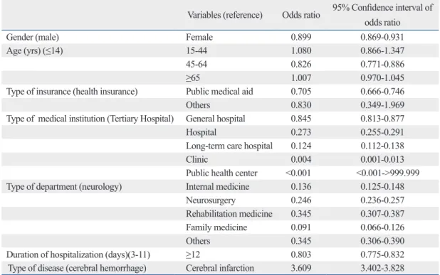
3). The OR for application of a complete study in females compared to males was 0.899 (95% CI, 0.869-0.931), and the OR of payment by public medical aid to that by health in- surance was 0.705 (95% CI, 0.666-0.746). Compared to ter- tiary hospitals, general hospitals, hospitals, long-term care hospitals, and clinics showed an OR for application of a complete study of 0.845 (95% CI, 0.813-0.877), 0.273 (95%
CI, 0.255-0.291), 0.124 (95% CI, 0.112-0.138), and 0.004 (95% CI, 0.001-0.013), respectively, and the OR for hospital- ization duration of less than 12 days compared to that of 12 days or longer was 0.803 (95% CI, 0.775-0.832). The OR of receiving a complete study for cerebral infarction compared to cerebral hemorrhage was 3.609 (95% CI, 3.402-3.828).
DISCUSSION
This study revealed that a complete study including both brain MRI and MRA as measures of diagnosing cerebro- ing to type of medical institution, tertiary hospitals showed
the highest level of complete studies (40.4%), followed by general hospitals (33.6%), hospitals (11.1%), long-term care facilities (5.3%), and clinics (0.2%) (p<0.001). The full-study application level for participants in the complete study group who were treated in departments of neurology was highest (51.9%), followed by those treated in departments of rehabil- itation medicine (18.5%), neurosurgery (13.9%), and internal medicine (6.4%) (p<0.001). The application level for the complete study group hospitalized for less than 12 days was 32.0%, and this was significantly higher than that for patients hospitalized for 12 days or longer (24.7%) (p<0.001). Cere- bral infarction also resulted in significantly higher application levels (34.2%) than did cerebral hemorrhage (8.9%) (p<0.001) (Table 2).
The factors identified in the multivariate logistic regression analysis as associated with patients’ receiving a complete study were gender, insurance type, medical institution type, department, duration of hospitalization, and diagnosis (Table
Table 2. Relationship between Levels of Application of Brain Imaging and Related Factors
Variables Complete study
group (%) Incomplete study
group (%) p value
Total 25,571 (28.5) 64,319 (71.5)
Gender Male 13,660 (31.3) 29,952 (68.7) 0.001
Female 11,911 (25.7) 34,367 (74.3)
Age (yrs) ≤14 121 (23.5) 394 (76.5)
15-44 1,541 (22.9) 5,199 (77.1)
45-64 9,077 (30.1) 21,069 (69.9)
0.001
≥65 14,832 (28.3) 37,657 (71.7)
Type of insurance Health insurance 23,419 (30.1) 54,511 (69.9)
0.001 Public medical aid 2,143 (18.0) 9,787 (82.0)
Others 9 (30.0) 21 (70.0)
Type of medical institution Tertiary Hospital 9,662 (40.4) 14,232 (59.6)
0.001 General Hospital 13,871 (33.6) 27,431 (66.4)
Hospital 1,546 (11.1) 12,340 (88.9)
Long-term Care Hospital 489 (5.3) 8,729 (94.7)
Clinic 3 (0.2) 1,540 (99.8)
Public health center 0 (0.0) 47 (100)
Type of department Neurology 19,191 (51.9) 17,765 (48.1)
0.001 Internal medicine 690 (6.4) 10,046 (93.6)
Neurosurgery 4,838 (13.9) 30,088 (86.1)
Rehabilitation medicine 431 (18.5) 1,904 (81.5) Family medicine 40 (2.2) 1,756 (97.8)
Others 381 (12.1) 2,760 (87.9)
Duration of hospitalization
(days) 3-11 14,768 (32.0) 31,343 (68.0)
0.001
≥12 10,803 (24.7) 32,976 (75.3)
Type of disease Cerebral hemorrhage 1,820 (8.9) 18,675 (91.1) 0.001
Cerebral infarction 23,751 (34.2) 45,644 (65.8)
produce accurate diagnoses. In particular, the department of neurology utilized brain imaging well. The more frequent use of full study in cerebral infarction than in cerebral hem- orrhage may be because examinations for cerebral infarc- tion are conducted more actively, as the disease is known to have a greater variety of causes and mechanisms than cere- bral hemorrhage.
Differences in the use of diagnostic measures and medi- cal service based on gender have been reported in many studies. According to a community-based research study, the frequency of X-ray was higher in females aged 65 years or older than in males of the same age range, while the fre- quency of CT and MRI was higher in males than in fe- males.16 Another study showed that females used intensive care units and selective life-supporting treatments less fre- quently than did males.17 Physicians are more likely to rec- ommend total knee arthroplasty for male patients than for female patients, with referral rates for men being nearly 22 times higher.18 Higher socioeconomic status is associated with increased use of MRI,16 and higher socioeconomic sta- tus has been demonstrated to be a significant predictor of an- giography use after acute myocardial infarction in Canada.19
This is the first study to use benefit claim data to assess the application level of MRI and MRA as measures for confirming the diagnosis in suspected first-attack cerebro- vascular disease patients observed in 2007 in Korea. This vascular disease was performed more frequently in men
than in women, in patients aged 45-64 years than in pa- tients aged less than 45 or over 65 years, in subjects cov- ered by health insurance than in those covered by public medical aid, in tertiary hospitals than in general hospitals and other institutions, in departments of neurology than in other departments, in subjects hospitalized for less than 12 days than in those hospitalized for 12 days or longer, and for cerebral infarction than for cerebral hemorrhage.
Incomplete studies were probably found more often among subjects aged 15 to 44 years because, given the association between stroke risk and age, many patients in this age range probably undergo examinations for other diagnoses, as they are not likely to be stroke patients. The difference in receipt of brain imaging according to insurance type is like- ly related to the economic burden to the hospital. The find- ing that the likelihood of a complete study was highest in tertiary hospitals, followed by general hospitals, hospitals, long-term care hospitals, clinics, and public health centers shows that relatively larger-scale medical institutions use higher-level tools to improve diagnostic accuracy of cere- brovascular disease. The departments of neurology, neuro- surgery, and rehabilitative medicine were more likely to use a complete study than were other departments, indicating that these three departments, which specialize in cerebro- vascular disease, use tools with high diagnostic value to
Table 3. Odds Ratio of Receiving a Complete Brain Imaging by Multivariate Logistic Regression Analysis of Associated Factors
Variables (reference) Odds ratio 95% Confidence interval of odds ratio
Gender (male) Female 0.899 0.869-0.931
Age (yrs) (≤14) 15-44 1.080 0.866-1.347
45-64 0.826 0.771-0.886
≥65 1.007 0.970-1.045
Type of insurance (health insurance) Public medical aid 0.705 0.666-0.746
Others 0.830 0.349-1.969
Type of medical institution (Tertiary Hospital) General hospital 0.845 0.813-0.877
Hospital 0.273 0.255-0.291
Long-term care hospital 0.124 0.112-0.138
Clinic 0.004 0.001-0.013
Public health center <0.001 <0.001->999.999
Type of department (neurology) Internal medicine 0.136 0.125-0.148
Neurosurgery 0.246 0.236-0.257
Rehabilitation medicine 0.345 0.307-0.387
Family medicine 0.091 0.066-0.126
Others 0.345 0.306-0.390
Duration of hospitalization (days)(3-11) ≥12 0.803 0.775-0.832
Type of disease (cerebral hemorrhage) Cerebral infarction 3.609 3.402-3.828
to know the true obstacles to a diagnostic approach to stroke and to find the solution for each problems.
Second, differences in accuracy of disease diagnosis and the ease of utilizing brain imaging based on the medical in- stitution were also not considered. The number of special- ists in cerebrovascular disease and the possession of equip- ment for brain imaging including brain MRI, MRA, and CT differ by medical institution. Therefore, whereas large- scaled medical institutions can diagnose a disease more ac- curately using medical staff specializing in a specific area, smaller institutions cannot do so. This is a large limitation of using benefit claim data. But this study presented the re- sults according to type of medical institution and type of department to overcome this limitation. It is also necessary to educate people to visit a medical institution capable of managing stroke when it is suspected. It is also necessary to establish the network between small and large medical in- stitutions. Recently, the establishment of nine regional re- ferral centers sepecializing in cardiac and cerebrovascular disease is in process to achieve the goal of managing all suspected stroke patients.
Third, brain CT and CTA play important roles along with MRI and MRA in diagnosing cerebrovascular disease, with CT being the primary modality of imaging method instead of MRI according to rough global guidelines by The Na- tional Institutes of Health Stroke Scale since 1995.23 We agree that many cases could have been evaluated with CT or CTA, but we were unable to get data for CT or CTA in this study. This study focused on the application level of MRI and MRA as diagnostic measures for first the attack of a suspected cerebrovascular disease. Next time, we should evaluate the diagnostic modalities for first attack of suspect- ed cerebrovascular diseases including CT, CTA or transcra- nial Doppler in addition to MRI and MRA.
ACKNOWLEDGEMENTS
This work was supported by Konkuk University in 2010.
REFERENCES
1. Korea National Statistical Office. Population Projections for Ko- rea. Seoul: 2006. p.1-56.
2. Bae HJ. Epidemiology of stroke: 2006 Update. Korean J Stroke 2007;9:5-10.
3. Rha JH. Stroke epidemiology 2007 Update. Korean J Stroke
study showed that application levels of MRI and MRA var- ied according to a variety of factors. Regardless of personal or institution-related factors, a medical environment condu- cive to the application of brain imaging to confirm suspect- ed disease is necessary to properly treat cerebrovascular disease. But it is necessary to study more accurate levels of CT, CTA, MRI or MRA as measures to diagnose cerebro- vascular disease. The designation and development of a re- gional heart and cerebrovascular disease center focusing on national university hospitals sponsored by the government would overcome the limitations reported here.20
Because this study used HIRA benefit claim data, the ac- curacy of diagnosis is not assured. However, to overcome this limitation, we excluded outpatients and inpatients who had visited medical institutions due to cerebrovascular dis- ease from 2003 to 2006, as well as patients hospitalized for a short time (1-2 days) for whom diagnosis and treatment of cerebrovascular disease were unclear. With such an oper- ational definition, the incidence of suspected cerebrovascu- lar disease cases in 2007 was estimated to be around 89,890 cases, which was lower than the estimation of Bae2 for 2004 (104,937 cases). Considering the characteristics of cerebrovascular disease, the possibility of visiting medical institutions when symptoms occur is very high, and the pos- sibility of omitting cerebrovascular disease as a primary or secondary diagnosis at first admission to medical institu- tions is very low. Similarly, the suspected incidence rate of cancer using HIRA data is not largely different from the real incidence rate in the central cancer registry database,21 and the approach used in this study included most patients with first-attack cerebrovascular disease in 2007.
This study also had the following limitations.
First, attention to a single diagnosis precluded recognition of variations in patient condition. For example, decreased consciousness, intractability, unstable vital signs, the pres- ence of medical devices such as a cardiac pacemakers, and low economic status can actually hinder MRI and MRA performance. There is no detailed data about factors hinder- ing MRI and MRA performance in Korea. However, signif- icantly higher relative risks (range 1.62-2.36) of diagnostic imaging utilization in the highest income quintile were found in pediatric and adult patient groups at all morbidity levels receiving MRI in Canada.22 Our study also identified the difference in MRI and MRA application according to type of insurance. It is also necessary to find more accurate fac- tors that may hinder MRI and MRA performance. We need
13. Fazekas F, Niederkorn K, Ebner F, Díez-Tejedor E. Relevance of neuroimaging in the evaluation of cerebral ischemia. Cerebrovasc Dis 2009;27 Suppl 1:1-8.
14. Korean Center for Disease Control and Prevention. Development of strategy for the role of community based cardio-cerebral dis- ease center. Seoul: 2009.
15. Korean Neurological Association. Role and future of neurology in geriatric medicine. Seoul: 2009.
16. Wang L, Nie JX, Tracy CS, Moineddin R, Upshur RE. Utilization patterns of diagnostic imaging across the late life course: a popu- lation-based study in Ontario, Canada. Int J Technol Assess Health Care 2008;24:384-90.
17. Fowler RA, Sabur N, Li P, Juurlink DN, Pinto R, Hladunewich MA, et al. Sex-and age-based differences in the delivery and out- comes of critical care. CMAJ 2007;177:1513-9.
18. Borkhoff CM, Hawker GA, Kreder HJ, Glazier RH, Mahomed NN, Wright JG. The effect of patients’ sex on physicians’ recom- mendations for total knee arthroplasty. CMAJ 2008;178:681-7.
19. Alter DA, Naylor CD, Austin PC, Chan BT, Tu JV. Geography and service supply do not explain socioeconomic gradients in an- giography use after acute myocardial infarction. CMAJ 2003;
168:261-4.
20. Korean Center for Disease Control and Prevention. Development of strategy for the role of community based cardio-cerebral dis- ease center. Seoul: 2009.
21. Yoon SJ, Bae SC, Lee SI, Chang H, Jo H, Sung J, et al. Measur- ing the burden of disease in Korea. J Korean Med Sci 2007;22:
518-23.
22. Demeter S, Reed M, Lix L, MacWilliam L, Leslie WD. Socioeco- nomic status and the utilization of diagnostic imaging in an urban setting. CMAJ 2005;173:1173-7.
23. Adams HP Jr, Adams RJ, Brott T, del Zoppo GJ, Furlan A, Gold- stein LB, et al. Guidelines for the early management of patients with ischemic stroke: A scientific statement from the Stroke Coun- cil of the American Stroke Association. Stroke 2003;34:1056-83.
2008;10:1-4.
4. Bryan RN, Levy LM, Whitlow WD, Killian JM, Preziosi TJ, Ro- sario JA. Diagnosis of acute cerebral infarction: comparison of CT and MR imaging. AJNR Am J Neuroradiol 1991;12:611-20.
5. Culebras A, Kase CS, Masdeu JC, Fox AJ, Bryan RN, Grossman CB, et al. Practice guidelines for the use of imaging in transient ischemic attacks and acute stroke. A report of the Stroke Council, American Heart Association. Stroke 1997;28:1480-97.
6. Fiebach JB, Schellinger PD, Gass A, Kucinski T, Siebler M, Villringer A, et al. Stroke magnetic resonance imaging is accurate in hyperacute intracerebral hemorrhage: a multicenter study on the validity of stroke imaging. Stroke 2004;35:502-6.
7. Kidwell CS, Chalela JA, Saver JL, Starkman S, Hill MD, Dem- chuk AM, et al. Comparison of MRI and CT for detection of acute intracerebral hemorrhage. JAMA 2004;292:1823-30.
8. Tsushima Y, Aoki J, Endo K. Brain microhemorrhages detected on T2*-weighted gradient-echo MR images. AJNR Am J Neuro- radiol 2003;24:88-96.
9. Dul K, Drayer BP. CT and MRI imaging of intracerebral hemor- rhage. In: Kase CS, Caplan LR, editors. Intracerebral Hemor- rhage. Boston, MA: Butterworth-Heinemann; 1994. p.73-93.
10. Alvarez-Linera J, Benito-León J, Escribano J, Campollo J, Gesto R. Prospective evaluation of carotid artery stenosis: elliptic centric contrast-enhanced MR angiography and spiral CT angiography compared with digital subtraction angiography. AJNR Am J Neu- roradiol 2003;24:1012-9.
11. Hirai T, Korogi Y, Ono K, Nagano M, Maruoka K, Uemura S, et al. Prospective evaluation of suspected stenoocclusive disease of the intracranial artery : combined MR angiogrphy and CT angiog- raphy compared with digital subtraction angiography. AJNR Am J Neuroradiol 2002;23:93-101.
12. Skutta B, Fürst G, Eilers J, Ferbert A, Kuhn FP. Intracranial steno- occlusive disease: double-detector helical CT angiography versus digital subtraction angiography. AJNR Am J Neuroradiol 1999;
20:791-9.