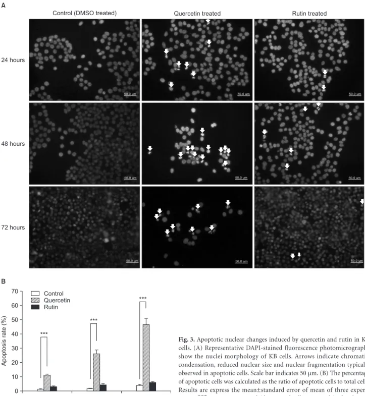
• 검색 결과가 없습니다.

Anticancer effects of quercetin on KB human oral cancer cells

N/A
N/A
Protected

Academic year: 2021

Share "Anticancer effects of quercetin on KB human oral cancer cells"

Copied!
10
0
0

로드 중.... (전체 텍스트 보기)

전체 글

(1)

Introduction

Oral cancer is the sixth most common cancer worldwide (Notani, 2000). Despite the introduction of novel therapeutic modalities, there has been only modest improvements in the long-term survival rates (Todd et al., 1997). Better knowledge of the underlying mechanisms of oral cancer are necessary to improve the survival rates, which, despite the earlier detection

of oral cancer, have not improved over the past two decades and remain among the worst of all cancer sites (Todd et al., 1997). Th e current standard of clinical care in several cancers including oral cancer involves primary surgery followed by cytotoxic chemotherapy, however undesirable side eff ects and recurrence are signifi cant problems (Tan et al., 2010).

In recent years, there has been a global trend toward the importance of naturally occurring phytochemicals in plants for the prevention and treatment of human diseases. Several of these phytochemicals have shown potential as cancer chemopreventive or therapeutic agents in the human body (Christou et al., 2001; Mukherjee et al., 2001; Pezutto, 1997;

van Poppel & van den Berg, 1997). Most of these bioactive phytochemicals exert their cancer chemotherapeutic activity

Anticancer eff ects of quercetin on KB human oral cancer cells

Seo-Yoon Kim 1 , Su-Gwan Kim 1 , Ji-Su Oh 1 , Young Ju Cha 2 , Yeon-Hee Moon 3 , Do Kyung Kim 4, *

1

Department of Oral and Maxillofacial Surgery, School of Dentistry, Chosun University, Gwangju,

2

Department of Coff ee & Chocolate, Koguryeo College, Naju,

3

Department of Dental Hygiene, Chodang University, Muan,

4

Department of Oral Physiology, School of

Dentistry, Chosun University, Gwangju, Korea

ABSTRACT

Purpose: The main aim of this study was to determine the effects of quercetin (3,5,7,3',4'-tetrahydroxyflavone) and rutin (quercetin-3-O-rutinoside) on the induction of cell growth and apoptosis in human oral cancer cells.

Materials and Methods: The effects of quercetin and rutin on cell growth and apoptosis induction in human oral cancer cells were examined using a MTT assay, nuclear staining with DAPI, immunoblotting and a determination of caspase activation in KB human oral cancer cells.

Results: Quercetin inhibited the growth of KB cells in a concentration- and time-dependent manner but rutin did not have a significant effect on the cell viability of KB cells. Treatment of KB cells with quercetin resulted in nuclear condensation and fragmentation, whereas rutin had little effect on nuclear condensation and fragmentation. Quercetin time-dependently promoted the proteolytic cleavage of procaspase-3 in KB cells with increasing amounts of cleaved caspase-3. On the other hand, rutin did not promote signifi cant proteolytic cleavage of procaspase-3. The proteolytic processing of caspase-7 in KB cells was increased by quercetin but not by rutin. Quercetin promoted the proteolytic cleavage of procaspase-9 with increases in the amount of cleaved caspase-9, whereas rutin did not. The activation of caspase-3 and -7 in living KB cells treated with quercetin was detected by fl uorescence microscopy but rutin did not lead to signifi cant caspase-3/-7 activation in the cells.

Conclusion: Quercetin strongly inhibits cell proliferation and induces apoptosis in KB human oral cancer cells, and might have potential properties for anti oral cancer drug discovery.

Key Words: Anticancer effects, Oral cancer cells, Quercetin, Rutin

Received Mar 13, 2012; Revised version received Apr 30, 2012 Accepted May 6, 2012

Corresponding author: Do Kyung Kim

Department of Oral Physiology, School of Dentistry, Chosun University, 375 Seosuk-dong, Dong-gu, Gwangju 501-759, Korea Tel: 82-62-230-6893, Fax: 82-62-224-3706

E-mail: [email protected]

(2)

by blocking cell cycle progression and triggering apoptotic cell death (Christou et al., 2001; Mukherjee et al., 2001; Pezutto, 1997; van Poppel & van den Berg, 1997). Therefore, the induction of apoptosis in cancer cells is an important indicator of the cancer treatment response by employing a bioactive substance to reduce and control human mortality due to cancer (Paschka et al., 1998; Smets, 1994).

Quercetin (3,5,7,3',4'-tetrahydroxyflavone), one of the active components of flavonoids derived from onions, fruits, buckwheat and Chinese herbs, has various benefi cial biological activities including anti-oxidant, anti-inflammation, anti- atherosclerosis and anti-tumor properties (Naderi et al., 2003; Mamani-Matsuda et al., 2006; Lin et al., 2008; Lotito et al., 2006). Some studies have shown that quercetin has anti- tumor effects through inhibiting of melanoma and prostate cancers (Zhang et al., 2000; Zhang et al., 2004). In particular, quercetin is also considered pharmacologically safe as it is a naturally occurring compound (Naderi et al., 2003; Mamani- Matsuda et al., 2006; Lotito & Frel, 2006). Quercetin in diet bioflavonoids often presents with its glycoside derivatives such as rutin (quercetin-3-O-rutinoside) (Naderi et al., 2003;

Mamani-Matsuda 2006; Lin et al., 2008; Lotito et al., 2006).

Rutin belongs to a class of plant secondary metabolites called flavonoids that are also known as rutoside, sophorin and quercetin-3-rutinoside (Janbaz et al., 2002; Joyeux et al., 1990;

Martin-Aragón et al., 2001; Liu, 1995; Ramos et al., 2008). In fact, biofl avonoids, such as quercetin, are quite identical to the composition of rutin and, hence, several experts agree that rutin and quercetin actually work in tandem by harmonizing each other (Janbaz et al., 2002; Joyeux et al., 1990; Martin- Aragón et al., 2001; Liu, 1995; Ramos et al., 2008). Although it has been shown to characterize anti-tumor activities in some tumors, their action mechanisms remain to be determined. In

addition, the eff ects and relationship of quercetin and rutin on oral cancer cells have not been analyzed yet.

In this study, therefore, the eff ect of quercetin and rutin on cell growth and mechanisms of cell death elicited by quercetin and rutin were examined in KB human oral cancer cells.

Materials and Methods

Materials

Th e KB human oral cancer cells were provided by American Type Culture Collection (ATCC, Rockville, MD, USA). Qu- ercetin, rutin (Fig. 1) and 3-[4,5-Dimethylthiazol-2-yl]-2,5- diphenyltetrazolium bromide (MTT) were supplied by Sigma (St. Louis, MO, USA). Anti-cleaved caspase-3, anti-cleaved caspase-7 and anti-cleaved caspase-9 antibodies were purchased from Cell Signaling Technology Inc. (Danvers, MA, USA).

Cell-permeable fluorogenic substrate PhiPhiLux-G1D2 was purchased from OncoImmunin Inc. (Gaithersburg, MD, USA).

Other analytical reagents were purchased based on the analytical grade.

Cell cultures

Th e KB human oral cancer cells were grown in MEM con tain- ing non-essential amino acids (NEAA) at a ratio of 100 : 1, and the media was supplemented with 5% fetal bovine serum (FBS) (Kim et al., 2008). Th e KB cells were maintained as monolayers in plastic culture plates at 37

o

C in a humidified atmosphere containing 5% CO

2

.

Inhibition of cell growth (MTT assay)

Th e cell viability test was performed according to the previ- ously described method with minor modifi cations (Hwang et al., 2007; Kim et al., 2010; Kwon et al., 2008). The cells were

Fig. 1. Chemical structures of querce-

tin (3,5,7,3',4'-tetrahydroxyflavone)

and rutin (quercetin-3-O-rutinoside).

(3)

seeded at a concentration of 5×10

3

cells/well in 24-well plates.

After 24 hours growth, the cells were treated with quercetin and rutin at various concentrations for 24-72 hours. The cell viability was assessed using MTT assay. Three separate experiments were performed for each concentration/exposure time combination.

Nuclear staining with DAPI

Nuclear staining with 40,60-diamidino-2-pheny lindole (DAPI) was performed to evaluate apoptosis. The KB cells were cultured in 24-well plates at a seeding density of 5×10

3

cells per well. Aft er 24 hours growth, the cells were treated with quercetin and rutin (0 and 30 μM) for 24-72 hours. Th e treated KB cells were fi xed with 1% paraformaldehyde for 30 minutes at room temperature and washed twice with PBS. Permeate the cells with ice-cold ethanol for 5 minutes at room temperature and washed twice with PBS. Th e fi xed cells were stained with DAPI (300 nM) for 5 minutes at room temperature in dark, washed twice with PBS and examined by fl uorescent inverted microscopy (IX71; Olympus, Tokyo, Japan).

Immunoblotting

Th e cells were treated with quercetin and rutin (0 and 30 μM) for 24 or 48 hours. Immunoblotting was performed using the previously described method with minor modifications (Choi

et al., 2010; Kim et al., 2010). Th e anti-cleaved caspase-3, anti- cleaved caspase-7 or anti-cleaved caspase-9 antibody (1 : 1,000 dilution; Cell Signaling Technology Inc.) was used as the primary antibody.

Determination of caspase activation

The activity of caspase-3/-7 was determined using the cell- permeable fluorogenic substrate PhiPhiLux-G

1

D

2

(OncoIm- munin Inc., Gaithersburg, MD, USA), which was used accor- ding to the manufacturer ’s instructions. Th e cells were treated with quercetin and rutin (30 μM) for 24 or 48 hours and incu- bated with PhiPhiLux-G

1

D

2

. The activity of caspase-3/-7 was visualized by fl uorescence microscopy (IX71).

Data analysis

All experiments were performed at least in triplicate. The

Fig. 2. Time- and concentration-dependent effects of quercetin and rutin on the cell viability in KB human oral cancer cells. The KB cells were treated with various concentrations of quercetin (A) and rutin (B) for 24 (circle), 48 (square) and 72 (triangle) hours. The cell viabilities were determined by the MTT assays. The percentage of cell viability was calculated as a ratio of A570nms of quercetin or rutin treated cells and un- treated control cells. Each data point represents the mean±standard error of mean of three experiments. **p<0.01 vs. control and ***p<0.001 vs.

control (the control cells measured in the absence of quercetin or rutin).

Table 1. Antiproliferative Effect of Quercetin in KB Cells

Time (hour) IC

50

(µM) 24

48 72

102.5±7.8 22.4±4.2 10.2±1.9

The IC

50

values represent the mean±standard error of mean for

three experiments.

(4)

results are reported as the mean±standard error of mean (SEM).

The statistical significance was analyzed using a Student's t-test for the two group comparisons and one way analysis of variance for the multi-group comparisons. A p-value <0.05 was considered signifi cant.

Results

Cytotoxic effect of quercetin and rutin in KB cells

To analyze and compare the eff ect of quercetin and rutin on the viability of KB cells, the cells were treated with quercetin

Fig. 3. Apoptotic nuclear changes induced by quercetin and rutin in KB cells. (A) Representative DAPI-stained fluorescence photomicrographs show the nuclei morphology of KB cells. Arrows indicate chromatin condensation, reduced nuclear size and nuclear fragmentation typically observed in apoptotic cells. Scale bar indicates 50 µm. (B) The percentage of apoptotic cells was calculated as the ratio of apoptotic cells to total cells.

Results are express the mean±standard error of mean of three experi-

ments. ***p<0.001 vs. control (the control cells measured in the absence

of quercetin).

(5)

and rutin at various concentrations for 24-72 hours, then MTT assay was performed. As shown in Fig. 2A, treatment of quercetin 0.1 and 0.3 μM did not significantly affect the cell viability of KB cells, but quercetin from 1 to 300 μM reduced KB cell viability. When the KB cells were treated with quercetin for 24-72 hours, quercetin inhibited the growth of KB cells in the concentration- and time-dependent manners, suggesting that quercetin induces KB cell death (Fig. 2A). Th e IC

50

values of quercetin on the cell viability aft er 24-72 hour treatments are shown in Table 1. Treatment of rutin from 0.1 to 300 μM for 24-72 hours had no signifi cant eff ect on the cell viability of KB cells (Fig. 2B).

Changes in nuclear morphology by quercetin and rutin

Th e nuclear morphological changes of KB cells by quercetin and rutin were assessed by DAPI staining. As shown in Fig.

3A, the control KB cell ’s nuclei have a normal regular and oval shape. Treatment with 30 μM quercetin for 24-72 hours resulted in nuclear condensation and fragmentation, characteristics of apoptosis. Rutin 30 μM for 24-72 hours scarcely induced nuclear condensation and fragmentation (Fig. 3A).

As quantifi ed in Fig. 3B, quercetin signifi cantly increased the apoptotic rate of KB cells to 11.0±0.89% (24 hours), 25.9±2.80%

(48 hours) and 46.7±3.98% (72 hours), whereas rutin did not cause a statistically signifi cant increase percentage of apoptotic rate (2.9±0.68%, 24 hours; 4.3±1.03%, 48 hours; 5.8±0.97%, 72 hours).

Activation of caspases by quercetin and rutin

Th e levels of cleaved caspase-3, caspase-7 and caspase-9 were examined by immunoblotting, and the activity of caspase-3/-7 was detected by fluorescence microscopy using a selective fl uorogenic substrate since caspase-3, caspase-7 and caspase-9 are eff ector caspases of apoptotic cell death (Cohen, 1997; Datta et al., 1997; Liu et al., 1997). Treatment with 30 μM quercetin for 24 or 48 hours signifi cantly promoted the proteolytic cleavages of procaspase-3 in a time-dependent manner in KB cells, with increases in the amount of cleaved caspase-3, but treatment with 30 μM rutin did not signifi cantly promote proteolytic cleavages of procaspase-3 (Fig. 4). Quercetin 30 μM for 24 or 48 hours also promoted the proteolytic cleavages of procaspase-7 in a time-dependent manner, with the increases in the amount of cleaved caspase-7, whereas rutin did not (Fig. 5). Treatment with 30 μM quercetin significantly promoted the proteolytic cleavages of procaspase-9 in a time-dependent manner, whereas treatment with 30 μM rutin did not significantly promote the

Fig. 4. Proteolytic cleavage of caspase-3 by treatments of quercetin and

rutin in KB cells. (A) Activity of cleaved caspase-3 by quercetin and rutin

in KB cells. The cells were treated with quercetin and rutin (30 µM) for

24 or 48 hours. The cell lysate was prepared and analyzed by immunob-

lotting as described in “Materials and Methods”. (B) Quantitative data

for (A) were analyzed by using Imagegauge 3.12 software after β-actin

normalization.

(6)

proteolytic cleavages of procaspase-9 (Fig. 6).

In addition, the activation of caspase-3/-7 in the quercetin- or rutin-treated KB cells was confirmed by fluorescence microscopy using fluorogenic substrate. As shown in Fig. 7,

quercetin treatment for 24 or 48 hours led to activation of caspase-3/-7 in a time-dependent manner in the living KB cells.

However, rutin 30 μM did not signifi cantly lead to activation of caspase-3/-7 in the living KB cells (Fig. 7).

Fig. 5. Proteolytic cleavage of caspase-7 by treatments of quercetin and rutin in KB cells. (A) Activity of cleaved caspase-7 by quercetin and rutin was measured in KB cells. (B) Quantitative data for (A) were analyzed by using Imagegauge 3.12 software after β-actin normalization.

Fig. 6. Proteolytic cleavage of caspase-9 by treatments of quercetin and

rutin in KB cells. (A) Activity of cleaved caspase-9 by quercetin and rutin

was measured in KB cells. (B) Quantitative data for (A) were analyzed by

using Imagegauge 3.12 software after β-actin normalization.

(7)

Discussion

Recent studies have shown that chemicals derived from natu- ral materials have been identified to elicit chemopreventive and therapeutic effects (Cheng et al., 2005; Park et al., 2005;

Tan et al., 2005). It has been reported that this effect alters various factors associated with the cell cycle and thereby induces the apoptotic cell death (Cheng et al., 2005; Hoshino et al., 1991; Park et al., 2005; Tan et al., 2005; Tian et al., 2006a).

Apoptosis, which is a major pathway of programmed cell death, plays an important role in regulating tissue development and homeostasis in eukaryotes (Green & Reed, 1998; Hengartner, 2000; Kaufmann & Hengartner, 2001). Apoptosis can occur via a death receptor-dependent extrinsic or a mitochondria- dependent intrinsic pathway, and is induced by a treatment with chemotherapeutic agents (Kaufmann & Earnshaw, 2000;

Reed, 2001).

There is a need to find new anti-cancer agents that can kill cancerous cells with minimal toxicity. Quercetin, founded from the citrus fruit such as buckwheat, onions and Chinese herbs, has been demonstrated significant anti-oxidant, anti- inflammation, anti-atherosclerosis and anti-tumor properties (Naderi et al., 2003; Mamani-Matsuda et al., 2006; Lin et al., 2008; Lotito et al., 2006). Several studies have suggested that quercetin has anti-tumor effects through inhibiting of melanoma and prostate cancers (Zhang, 2000; Zhang et al., 2004). Rutin, one of the glycoside derivatives of quercetin,

belongs to a class of plant secondary metabolites that are also known as rutoside, sophorin and quercetin-3-rutinoside (Janbaz et al., 2002; Joyeux et al., 1990; Martin-Aragón et al., 2001; Liu, 1995; Ramos et al., 2008). Although quercetin has been shown to characterize anti-tumor activities in some tumors, the mechanisms of action remain to be elucidated.

In addition, the eff ect and relationship of quercetin and rutin on oral cancer cells are not clearly established. In this study, therefore, the cytotoxic effect of quercetin and rutin and the mechanism of cell death exhibited by quercetin and rutin were examined in KB human oral cancer cells.

In MTT assay, quercetin inhibited the growth of KB cells in a concentration- and time-dependent manners, whereas rutin did not signifi cantly aff ect the cell viability of KB cells (Fig. 2).

In this study, quercetin but not rutin showed anti-proliferative eff ects in KB cells. Th ese are in agreement with previous reports that showed a generalized growth inhibitory eff ect of quercetin on several cancer cell lines (Alía et al., 2006; Nichenametla et al., 2006; Novotný et al., 2001; Tian et al., 2006b). Also, this corresponded with the results of rutin that has not anti- proliferative eff ects in vitro situation in several types of cancer cells (Lima et al., 2006; Ramos et al., 2008). These results speculated that quercetin has cytotoxicity for oral cancer cells.

In addition, these results indicated that it has potential value for anti-cancer drug discovery.

Apoptosis is an important way to maintain cellular homeo-

stasis between cell division and cell death (Green & Reed,

Fig. 7. Activation of caspase-3/-7 by treatments of quercetin and rutin in living KB cells. The cells were treated with quercetin and rutin (0 and 30

µM) for 24 or 48 hours and added specifi c cell-permeable substrate Phiphilux G

1

D

2

. Active of caspase-3/-7 was visualized by fl uorescence micros-

copy.

(8)

1998; Hengartner, 2000; Kaufmann & Hengartner, 2001). Th e induction of apoptosis in cancer cells is one of useful strategies for anti-cancer drug development (Hu & Kavanagh, 2003). So, many studies were performed for screening of apoptosis from natural material-derived compounds (Christou et al., 2001;

Mukherjee et al., 2001; Pezutto, 1997; van Poppel & van den Berg, 1997). In this study, treatment with quercetin induced nuclear condensation and fragmentation in KB cells and signifi cantly increased the apoptotic rate of KB cells, suggesting apoptotic cell death (Fig. 3). In the case of rutin, although rutin induced nuclear condensation and fragmentation scarcely, rutin did not cause a statistically signifi cant increase percentage of apoptotic rate (Fig. 3). Th ese are in agreement with the MTT assay results of this study that showed the growth inhibitory effects of quercetin but not rutin on KB cells. These results indicated that quercetin inhibits the growth of KB cells by activating cell apoptosis.

Th e activation of a family of intracellular cysteine proteases, called caspases, plays an important role in the initiation and execution of apoptosis induced by various stimuli (Datta et al., 1997; Liu et al., 1997). Among the caspases identified in mammalian cells, caspase-3, caspase-7 and caspase-9 may serve as eff ector caspases of apoptotic cell death (Cohen, 1997; Datta et al., 1997; Liu et al., 1997). Caspase-3, caspase-7 and caspase-9 are synthesized as inactive proenzymes (of sizes 32 kDa, 35 kDa and 47 kDa, respectively), which require proteolytic activation to cleaved enzymes (of sizes 19 kDa, 20 kDa and 37 kDa, respectively) (Cohen, 1997; Datta et al., 1997; Liu et al., 1997). The results of this study showed that low levels of cleaved capase-3, -7 and -9 were present in quercetin-untreated KB cells, and quercetin significantly promoted proteolytic cleavages of procaspase-3, -7 and -9 in a time-dependent manner in the KB cells, with increases in the amount of cleaved caspase-3, -7 and -9 (Figs. 4-6). Rutin, on the other hand, did not signifi cantly promote the proteolytic cleavages of procaspase-3, -7 and -9 (Figs. 4-6). In addition, the activity of caspase-3/-7 was increased by quercetin treatment in living KB cells compared to the DMSO treatment as a control, however, rutin did not significantly lead to activate the caspase-3/-7 in the KB cells (Fig. 7). Th ese corresponded with the MTT assay results and the nuclear morphological assay results of this study that showed the activation of cell apoptosis by quercetin but not rutin in KB cells. Th ese results suggest that quercetin

induces apoptotic cell death through caspase-3-, caspase-7- and caspase-9-dependent processing in the KB cells.

In this study, quercetin induced apoptotic cell death of KB cells, while rutin did not affect the cell viability of KB cells and activation of cell apoptosis. These results may be due to the lower hydrophobicity and bioavailability of rutin (Ramos et al., 2008). It has been reported that the estimated log P for quercetin is 1.48, whereas -2.02 is the value estimated for rutin in the estimates of a compound ’s hydrophobicity by using the KowWin (LogKow) software (Lima et al., 2006). Although rutin did not aff ect the cell viability of KB cells in an in vitro situation, dietary rutin can play an important role in the chemoprevention in an in vivo situation, since it is known that it can be deglycosylated to yield quercetin in the intestine by colon microflora (Kuo, 1996). However, the mechanisms of apoptosis induced by quercetin and rutin in KB cells are not yet completely understood. Further studies will reveal the precise cellular and molecular mechanisms of apoptosis induced by quercetin and rutin.

In conclusion, these in vitro results indicate that the quercetin highly inhibits cell proliferation and induces apoptosis in KB human oral cancer cells, whereas Rutin does not. Moreover, these results suggest that quercetin could be a model compound for further development of natural product-derived anti oral cancer agents. However, to elaborate this nascent possibility, further investigation of its activity including in vivo and purifi cation of bioactive compounds is now in progress.

Acknowledgments

This study was supported by research fund from Chosun University, 2012.

References

Alía M, Mateos R, Ramos S, Lecumberri E, Bravo L, Goya L:

Influence of quercetin and rutin on growth and antioxidant defense system of a human hepatoma cell line (HepG2). Eur J Nutr 45:19-28, 2006.

Cheng YL, Lee SC, Lin SZ, Chang WL, Chen YL, Tsai NM, Liu YC, Tzao C, Yu DS, Harn HJ: Anti-proliferative activity of Bupleurum scrozonerifolium in A549 human lung cancer cells in vitro and in vivo. Cancer Lett 222:183-193, 2005.

Choi HS, Cho MC, Lee HG, Yoon DY: Indole-3-carbinol induces

apoptosis through p53 and activation of caspase-8 pathway in

(9)

lung cancer A549 cells. Food Chem Toxicol 48:883-890, 2010.

Christou L, Hatzimichael E, Chaidos A, Tsiara S, Bourantas KL:

Treatment of plasma cell leukemia with vincristine, liposomal doxorubicin and dexamethasone. Eur J Haematol 67:51-53, 2001.

Cohen GM: Caspases: the executioners of apoptosis. Biochem J 326:1-16, 1997.

Datta R, Kojima H, Yoshida K, Kufe D: Caspase-3-mediated cleavage of protein kinase C theta in induction of apoptosis. J Biol Chem 272:20317-20320, 1997.

Green DR, Reed JC: Mitochondria and apoptosis. Science 281:1309-1312, 1998.

Hengartner MO: Th e biochemistry of apoptosis. Nature 407:770- 776, 2000.

Hoshino T, Hara A, Inoue M, Honda J, Imai Y, Oizumi K, Yokoyama MM: Flow cytometric measurement of NK cell cytotoxicity. J Clin Lab Immunol 36:39-43, 1991.

Hu W, Kavanagh JJ: Anticancer therapy targeting the apoptotic pathway. Lancet Oncol 4:721-729, 2003.

Hwang JH, Kim JY, Cha MR, Park HR: Eff ect of methanolic extract from silkworm droppings on proliferation and caspase activity in HT-29 human colon cancer cells. J Med Food 10:467-472, 2007.

Janbaz KH, Saeed SA, Gilani AH: Protective effect of rutin on paracetamol- and CCl4-induced hepatotoxicity in rodents.

Fitoterapia 73:557-563, 2002.

Joyeux M, Rolland A, Fleurentin J, Mortier F, Dorfman P: tert-Butyl hydroperoxide-induced injury in isolated rat hepatocytes: a model for studying anti-hepatotoxic crude drugs. Planta Med 56:171-174, 1990.

Kaufmann SH, Earnshaw WC: Induction of apoptosis by cancer chemotherapy. Exp Cell Res 256:42-49, 2000.

Kaufmann SH, Hengartner MO: Programmed cell death: alive and well in the new millennium. Trends Cell Biol 11:526-534, 2001.

Kim CS, Cho SH, Chun HS, Lee SY, Endou H, Kanai Y, Kim do K: BCH, an inhibitor of system L amino acid transporters, induces apoptosis in cancer cells. Biol Pharm Bull 31:1096- 1100, 2008.

Kim H, Moon JY, Mosaddik A, Cho SK: Induction of apoptosis in human cervical carcinoma HeLa cells by polymethoxylated flavone-rich Citrus grandis Osbeck (Dangyuja) leaf extract.

Food Chem Toxicol 48:2435-2442, 2010.

Kuo SM: Antiproliferative potency of structurally distinct dietary fl avonoids on human colon cancer cells. Cancer Lett 110:41- 48, 1996.

Kwon JI, Kim GY, Park KY, Ryu CH, Choi YH: Induction of apoptosis by linoleic acid is associated with the modulation of Bcl-2 family and Fas/FasL system and activation of caspases in AGS human gastric adenocarcinoma cells. J Med Food 11:1-8, 2008.

Lima CF, Fernandes-Ferreira M, Pereira-Wilson C: Phenolic compounds protect HepG2 cells from oxidative damage:

relevance of glutathione levels. Life Sci 79:2056-2068, 2006.

Lin CW, Hou WC, Shen SC, Juan SH, Ko CH, Wang LM, Chen YC: Quercetin inhibition of tumor invasion via suppressing PKC delta/ERK/AP-1-dependent matrix metalloproteinase-9 activation in breast carcinoma cells. Carcinogenesis 29:1807- 1815, 2008.

Liu J: Pharmacology of oleanolic acid and ursolic acid. J Ethno- pharmacol 49:57-68, 1995.

Liu X, Zou H, Slaughter C, Wang X: DFF, a heterodimeric protein that functions downstream of caspase-3 to trigger DNA fragmentation during apoptosis. Cell 89:175-184, 1997.

Lotito SB, Frei B: Dietary flavonoids attenuate tumor necrosis factor alpha-induced adhesion molecule expression in human aortic endothelial cells. Structure-function relationships and activity after first pass metabolism. J Biol Chem 281:37102- 37110, 2006.

Mamani-Matsuda M, Kauss T, Al-Kharrat A, Rambert J, Fawaz F, Thiolat D, Moynet D, Coves S, Malvy D, Mossalayi MD:

Therapeutic and preventive properties of quercetin in experimental arthritis correlate with decreased macrophage infl ammatory mediators. Biochem Pharmacol 72:1304-1310, 2006.

Martin-Aragón S, de las Heras B, Sanchez-Reus MI, Benedi J:

Pharmacological modification of endogenous antioxidant enzymes by ursolic acid on tetrachloride-induced liver damage in rats and primary cultures of rat hepatocytes. Exp Toxicol Pathol 53:199-206, 2001.

Mukherjee AK, Basu S, Sarkar N, Ghosh AC: Advances in cancer therapy with plant based natural products. Curr Med Chem 8:1467-1486, 2001.

Naderi GA, Asgary S, Sarraf-Zadegan N, Shirvany H: Anti-oxidant eff ect of fl avonoids on the susceptibility of LDL oxidation. Mol Cell Biochem 246:193-196, 2003.

Nichenametla SN, Taruscio TG, Barney DL, Exon JH: A review of the effects and mechanisms of polyphenolics in cancer. Crit Rev Food Sci Nutr 46:161-183, 2006.

Notani PN: Epidemiology and prevention of head and neck cancer:

a global view. In: Saranath D, ed. Contemporary issues in oral cancer, pp. 1-29, Oxford University Press, New Delhi, 2000.

Novotný L, Vachálková A, Biggs D: Ursolic acid: an anti-tumo- rigenic and chemopreventive activity. Minireview. Neoplasma 48:241-246, 2001.

Park DI, Lee JH, Moon SK, Kim CH, Lee YT, Cheong J, Choi BT, Choi YH: Induction of apoptosis and inhibition of telomerase activity by aqueous extract from Platycodon grandifl orum in human lung carcinoma cells. Pharmacol Res 51:437-443, 2005.

Paschka AG, Butler R, Young CY: Induction of apoptosis in prostate cancer cell lines by the green tea component, (-)-epigallocatechin-3-gallate. Cancer Lett 130:1-7, 1998.

Pezzuto JM: Plant-derived anticancer agents. Biochem Pharmacol 53:121-133, 1997.

Ramos AA, Lima CF, Pereira ML, Fernandes-Ferreira M, Pereira-

Wilson C: Antigenotoxic eff ects of quercetin, rutin and ursolic

(10)

acid on HepG2 cells: evaluation by the comet assay. Toxicol Lett 177:66-73, 2008.

Reed JC: Apoptosis-regulating proteins as targets for drug discovery. Trends Mol Med 7:314-319, 2001.

Smets LA: Programmed cell death (apoptosis) and response to anti-cancer drugs. Anticancer Drugs 5:3-9, 1994.

Tan ML, Sulaiman SF, Najimuddin N, Wang S, Muhammad TS:

Methanolic extract of Pereskia bleo (Kunth) DC. (Cactaceae) induces apoptosis in breast carcinoma, T47-D cell line. J Ethnopharmacol 96:287-294, 2005.

Tan X, Sidell N, Mancini A, Huang RP, Wang S, Horowitz IR, Liotta DC, Taylor RN, Wieser F: Multiple anticancer activities of EF24, a novel curcumin analog, on human ovarian carcinoma cells. Reprod Sci 17:931-940, 2010.

Tian Z, Chen S, Zhang Y, Huang M, Shi L, Huang F, Fong C, Yang M, Xiao P: Th e cytotoxicity of naturally occurring styryl lactones.

Phytomedicine 13:181-186, 2006a.

Tian Z, Lin G, Zheng RX, Huang F, Yang MS, Xiao PG: Anti- hepatoma activity and mechanism of ursolic acid and its derivatives isolated from Aralia decaisneana. World J Gastroenterol 12:874-879, 2006b.

Todd R, Donoff RB, Wong DT:The molecular biology of oral carcinogenesis: toward a tumor progression model. J Oral Maxillofac Surg 55:613-623, 1997.

van Poppel G, van den Berg H: Vitamins and cancer. Cancer Lett 114:195-202, 1997.

Zhang X, Xu Q, Saiki I: Quercetin inhibits the invasion and mobility of murine melanoma B16-BL6 cells through inducing apoptosis via decreasing Bcl-2 expression. Clin Exp Metastasis 18:415-421, 2000.

Zhang XM, Huang SP, Xu Q: Quercetin inhibits the invasion of

murine melanoma B16-BL6 cells by decreasing pro-MMP-9

via the PKC pathway. Cancer Chemother Pharmacol 53:82-88,

2004.

수치

Fig. 1. Chemical structures of querce- querce-tin (3,5,7,3',4'-tetrahydroxyflavone)  and rutin (quercetin-3-O-rutinoside).
Table 1. Antiproliferative Effect of Quercetin in KB Cells
Fig. 3. Apoptotic nuclear changes induced by quercetin and rutin in KB  cells. (A) Representative DAPI-stained fluorescence photomicrographs  show the nuclei morphology of KB cells
Fig. 4. Proteolytic cleavage of caspase-3 by treatments of quercetin and  rutin in KB cells
+2

참조

관련 문서

This study was conducted from 24 May to 3 November in Cheju to investigate the effects of planting date and plant distance on the grain yield of black

The purpose of this study is to analyze the effects of sports marketing strategy fit on organizational attitude and organizational citizenship behavior

Inhibitory effects of classified methanol extracts of Smilax china L on the COX-2 and iNOS expression of human colorectal cancer cell lines... Inhibitory

The purpose of this study was to examine the parents' perceptions on the current status, effects and satisfaction of personal assistant service for students

In this study, two experiments were conducted to understand the effects of additional charge on the detailed growth mechanism of Alq 3 and to determine the effect of

Fig 4 : Inhibitory effects of classified methanol extracts of Acalypha australis the COX-2 protein expression of the human oral cavity carcinoma KB cells....

The purpose of this study was to investigate the effects of sound-finding strategies based on phonological awareness on word recognition, oral reading

In the present study we investigated the proliferation effect of human oral cancer KB cell treated with pulsatilla koreana extract.. We analyzed the effects of this