원 저 Korean Circulation J 2003;33(1):58-62
심실 재분극에 대한 흡연의 급성 효과
원광대학교 의과대학 내과학교실
김남호·오석규·정진원
The Acute Effects of Cigarette Smoking on the Heterogeneity of Ventricular Repolarization in Healthy Subjects
Nam-Ho Kim, MD, Seok Kyu Oh, MD and Jin-Won Jeong, MD
Departments of Internal Medicine, Wonkwang University School of Medicine, Iksan, Korea
ABSTRACT
Background and Objectives:Although cigarette smoking is a known risk factor of sudden cardiac death, little is known about the effects of smoking on the heart. QT interval and QT dispersion prolongation have been associated with a lowered ventricular fibrillation threshold and the occurrence of sudden cardiac death.
The aim of this study was to clarify the acute effects of cigarette smoking on the QT interval and dispersion.
Subjects and Methods:The study population consist of 30 young male cigarette smokers, (with a mean age, and body mass index of 26±2 years;and 22.3±2.1 kg/m2) respectively. Standard 12-lead surface electrocar- diograms were recorded, at immediately before, during, and 5 minutes after, smoking 3 cigarettes. The RR interval, maximum and minimum QT intervals, QT dispersion (QT maximum-QT minimum), and the rate- corrected QT dispersion were all evaluated. Results:The RR interval was significantly reduced after 1 cigarette smoking (885±200 vs. 730±110 ms, p<0.01). The maximal QTBc interval was significantly longer after smoking 1 cigarette (415±21 vs. 442±21 ms, p<0.01), but the minimum QTBc interval showed no significant changes (369±23 vs. 376±22 ms, p=0.11). The QTBc dispersion (40±14 vs. 52±12 ms, p<0.01) and QTFc dispersion (42±14 vs. 58±14 ms, p<0.01) were significantly increased after smoking. Conclusion:In young males, smoking acutely increases the maximum QT interval and QT dispersion. These changes may explain the higher risk of sudden cardiac death. (Korean Circulation J 2003;33(1):58-62)
KEY WORDS:QT dispersion;Smoking;Death, sudden, cardiac.
서 론
심실 세동이나 돌연사와 같은 급성 심장 사고는 흡연 에 의해 증가되며, 특히 관상동맥 질환이 있는 경우에 더욱 증가된다.1) 그러나 그 유발기전은 잘 알려져 있지
않다.
QT 분산은 심실 재분극의 불균일성 지표자로 표준 12유도 심전도상 간단히 기록할 수 있다.2-4) QT 간격 및 분산의 증가는 교감신경계의 심장에 대한 불균형 활 성화와 연관이 있으며, 자율신경계의 긴장 정도가 중요 한 결정인자이다. 흡연은 자율신경계의 균형에 영향을 주며 장기간의 노출은 QT 간격과 QT 분산을 변화시 킨다.5-8) Ileri 등9)은 흡연자에서 비흡연자에 비하여 QT 분산이 증가함을 관찰하였다. 그러나 QT 간격 및 분산에 대한 흡연의 급성 효과는 아직까지 보고된 바 논문접수일:2002년 09월 23일
심사완료일:2002년 11월 19일
교신저자:정진원, 570-711 전북 익산시 신용동 344-2 원광대학교 의과대학 내과학교실
전화:(063) 850-1070·전송:(063) 852-8480 E-mail:[email protected]
없다. 본 연구는 건강 성인에서 흡연이 QT 간격과 분 산에 미치는 급성 효과를 알아보고자 하였다.
대상 및 방법
대상 및 방법
30명의 건강한 성인 남자(남:30, 평균연령:26±
2세, 체중질량지수:22.3±2.1 kg/m2)를 대상으로 하 였으며, 평균 5±3 갑년의 흡연력을 가지고 있었다. 당 뇨, 고혈압, 구조적인 심장 질환을 갖는 환자는 대상에 서 제외하였다. 모든 대상자들은 검사 당일에 커피나 차 등의 카페인이 포함된 음료를 제외한 가벼운 아침식 사를 실시후 평소와 같이 일상생활을 하나 무리한 신체 적 운동은 피하였고, 오전 11시에서 오후 1시 사이에 검사를 시행하였다. 담배는 1개비당 타르 7.0 mg, 니 코틴 0.75 mg을 포함하고 있는 디스®을 사용하였으며, 15분에서 20분에 걸쳐 연속하여 3개의 담배를 피우도 록 하였다.
심전도
적절한 실내온도(22~25℃)를 유지한 조용한 방에서 심전도를 기록하였다. 표준 12 유도 심전도는 25 mm/
sec의 속도로 흡연하기 직전, 1개비 흡연후, 2개비 흡연 후, 3개비 흡연후, 흡연후 5분에 각각 기록하였다. 심전 도는 검사의 목적을 알지 못하는 한 명의 순환기 의사 가 분석하였으며, 각각의 심전도는 RR 간격, QRS 축, QRS voltage(Cornell voltage10):RaVL+SV3, Soko- low-Lyon voltage11):SV1+RV5), QT 간격, 심박수 교정 QT 간격 (Bazett’s formula12):QTBc=QT/RR 1/2, Fridericia’s formula13):QTFc=QT/RR1/3)을 분석하 였다.
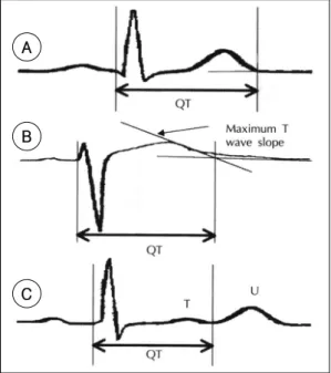
심전도 측정시 T파의 끝점은 T파가 등전위선(iso- electric line)과 만나는 점으로 정의하였고, T파의 끝 이 등전위선과 만나는 점이 명확하지 않는 경우에는 T 파의 중요 하강선과 등전위선이 만나는 점으로 하였다.
또한 U파와 T파가 겹쳐 있는 경우에는 T파와 U파의 교차점으로 정의하였다(Fig. 1).
통계분석
모든 측정값은 평균±표준오차로 기록하였으며, 통계 처리는 SPSS win 9.0을 이용하여 흡연 전·후의 연속
형 자료의 차이 검정은 paired samples t-test를 이용 하였다. p값이 0.05 미만인 경우를 통계적으로 유의한 것으로 판정하였다.
결 과
RR 간격은 흡연 전과 비교하여 1개비 흡연 후 통계 학적으로 유의하게 짧아졌으며(885±200 vs 730±
110 ms, p<0.01), 이러한 변화는 흡연 후 5분까지 지 속되었다. 그러나 담배 개수에 따른 차이는 없었다. QRS 축과 voltage의 변화는 흡연 전·후에 차이가 없었다.
최대 QTBc 간격(415±21 vs 442±21 ms, p<0.01) 및 QTFc 간격(405±14 vs 418±18 ms, p<0.01) 모 두 1개비 흡연 후 유의하게 길어졌으며, QTBc 분산(40
±14 vs 52±12 ms, p<0.01) 및 QTFc 분산(42±14 vs 58±14 ms, p<0.01) 또한 유의하게 증가하였으며, 흡연후 5분까지 지속되었다. 담배 개수에 따른 차이는 없었다. 최소 QT 간격은 흡연 전·후에 차이가 없었다 (Table 1)(Fig. 2).
Fig. 1. Main QT measurement techniques. A:from beginning of the earliest QRS complex to return of the repolarization trace to the isoelectric line, B:from beginning of the earliest QRS complex to the inte- rsection of a tangent to the downslpoe of the major repolarization wave with the isoelectric line, C:from beginning of the earliest QRS complex to the nadir between the T wave and the following U wave.
A
B
C
담배 1개비 흡연 후 QTBc 및 QTFc 분산은 각각 18
±12 ms, 16±12 ms 증가하였다. 이러한 QT 분산의 증가에 영향을 주는 인자를 분석하기 위하여 나이, 흡연
력, 흡연 전·후의 RR 간격, QRS 축, QRS voltage, 최 대 QT 간격, 그리고 최소 QT 간격의 변화를 독립변수 로 하여 다형회귀분석을 실시하였다. 이중 최대 QT 간 격 및 최소 QT 간격이 독립적인 인자로 작용하였다 (Table 2).
고 찰
QT 간격은 심실의 탈분극과 재분극 시간을 심전도 상 측정하는 것이다. 심박수로 교정된 QT 간격의 연장 은 심각한 심실 부정맥과 관련이 있고, 돌연심장사의 독립적인 위험인자로 알려져있다.14) QT 분산은 심실 재분극의 불균일성을 측정하는 하나의 방법으로 알려 져 있으며, 이러한 심실 재분극의 불균일성은 심실 빈 맥 또는 심실 세동 발생의 중요한 작용인자로 알려져왔 다.2-4) QT 분산의 증가는 심근 경색후 사망률의 증가 와 관련이 있고, 그 외에도 당뇨병, 심근허혈, 알코올중 독, 만성 간질환, 만성 신부전, 고혈압, 흡연자 등에서 QT 간격 및 분산의 증가가 보고된 바 있다.9)15-20)
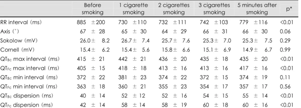
흡연은 심혈관계질환에서 조절 가능한 위험인자 중 의 하나이다. 흡연은 동맥경화과정을 악화시킬 뿐 아니 라 심실세동과 돌연사와 같은 급성 심장 사고를 유발하 며, 특히 관상동맥질환이 있는 경우에는 현저히 증가한 다고 알려져왔으나 그 발생기전은 아직 명확하지 않다.21) 심혈관계에서 흡연의 급성 효과는 주로 초기에 노르에 피네프린(norepinephrine)이 증가하고 이후로 에피네 프린이 증가하는 것으로 보아 아드레날린성 신경에서 Table 1. Mean changes of ECG parameters after smoking
Before smoking
1 cigarette smoking
2 cigarettes smoking
3 cigarettes smoking
5 minutes after smoking p*
RR interval (ms) 885.0±200 730.0±110 732.0±111 742.0±103 779.0±116 <0.01 Axis (°) 067.0±028 065.0±030 064.0±029 066.0±031 066.0±030 0.06 Sokolow (mV) 026.0±008.2 026.7±0.7.4 . 25.7±0.7.6 025.3±0.7.0 . 25.3±007.5 0.29 Cornell (mV) 015.4±006.2 015.4±0.5.6 . 15.8±0.6.6 015.1±0.6.9 . 14.9±006.7 0.99 QTBc max interval (ms) 415.0±021 442.0±021 436.0±020 435.0±018 435.0±020 <0.01 QTFc max interval (ms) 405.0±015 418.0±018 413.0±016 413.0±016 417.0±016 <0.01 QTBc min interval (ms) 372.0±022 381.0±023 374.0±022 372.0±015 374.0±019 0.11 QTFc min interval (ms) 363.0±018 360.0±021 355.0±023 354.0±017 357.0±017 0.56 QTBc dispersion (ms) 040.0±014 . 52.0±.12 . 52.0±. 16 054.0±. 15 055.0±. 14 <0.01 QTFc dispersion (ms) 042.0±014 . 58.0±.14 . 58.0±. 19 060.0±. 18 . 60.0±. 16 <0.01 Data are expressed as mean±SD. *:corresponds to a two-tailed paired t-test comparing subjects pre- and postsmoking of 1 cigarette, QTBc:corrected QT interval using Bazett's formula, QTFc:corrected QT interval using Fridericia’s formula, min:minimum, max:maximum
Table 2. Multiple regression analysis with QT Bc dispersion postsmoking as dependent variable
Variable B
Standard error of estimate
Beta Significance
Age -1.88 0.23 -0.004 0.939
ΔRR interval -2.40 0.78 -0.026 0.775
ΔQTBcmax -1.03 0.57 -1.848 0.001
ΔQT Bcmin -1.02 0.04 -1.504 0.001 Multiple γ =0.999, γ2=0.998, standard error of esti- mate 1.25, p=0.001. Beta:standardized coefficient of regression, B:regression coefficient, ΔQTBcmax:differe- nce maximum QT interval using Bazett's formula bet- ween pre- and postsmoking of 1 cigarette, ΔQTBc min: difference minimum QT interval using Bazett’s formula between pre- and postsmoking of 1 cigarette, ΔRR interval:difference of RR interval between pre- and postsmoking of 1 cigarette
Fig. 2. Linear graphs showing average changes of QT dispersion.
즉각적인 카테콜아민의 분비에 이어 부신수질에서 전 신적인 분비가 이루어지며 작용이 일어나는 것으로 생 각된다. 심근수축력과 심박동수는 베타1-수용체 자극 의 결과로 증가되어진다.22)23) 따라서 흡연은 관상동맥 의 이완능력을 급격히 감소시켜 허혈성 역치를 낮추고 이에 따라 관상동맥질환이 유발되거나 급성 심장사의 위험을 증가시키는 것으로 생각된다.24) 습관적인 흡연 은 동방결절에 대한 신경조절의 현저한 장애를 유발한 다. 흡연가는 휴식기에 미주 신경 조절의 감소와 압반 사(baroreflex) 약화, 그리고 교감신경계의 증가가 특 징이다.5-8) 이러한 것들은 관상동맥의 혈관긴장도를 조 절하는 인자인데, 좌심실에 균일하게 분포되어 있는 것 이 아니므로 흡연에 의한 자율신경계의 변화는 심근 관 류와 재분극의 불균일성을 유발시킨다. 이러한 교감신 경계의 부정맥 유발 효과는 흡연이 심혈관질환의 위험 을 증가시키는 데 기여한다. 또한 QT 분산은 특히 관 상동맥질환을 갖고 있는 환자에서 주로 아침에 증가하 는 일중변화를 보이는데 이러한 소견은 아침에 급성 심 장사나 급사의 발생에 교감신경계가 중요한 역할을 한 다는 것을 암시한다.25)
본 연구에서는 심박동수로 교정된 QT 간격을 2가지 공식에 의해서 각각 구하였다. 가장 널리 사용되어지는 공식이 Bazett 공식인데 이는 심박동수가 느리거나 빠 른 경우에는 오차가 심하여진다.26) 그래서 Fridericia 공식을 같이 사용하였다. 본 연구에서 심박동수는 대체 로 60~100/분 사이에 포함되어 있었으나 각각의 공식 에 의하여 측정된 값은 10 ms에서 20 ms에 이르는 차이가 발생하였다. 이와 같은 오차를 감안하더라도 2 가지 공식으로 교정된 QT 간격 및 분산은 흡연 직후에 똑같이 증가하는 결과를 보였다. 흡연 후 발생한 RR 간격의 감소는 흡연이 교감신경계를 항진시킴으로써 심박동수를 증가시킨 것으로 생각되어진다. 이러한 교 감신경계의 항진이 심실내 재분극의 불균일성에 영향 을 미쳤는지에 대한 연구는 아직 미비하다. 또한 흡연 후 최소 QT 간격은 큰 변화가 없으나 최대 QT 간격 의 증가가 현저하였으며, 이러한 변화는 흡연에 의한 QT 분산 증가의 주된 기전이 심실 재분극의 지연에 의 해 이루어진 것으로 생각할 수 있다. 하지만, 본 연구는 몇 가지의 제한점이 있다. 첫째, 본 연구는 오직 젊은 성인을 대상으로 한 소규모의 연구였다. 관상동맥질환 자에서 흡연은 강력한 위험인자이고, 돌연사 또한 건강
한 성인보다 관상동맥질환자에서 더 잘 발생하는 것으 로 알려져있다. 추후에는 관상동맥질환자를 포함한 대 규모의 연구가 이루어져야 할 것이다. 둘째, QT 간격 및 분산의 증가가 흡연에 의한 어떠한 기전에 의하여 이루어지는지에 대하여는 연구하지 못하였다. 즉, 흡연 이 교감신경의 항진과 관련하여 심박동수 증가가 이루 어졌는지, 아니면 저산소증에 의해 이와 같은 현상이 발생하였는지 등의 연구가 병행되어야 할 것이다. 셋째, 1개비 흡연후 증가된 RR 간격, 최대 QT 간격, QT 분 산이 2개비, 3개비로 담배의 개수를 늘려도 더 이상 변 화가 없는데 이의 기전이 명확하지 않다. 이와 같은 제 한점에도 불구하고 본 연구는 건강한 성인에서 흡연은 QT 간격 및 분산을 증가시키는 작용이 있고, 이는 심 실세동이나 돌연사와 같은 급성 심장 사고의 중요한 위 험인자로 작용할 수 있을 것으로 생각된다.
요 약
배경 및 목적:
심실세동이나 돌연사와 같은 급성 심장 사고는 흡연 에 의해 증가될되어지나 그 유발기전은 잘 알려져 있지 않다. QT 간격 및 분산의 증가는 심실세동 역치를 감 소시키며 돌연심장사의 발생과 관련이 있다. 본 연구는 건강한 성인에서 흡연이 QT 간격 및 분산에 미치는 영 향을 보고자 하였다.
방 법:
30명의 건강한 성인 남자를 대상으로 오전 11시에 서 오후 1시 사이에 심전도를 기록하였다. 표준 12유 도 심전도를 흡연직전, 1개비 흡연후, 2개비 흡연후, 3 개비 흡연후, 흡연 후 5분에 각각 기록하여 분석하였다.
QT 분산은 12유도에서 최대 QT 간격과 최소 QT 간 격의 차이로 정의하였으며, 심박동수로 교정된 QT 간 격은 Bazett 공식(QTBc=QT/RR1/2)과 Fridericia 공 식(QTFc=QT/RR1/3)을 이용하였다.
결 과:
RR 간격은 흡연전과 비교하여 1개비 흡연후부터 유의 하게 짧아졌으며(885±200 vs. 730±110 ms, p<0.01), 이는 흡연 후 5분까지도 지속되었다. 최대 QTBc 간격 (415±21 vs. 442±21 ms, p<0.01) 및 QTFc 간격 (405±14 vs. 418±18 ms, p<0.01) 모두 1개비 흡 연 후 유의하게 길어졌으며, QTBc 분산(40±14 vs.
52±12 ms, p<0.01) 및 QTFc 분산(42±14 vs. 58±
14 ms, p<0.01) 또한 유의하게 증가하였다.
결 론:
건강성인에서 흡연은 QT 간격 및 분산을 증가시키 고, 이의 증가는 심실세동이나 돌연심장사와 같은 급성 심장 사고의 위험 인자로 작용할 수 있을 것으로 생각 된다.
중심 단어:QT 분산;흡연;심인성급사.
이 논문은 2001년도 원광대학교의 교비 지원에 의해서 수 행됨.
REFERENCES
1) Hallstrom AP, Cobb LA, Ray R. Smoking as a risk factor for recurrence of sudden cardiac arrest. N Engl J Med 1986;
314:271-5.
2) Higham PD, Campbell RW. QT dispersion. Br Heart J 1994;71:508-10.
3) Pye M, Quinn AC, Cobbe SM. QT interval dispersion: a noninvasive marker of susceptibility to arrhythmia in patients with sustained ventricular arrhythmias? Br Heart J 1994;
71:511-4.
4) Coumel P, Maison-Blanche P, Badilini F. Dispersion of ventricular repolarization. Circulation 1998;97:2491-3.
5) Narkiewicz K, van de Borne PJ, Hausberg M, Cooley RL, Winniford MD, Davision DE, Somers VK. Cigarette smo- king increases sympathetic outflow in humans. Circulation 1998;98:528-34.
6) Niedermaier ON, Smith ML, Beightol LA, Zukowska-Gro- jec Z, Goldstein DS, Eckberg DL. Influence of cigarette smoking on human autonomic function. Circulation 1993;
88:562-71.
7) Celermajer DS, Sorensen KE, Georgakopoulos D, Bull C, Thomas O, Robinson J, Deanfield JE. Cigrette smoking is associated with dose-related and potentially reversible im- pairment of endothelium-dependent dilation in healthy you- ng adults. Circulation 1993;88:2149-55.
8) Lucini D, Bertocchi F, Malliani A, Pagani M. A controlled study of the autonomic changes produced by habitual ciga- rette smoking in healthy subjects. Cardiovasc Res 1996;
31:633-9.
9) Ileri M, Yetkin E, Tandogan I, Hisar I, Atak R, Senen K, Cehreli S, Demirkan D. Effect of habitual smoking on QT interval duration and dispersion. Am J Cardiol 2001;88:
322-5.
10) Casale PN, Devereux RB, Alonso DR, Campo E, Kligfield P. Improved sex-specific criteria of left ventricular hypertrophy for clinical and computer interpretation of electrocardiogr- ams: validation with autopsy findings. Circulation 1987;
75:565-72.
11) Sokolow M, Lyon TP. The ventricular complex in left ventr-
icular hypertrophy as obtained by unipolar precordial and limb leads. Am Heart J 1949;67:161-86.
12) Bazett HC. An analysis of the time-relations of electrocar- diograms. Heart 1918;7:353-70.
13) Fridericia LS. Die systolendauer im elektrokardiogramm bei normalen menchen und bei Herzkranken. Acta Med Scand 1920;53:469-86.
14) Schouten EG, Dekker JM, Meppelink P, Kok FJ, Vanden- broucke JP, Pool J. QT interval prolongation predicts cardi- ovascular mortality in an apparently healthy population.
Circulation 1991;84:1516-23.
15) Statters DJ, Malik M, Ward DE, Camm AJ. QT dispersion:
problem of methodology and clinical significance. J Cardi- ovasc Electrophysiol 1994;5:672-85.
16) Mayet J, Shahi M, McGrath K, Poulter NR, Server PS, Foale RA, Thom SA. Left ventricular hypertrophy and QT dispersion in hypertension. Hypertension 1996;28:791-6.
17) Zabel M, Klingenheben T, Franz MR, Hohnloser SH.
Assessment of QT dispersion for prediction of mortality or arrhythmic events after myocardial infarction. Circulation 1998;97:2543-50.
18) de Bruyne MC, Hoes AW, Kors JA, Hofman A, van Bem- mel JH, Grobbe DE. QTc dispersion predicts cardiac mort- ality in the elderly. Circulation 1998;97:467-72.
19) Okin PM, Devereux RB, Howard BV, Fabsitz RR, Lee ET, Welty TK. Assessment of QT interval and QT dispersion for prediction of all-cause and cardiovascular mortality in am- erican indians. Circulation 2000;101:61-6.
20) Kim NH, Cho JG, Lee SH, Lee SU, Park HW, Kang KT, Kim KH, Kim W, Cho JH, Ahn YK, Jeong MH, Park JC, Kang JC. Value of QT dispersion as a predictor of coronary artery stent restenosis. Korean Circ J 2000;30:555-62.
21) Hung J, Lam JY, Lacoste L, Letchacovski G. Cigarette smoking acutely increases platelet thrombus formation in patients with coronary artery disease taking aspirin. Cir- culation 1995;92:2432-6.
22) Cryer PE, Haymond MW, Santiago JV, Shah SD. Norepine- phrine and epinephrine release and adrenergic mediation of smoking associated hemodynamic and metabolic events.
N EnglJ Med 1976;295:573-7.
23) Benowitz NL. Drug therapy: pharmacologic aspects of cig- arette smoking and nicotine addiction. N EnglJ Med 1988;
319:1318-30.
24) Czernin J, Sun K, Bruken R, Bttcher M, Phelps M, Sche- lbert H. Effect of acute and long-term smoking on myocardial blood flow and flow reserve. Ciculation 1995;91:2891-7.
25) Batur MK, Aksoyek S, Oto A, Yildirir A, Ozer N, Atalar E, Aytemir K, Kabakci G, Ovunc K, Ozmen F, Kes S. Circa- dian variation of QTc dispersion: is it a clue to morning increase of sudden cardiac death? Clin Cardiol 1999;22:
103-6.
26) Karjalainen J, Viitasalo M, Manttari M, Manninen V. Rela- tion between QT intervals and heart rates from 40 to 120 beats/min in rest electrocardiograms of men and a simple method to adjust QT interval values. J Am Coll Cardiol 1994;23:1547-53.