A CASE OF SQUAMOUS CELL CARCINOMA OF THE
UTERINE CERVIX WITH DIFFUSE HEMATOGENOUS LUNG METASTASIS IN A 36-YEAR-OLD VIRGIN
A-Ra Shim, MD
1, Eun-Kyoung Choi, MD
2, Sung Hyun Yun, MD
1, Maria Lee, MD
1, Mi Kyung Lee, MD
3, Sang Wun Kim, MD
11Division of Gynecologic Oncology, Department of Obstetrics and Gynecology, Yonsei University College of Medicine, Seoul; Departments of 2Obstetrics and Gynecology, 3Diagnostic Pathology, The National Health Insurance Corporation Ilsan Hospital, Koyang, Korea
The carcinoma of the uterine cervix in virgins is extremely rare. A 36-year-old virgin presented with a 4.8 × 2.2 cm sized endocervical mass with bleeding on sonography. Biopsy revealed a squamous cell carcinoma arising in the uterine cervix without human papilloma virus (HPV) DNA. Positron emission tomography revealed cervical cancer with multiple hematogenous lung metastases. After 3 cycles of preoperative chemotherapy, she underwent modified radical hysterectomy and 6 cycles of postoperative chemotherapy. After the treatment, complete resolution was obtained, and she has been followed without recurrence for 4 years. Although > 90% of cervical cancer is caused by HPV infection, we cannot completely deny the possibility of HPV negative cervical carcinoma.
Keywords:
Squamous cell carcinoma; Virgin; Human papilloma virus DNA negative
Received: 2011. 5.25. Accepted: 2011. 7.11.
Corresponding author: Sang Wun Kim, MD, PhD
Division of Gynecologic Oncology, Department of Obstetrics and Gynecology, Yonsei University College of Medicine, 250 Seongsan- ro, Seodaemun-gu, Seoul 120-752, Korea
Tel: +82-2-2228-2230 Fax: +82-2-313-8357 E-mail: [email protected]
Cervical cancer is one of the most prevalent cancer in women in South Korea, and infection with oncogenic human papilloma virus (HPV) is believed as the direct causative factor [1]. The Interna- tional Biological Study on Cervical Cancer (IBSCC) conducted a study of invasive cervical cancers and reported a worldwide HPV prevalence of 93 percent [2]. Then, Walboomers et al. [3] reana- lyzed HPV-negative cases from the IBSCC study and claimed that the worldwide HPV prevalence in cervical carcinomas was 99.7 percent. However, even with this prevalence, the possibility of HPV unrelated carcinomas still needs to be considered.
We performed a literature search in Ovid MEDLINE and PubMed with key words, “cervical cancer, and/or virgin, and/or HPV nega- tive” and identifi ed only 4 cases reported between January 1966 and July 2008 [4,5]. Here we report one case of HPV negative, squamous cell carcinoma of the uterine cervix with diffuse he- matogenous lung metastasis in a 36-year-old woman whose hy- men was completely intact.
Case Report
In November 2007, a 36-year-old, never married nulligravida was
http://dx.doi.org/10.5468/KJOG.2011.54.10.634pISSN 2233-5188 · eISSN 2233-5196
referred to the National Health Insurance Corporation Ilsan Hos- pital for further evaluation and treatment of intractable vaginal bleeding which lasted for about 20 days. Because she claimed that she never had sexual experience and her hymen was com- pletely intact on physical examination, we performed transrectal ultrasonography to investigate the source of the vaginal bleeding.
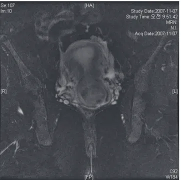
The ultrasonography and magnetic resonance imaging with con- trast enhancement showed a 4.8 × 2.2 cm sized mass, dilating the endocervical canal, and it was suspected to be endometrial carci-
Th is is an Open Access article distributed under the terms of the Creative Commons Attribution Non-Commercial License (http://creativecommons.org/licenses/
by-nc/3.0/) which permits unrestricted non-commercial use, distribution, and reproduction in any medium, provided the original work is properly cited.
Copyright © 2011. Korean Society of Obstetrics and Gynecology
noma (Fig. 1).
The medical, surgical, and menstrual history of the patient was unremarkable. She had been well and healthy until the bleeding started in October 2007. After the bleeding started, she fi rst vis- ited a private clinic where an endometrial mass measured by 5 × 2 cm was noted on transrectal ultrasonography. She was prescribed oral contraceptives, but the symptom persisted.
Although she was a virgin, we decided to carry out endometrial curettage because a malignant lesion was so strongly suspected that histological confi rmation was warranted. Moreover, her bleed- ing had not stopped. Endometrial curettage was performed under the patient’s consent. During the curettage, we noticed that her uterine cervix on the speculum was orange colored, easily bleed, and fragile, which are all suggestive of classical squamous cell car- cinoma of the cervix. A frozen section of the fractional curettage confirmed that she had squamous cell carcinoma of the uterine cervix, not endometrial adenocarcinoma.
She was admitted for the baseline workup, including positron emission tomography (PET), colonoscopy, intravenous pyelogram (IVP), and cystoscopy. PET scan revealed 5 cm sized carcinoma with multiple hematogenous lung metastases (Fig. 2). However, there were no lesions suggestive of lymph node metastasis or lo- cal pelvic extension. The cancer mass appeared to be absolutely confined to the uterine cavity except the hematogenous lung
metastasis. Cystoscopy, IVP, and colonoscopy results were not re- markable.
After three cycles of preoperative adjuvant chemotherapy with cisplatin (50 mg/m
2) and vincristine (1 mg/m
2) per week, she underwent modified radical hysterectomy, ovarian transposition, bilateral pelvic lymph node dissection, and para-aortic lymph node sampling. Grossly, the cervix revealed a large tumorous mass, measuring 4.5 × 3.2 cm in size at the posterior wall. The cut sec- tion of the mass was yellow-gray, fi rm, and necrotic. The mass was
Fig. 1. Magnetic resonance imaging demonstrated a 4.8 × 2.2 cm sizedmass and it was suspected to be endometrial carcinoma.
Fig. 2. Positron emission tomography scan revealed cancer mass con- fi ned to the uterine cavity with multiple hematogenous lung metastases.
Fig. 3. Gross fi ndings.
extended up to the serosa of the posterior wall and focally into endometrial cavity (Fig. 3). The uterine cavity measured 3 cm long, and the endometrium mass irregularly thickened. Microscopically, the tumor mass showed histologic features of typical squamous cell carcinoma with individual keratinization (Fig. 4A). Tumor cells reached the endometrium but were confined to the uterus. No lymph node metastasis or parametrial involvement was found. Tu- mor cells were strongly positive for p53 in immunohistochemical assay (Fig. 4B).
The patient had no postoperative complications and underwent six cycles of postoperative adjuvant chemotherapy with carbopla- tin and paclitaxel. Surprisingly, her multiple hematogenous lung metastatic nodules were completely resolute after three cycles of chemotherapy, and she recovered complete resolution after com- pleting all six cycles of chemotherapy. She has been followed in the out-patient department without recurrence for 4 years.
Discussion
This HPV genome negative squamous cell carcinoma developed in a virgin is a very interesting case in terms of its extreme rarity and atypical clinical course.
This case is directly contrary to our belief that virtually all cervical car- cinoma is caused by sexually transmitted HPV. A few possible carcino- genetic mechanisms could be suggested to explain this rare case.
First, this may be a case with a failure to identify HPV. Integration of HPV DNA in the host cell genome could result in disruption,
combined with deletions, of HPV DNA at integration sites [6].
However, modern PCR technologies are too advanced to detect HPV DNA at the single copy level even in an overwhelming back- ground of human DNA. Therefore, this explanation is not likely to be valid in our case.
Second, there are pathways which might give rise to cervical car- cinoma in which HPV is transiently involved, thus called a “hit- and-run” mechanism. However, the “hit-and-run” mechanism is unlikely, because transcription of E6 and E7 oncoproteins of HR- HPV is required for the maintenance of epithelial transformation of the uterine cervix [3,7,8]. Actually, only one case appeared to be related with this mechanism [9].
Finally, carcinoma of the cervix might develop through truly HPV- independent pathways. This speculation is supported by an epide- miological fi nding that patients with HPV-negative cervical intra- epithelial neoplasia (CIN) have a different spectrum of risk factors from those with HPV-positive CIN [9]. One of the target molecule related with this HPV-independent carcinogenesis of uterine cer- vix is p53. Although most p53 dysfunctions in cervical cancer is related with E6 oncoprotein of high risk (HR)-HPV, other mecha- nisms such as point mutation or genetic polymorphism [10,11]
could occur through a HPV-independent process, and initiate the malignant transformation. In our case, p53 mutation was shown by immunohistochemical study (Fig. 4B). Based on this fi nding, we speculated that this HPV-negative cervical cancer arose through de novo mutation of p53, without HR-HPV DNA.
Moreover, this case had diffuse hematogenous lung metastasis.
Generally, the cervical cancer progresses in a “level by level” fash-
Fig. 4. (A) Most tumor cells show squamous cell carcinoma, large cell non-keratinizing (H&E, × 200). (B) Positive immunoreactivities for P53 (Immuno- histochemistry, × 200).A B
ion to regional nodes through the lymphatic channels, and also to extra-nodal sites via the hematogenous stream [12]. However, if it was for pulmonary metastasis, the clinical stage of this case was just Ib2. The lesion was strictly confi ned to the uterine cervix, and there was no demonstrable pelvic and paraaortic lymph node me- tastasis. Our case showed a very early and unique (hematogenous, rather than lymphatic) pattern of metastasis compared with those of typical carcinoma of the cervix. It might be attributed to de novo p53 mutation that was already described. In many other ma- lignancies, the p53 mutation is related with poor prognosis such as locally advanced lesion, multiple lymph node metastasis, and distant solid organ metastasis. However, the value of p53 muta- tion as a prognostic marker in cervical cancer is not so useful [13]
because the E6 of the HR-HPV basically initiates carcinogenesis by ubiquitin-dependent degradation of p53 [8,14,15]. However, if this p53 overexpression is initiated by a de novo process, the prognosis could be different from that of typical HPV-related cervi- cal cancer.
Generally, it is difficult to recommend transvaginal procedures such as PAP smear, colposcopy or curettage to patients who claim to have never had sexual intercourse. Many clinicians regard cervi- cal cancer as virtually nonexclusively caused by HR-HPV infection.
In these aspects, cervical cancer in virgins might be neglected and diagnosed in late stage. Therefore, clinicians should make a clear decision over when to use invasive procedures, and always have suspicion about the possibilities of rare occasions.
References
1. Bosch FX, de Sanjosé S. The epidemiology of human pap- illomavirus infection and cervical cancer. Dis Markers 2007;23:213-27.
2. Bosch FX, Manos MM, Muñoz N, Sherman M, Jansen AM, Peto J, et al. Prevalence of human papillomavirus in cervical cancer: a worldwide perspective. International biological study on cervical cancer (IBSCC) Study Group. J Natl Cancer Inst 1995;87:796-802.
3. Walboomers JM, Jacobs MV, Manos MM, Bosch FX, Kummer JA, Shah KV, et al. Human papillomavirus is a necessary cause of invasive cervical cancer worldwide. J Pathol 1999;189:12-9.
4. Wang IT, Chang CW, Tang WL, Chen WY, Liu WM. Keratinizing squamous cell carcinoma of the cervix in a sexually inexperi- enced woman: a case report. J Reprod Med 2006;51:745-6.
5. Bielecki A, Borowicz K, Misiewicz L. Adenocarcinoma of the cervix uteri in a 37-year-old virgin with coexisting cancer of the body of the uterus. Am J Obstet Gynecol 1963;87:135-6.
6. Schwarz E, Freese UK, Gissmann L, Mayer W, Roggenbuck B, Stremlau A, et al. Structure and transcription of human papillomavirus sequences in cervical carcinoma cells. Nature 1985;314:111-4.
7. Narisawa-Saito M, Kiyono T. Basic mechanisms of high-risk human papillomavirus-induced carcinogenesis: roles of E6 and E7 proteins. Cancer Sci 2007;98:1505-11.
8. Wise-Draper TM, Wells SI. Papillomavirus E6 and E7 proteins and their cellular targets. Front Biosci 2008;13:1003-17.
9. Burger MP, Hollema H, Pieters WJ, Schröder FP, Quint WG.
Epidemiological evidence of cervical intraepithelial neoplasia without the presence of human papillomavirus. Br J Cancer 1996;73:831-6.
10. Park DJ, Wilczynski SP, Paquette RL, Miller CW, Koeffler HP.
p53 mutations in HPV-negative cervical carcinoma. Oncogene 1994;9:205-10.
11. Min-min H, Ming-rong X, Ze-yi C, Kai-xuan Y, Zhi-lin S. Analy- sis of p53 codon 72 polymorphism and its association with human papillomavirus 16 and 18 E6 in Chinese cervical le- sions. Int J Gynecol Cancer 2006;16:2004-8.
12. Belhocine T, Kridelka F, Thille A, De Barsy C, Foidart-Willems J, Hustinx R, et al. Staging of primary cervical cancers: the role of nuclear medicine. Crit Rev Oncol Hematol 2003;46:275-84.
13. Lindstrom AK, Stendahl U, Tot T, Lidström BM, Hellberg D.
Predicting the outcome of squamous cell carcinoma of the uterine cervix using combinations of individual tumor marker expressions. Anticancer Res 2007;27:1609-15.
14. Braun K, Ehemann V, Waldeck W, Pipkorn R, Corban-Wilhelm H, Jenne J, et al. HPV18 E6 and E7 genes affect cell cycle, pRB and p53 of cervical tumor cells and represent prominent can- didates for intervention by use peptide nucleic acids (PNAs).
Cancer Lett 2004;209:37-49.
15. Nair S, Pillai MR. Human papillomavirus and disease
mechanisms: relevance to oral and cervical cancers. Oral Dis
2005;11:350-9.
성경험이 없는 36세 여성 자궁경부에서 발생한 편평세포암 1예
1연세대학교 의과대학 산부인과학교실, 2국민건강보험공단 일산병원 산부인과학교실, 3국민건강보험공단 일산병원 병리학교실 심아라1, 최은경2, 윤성현1, 이마리아1, 이미경3, 김상운1
자궁경부암은 한국에서 가장 흔한 부인암중 하나이며 그 발생 원인으로 인유두종바이러스 감염이 매우 밀접한 연관성을 가지고 있어 성 경험이 없는 여성에서의 발병은 극히 드물다. 본원에서 36세의 성경험이 없으며 인유두종 바이러스 DNA 음성인 여성에서 발생한 폐 전이 까지 동반한 자궁경부 편평상피암을 경험하였기에 간단한 문헌 고찰과 함께 보고하는 바이다.
중심단어: 편평상피암, 자궁경부암, 처녀, 인유두종바이러스 DNA 음성