© 2020 The Korean Ophthalmological Society
This is an Open Access article distributed under the terms of the Creative Commons Attribution Non-Commercial License (http://creativecommons.org/licenses /by-nc/3.0/) which permits unrestricted non-commercial use, distribution, and reproduction in any medium, provided the original work is properly cited.
90
Korean J Ophthalmol Vol.34, No.1, 2020
Pigmented Paravenous Retinocho- roidal Atrophy
Dear Editor,
We experienced a case of pigmented paravenous retino- choroidal atrophy (PPRCA), an unusual retinal degenera- tion characterized by perivenous aggregations of pigment clumps that are distributed along the retinal veins [1]. PPR- CA is extremely rare; to our knowledge, only two cases have been reported in Korea [2].
An 8-year-old girl with history of congenital glaucoma and glaucoma surgery reported to our clinic for further evaluation. At the age of five, she had undergone strabis- mus surgery for exotropia. Her best-corrected visual acuity was 20 / 150 in the right eye and 20 / 33 in the left eye. The anterior segment exam showed a patent peripheral iridec- tomy site in her right eye. Intraocular pressure, anterior chamber reaction, shape of the optic disc, and pupillary light reflex were all normal. She had right exotropia, but her ocular movement was not limited.
The rod and cone response was detectable but moderate- ly decreased on electroretinography. The fundus showed bilateral bone-spicule pigmentation and retinochoroidal at- rophy in the peripheral paravenous distribution without definite macula involvement. No active inflammatory re- sponses of the retina or vitreous, such as vitreous cells, vit- reous opacity, retinal infiltration, or hemorrhage, were ob- served. The Goldmann perimetry test demonstrated a visual field of about 40 to 50 degrees with arcuate scotoma and an enlarged blind spot corresponding to the lesion.
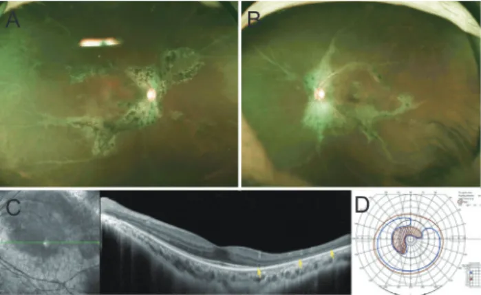
On her last visit, 11 years after the initial presentation, her corrected visual acuity was unchanged: 20 / 200 in the right eye and 20 / 33 in the left eye. There was no signifi- cant change in the area of retinal degeneration around the blood vessels, but bone-spicule pigmentation had increased on fundus examination. The area of retinal degeneration
was well defined, and diffuse disruption of the outer retina layer was observed in spectral domain optical coherence tomography. The microstructure of the macula remained intact, and the Goldmann perimetry examination was al- most stationary (Fig. 1A-1D).
PPRCA has a non-progressive, slow, or subtly progres- sive course [3]. The etiology of PPRCA is not clearly un- derstood. The majority of PPRCA cases occur sporadically, although there have been specific cases of familial occur- rence. Since some patients have bilateral PPRCA with macular coloboma, ophthalmologists have postulated that it is a developmental abnormality and, thus, a disorder of the retinal blood vessels during the embryonic stages of eye development. In the present case, the patient’s history of congenital glaucoma suggested that PPRCA may have been caused or accompanied by a developmental problem.
On the other hand, a number of inflammatory PPRCA cas- es has been reported, leading to a different hypothesis cit- ing an inflammatory etiology, including Behcet’s disease, measles, rubella, uveitis, and other unknown causes of in- flammation [4,5]. These suggest that PPRCA may be a set of morphologically similar diseases derived from heterog- enous causes.
Korean J Ophthalmol 2020;34(1):90-91 https://doi.org/10.3341/kjo.2019.0078
Received: July 4, 2019 Final revision: July 27, 2019 Accepted: August 6, 2019
Fig. 1. The bony-spicule shaped retinochoroidal atrophy with pigmentation along retinal veins: wide fundus photography of the patient’s (A) right eye and (B) left eye. (C) Optical coherence to- mography of the patient’s left eye. Diffuse disruption of the outer retina layer was observed while the microstructure of the macula remains intact. (D) Goldman perimetry examination of patient’s left eye. Visual field is about 40 to 50 degrees; arcuate scotoma with enlarged blind spot corresponding to pigmented paravenous retinochoroidal atrophy presents.
A
C
B
D
91 Most PPRCA patients are asymptomatic, although some
complain of mild blurred vision. Certain patients, who complain of poor dark adaptation, poor night vision or a blind eye, do not suffer from PPRCA but instead have reti- nitis pigmentosa (RP) or pseudo PPRCA. PPRCA is com- monly bilateral and symmetric. The visual field may be normal, or changes may be variable with topography of pigmentation and atrophy [4].
PPRCA is diagnosed by the typical funduscopic fea- tures of bilaterally symmetrical accumulation of pigment and retinochoroidal atrophy along the retinal veins, invari- ably beginning a distance from the optic nerve head. Peri- papillary pigmentary changes may be observed as well as areas of chorioretinal atrophy adjacent to the perivenular pigmentary changes. The unaffected retinal areas appear normal. The pigmentation is typical of bone corpuscle pig- mentation, coarse pigment clumps, and fine pigmentary changes. This perivenular appearance is clearly different from RP, which typically shows bony spicule change in the whole retina or a certain sector. As in our case, optical co- herence tomography shows degeneration of the outer retina photoreceptor without affecting the inner retina. This characteristic distinguishes PPRCA from vasculitis. Fluo- rescein angiography, indocyanine green angiography, and electrophysiological tests may be used to confirm the diag- nosis [4].
In conclusion, we report a case of PPRCA, a disease that is very rare worldwide. The diagnosis of PPRCA is often neglected due to the paucity of cases and the similarity with RP. Since the disease course and its treatment are dif- ferent from those of RP or active vasculitis, PPRCA should be distinguished from other similar diseases.
Won Jong Choi
Department of Ophthalmology, Seoul National University Bundang Hospital, Seoul National University College of Medicine, Seongnam; Seoul National University Hospital, Seoul National University College of Medicine, Seoul, Korea
Kwangsic Joo, Kyu Hyung Park
Department of Ophthalmology, Seoul National University Bundang Hospital, Seoul National University College of Medicine, Seongnam, Korea
E-mail (Kwangsic Joo): [email protected]
Conflict of Interest
No potential conflict of interest relevant to this article was reported.
References
1. Shen Y, Xu X, Cao H. Pigmented paravenous retinochoroi- dal atrophy: a case report. BMC Ophthalmol 2018;18:136.
2. Park HS, Yang JY, Park HJ. A case of unilateral focal pig- mented paravenous retinochoroidal atrophy. J Korean Ophthalmol Soc 2018;59:1190-4.
3. Choi JY, Sandberg MA, Berson EL. Natural course of ocu- lar function in pigmented paravenous retinochoroidal atro- phy. Am J Ophthalmol 2006;141:763-5.
4. Huang HB, Zhang YX. Pigmented paravenous retinocho- roidal atrophy (review). Exp Ther Med 2014;7:1439-45.
5. Traboulsi EI, Maumenee IH. Hereditary pigmented para- venous chorioretinal atrophy. Arch Ophthalmol 1986;104:1636- 40.