© 2014 The Korean Ophthalmological Society
This is an Open Access article distributed under the terms of the Creative Commons Attribution Non-Commercial License (http://creativecommons.org/licenses /by-nc/3.0/) which permits unrestricted non-commercial use, distribution, and reproduction in any medium, provided the original work is properly cited.
Original Article
Bone Marrow-derived Mesenchymal Stem Cells Affect Immunologic Profiling of Interleukin-17-secreting Cells in a
Chemical Burn Mouse Model
Ja Young Lee1,2, Hyun Jeong Jeong2, Mee Kum Kim1,2, Won Ryang Wee1,2
1Department of Ophthalmology, Seoul National University College of Medicine, Seoul, Korea
2Laboratory of Ocular Regenerative Medicine and Immunology, Seoul Artificial Eye Center, Seoul National University Hospital Biomedical Research Institute, Seoul, Korea
Purpose: This study investigated interleukin (IL)-17-secreting cell involvement in sterile inflammation, and evalu- ated the effect of mesenchymal stem cells (MSCs) on IL-17-secreting cell immunologic profiling.
Methods: Twenty mice were sacrificed at time points of 6 hours, 1 day, 1 week, and 3 weeks (each group, n = 5) after the cornea was chemically injured with 0.5N NaOH; IL-17 changes in the cornea were evaluated using enzyme-linked immunosorbent assay. Further, IL-17 secreting cells were assessed in the cervical lymph nodes by a flow cytometer. Rat MSCs were applied intraperitoneally in a burn model (n = 10), IL-17-secreting T helper 17 (Th17) cell and non-Th17 cell changes were checked using a flow cytometer in both cornea and cervical lymph nodes at 1week, and compared with those in the positive control (n = 10).
Results: IL-17 was highest in the cornea at 1 week, while, in the cervical lymph nodes, IL-17-secreting cells showed early increase at 6 hours, and maintained the increase through 1 day to 1 week, and levels returned to the basal level at 3 weeks. Specifically, the non-Th17 cells secreted IL-17 earlier than the Th17 cells. When the MSCs were applied, IL-17 secretion was reduced in CD3(+)CD4(-)CD8(-), CD3(+)CD4(+)CD8(-), and CD3(+) CD4(-)CD8(+) cells of the cervical lymph nodes by 53.7%, 43.8%, and 50.8%, respectively. However, in the cornea, IL-17 secretion of CD3(+)CD4(-)CD8(-) cells was completely blocked.
Conclusions: The results indicated that both IL-17-secreting non-Th17 and Th17 cells were involved in the chemical burn model, and MSCs appeared to mainly modulate non-Th17 cells and also partially suppress the Th17 cells.
Key Words: Chemical burns, Interleukin-17, Mesenchymal stromal cells, Th17 cells
Mesenchymal stem cells (MSCs) are reported to have self-renewal capacity for multipotent differentiation, and immunomodulatory properties. MSCs have been studied for therapeutic purposes in various fields. MSCs have been
known to modulate both innate and adaptive immunity, through the suppression of T cells, B cells, dendritic cells, natural killer (NK) cells, macrophages and neutrophils, and they have been investigated as therapeutic targets be- cause of their ability to intervene in autoimmune disease or inflammatory disease [1-4].
The T helper 17 (Th17) cell was extensively investigated as a critical factors that can cause various autoimmune diseases or inflammatory diseases [5-8]. In addition, recent studies highlighted the function of innate and adaptive
Received: March 26, 2013 Accepted: June 14, 2013
Corresponding Author: Mee Kum Kim, MD, PhD. Department of Oph- thalmology, Seoul National University College of Medicine, #103 Dae- hak-ro, Jongno-gu, Seoul 110-799, Korea. Tel: 82-2-2072-2665, Fax: 82-2- 741-3187, E-mail: [email protected]
interleukin (IL)-17-secreting cells with variable cell sourc- es in various inflammatory diseases [9]. The IL-17 receptor is ubiquitously expressed throughout the body, resulting in pleiotropic actions of IL-17 on a wide range of cell types.
One of the well-known biological outcomes of IL-17 sig- naling is the induction of proinflammatory cytokines and chemokines from target cells, such as fibroblasts and epi- thelial cells, leading to the recruitment of neutrophils [10].
The primary function of activated neutrophils is to fight against invading pathogens, but this can damage the surrounding tis- sues. There have been numerous research studies to determine the immune-modulatory role of MSCs on the Th17 cells [11-15], however, only a few studies had investigated the effect of MSCs on other sources of IL-17-secreting cells, including in- nate lymphoid cells or gammadelta T-cells [16].
The MSC-based cellular therapy could be applied to treat graft versus host disease and autoimmune disease. In addition, MSCs have been exploited for many ocular dis- ease treatments, such as corneal disease, uveitis and retinal diseases. In functional disabilities, such as ocular blind- ness, chemical burns often occur as an occupational haz- ard and can induce severe inflammation through both in- nate and adaptive immunity, including IL-17-secreting cells [17]. In fact, the anti-inflammatory effect of MSCs has recently been studied in chemically injured eyes [18- 22]. However, the function of the innate and adaptive IL-17-secreting cells in inflammation has not yet been de- termined in a chemical burn model, and the effects of MSCs on the innate and adaptive IL-17-secreting cells have not been investigated in this model either.
Therefore, we hypothesized that both innate and adap- tive IL-17-secreting cells may be involved in the inflamma- tion that results after chemical injury to the cornea. First, we investigated time-dependent immunologic profiling of both innate and adaptive IL-17-secreting cells in a chemi- cally-injured animal model, and then we determined the effect of bone-marrow derived MSCs on both innate and adaptive IL-17-secreting cells in this model.
Materials and Methods
Animals
Five-week-old male C57BL/6 mice, weighing 30 g, were purchased from the Orient Bio (Seongnam, Korea). All an-
imal experiments were conducted in accordance with the ARVO Statement for the Use of Animals in Ophthalmic and Vision Research. The experimental protocols were ap- proved by the Institutional Animal Care and Use Commit- tee of the Seoul National University Hospital (permit num- ber 11-0212).
Corneal chemical burn model and time-dependent changes of interleukin-17 in the model
Mice were anesthetized with an intraperitoneal injection of zolazepam (Zoletil; Virbac Laboratories, Carros, France;
0.006 mL/10 g) and xylazine hydrochloride (Rompun;
Bayer, Frankfurt, Germany; 0.004 mL/10 g). To create the chemical injury, a 6-mm filter paper soaked in 100% etha- nol was applied to the whole cornea of the left eye, includ- ing the limbus for 30 seconds, followed by a rinse with 10- mL of balanced salt solution, then the corneal and limbal epithelium were removed with a surgical blade. Next, a 6-mm filter paper soaked in 0.5N NaOH was applied to the corneal surface for 15 seconds, followed by rinsing with 20-mL of balanced salt solution. Ofloxacin ophthalmic ointment was instilled immediately.
To investigate the changes of IL-17, twenty mice, which had undergone the cornea chemical burn procedure, were sacrificed at times point of 6 hours (n = 5), 1 day (n = 5), 1 week (n = 5), and 3 weeks (n = 5) after injury. To evaluate the inflammatory cytokines changes in the cornea, all in- jured corneas were harvested and were pooled to evaluate cytokine changes in enzyme-linked immunosorbent assay (ELISA). The cervical lymph nodes were evaluated to as- sess IL-17-secreting cell changes using a fluorescent- activated cell sorter (FACS) analysis at each time point.
Mesenchymal stem cells preparation and application Rat primary MSC lines (cryopreserved rat mesenchymal stem cells, part no. 2004005; Millipore, Temecula, CA, USA) were thawed, according to the manufacturer’s in- structions. The cells were resuspended and cultured in alpha minimum essential medium (Invitrogen-GIBCO, Carlsbad, CA, USA), supplemented with 10% fetal bovine serum (Atlanta Biological, Lawrenceville, GA, USA), Pen- icillin/Streptomycin (100 U/mL, Invitrogen-GIBCO) and 200 mM L-glutamine (Invitrogen-GIBCO) for 72 hours in a T75 flask. The cells were plated in a flask at a density of
1 × 10⁶ cells/cm2 and incubated at 37°C with 5% CO2. The media was exchanged every two days, and after 10 to 14 days, the non-adherent cells and supernatant were discard- ed. Adherent cells were treated with Accutase (Millipore) for three minutes and replated in a new flask at 5 × 10³ cells/cm2. Adherent cells, obtained after the second subcul- ture, were used for the experiment.
Thirty mice were randomly divided into three groups (each group, n = 10). The first group was the positive con- trol group that received chemical injury to the cornea by the method mentioned above and the negative control group did not receive chemical injury. The second group was the treatment group, and the mice were given intra- peritoneal injections of the media, including rat MSCs (5 × 10⁶/200 μL) after the chemical injury. All the mice were sacrificed at 1 week after injury.
Cytokine quantification by enzyme-linked immunosorbent assay
To investigate the time-dependent change of inflamma- tory cytokines in the cornea based on the chemical burn model, the corneal samples at different time points after injury were lysed in protein extraction buffer (PRO- PREPTM Solution; Intron Biotechnology, Seongnam, Ko- rea), and were centrifuged at 2,500 rpm for 20 minutes. An aliquot of each supernatant was assayed. The concentra- tions of cytokines were measured using an ELISA, accord- ing to the manufacturer’s recommendations (R&D sys- tems, Minneapolis, MN, USA) at 450 nm that included IL- 6, IL-17, tumor necrosis factor alpha (TNF-α) and interferon gamma (IFN-γ). The analysis was performed with the pooling of the cornea at each time point with du- plicates.
Fluorescent-activated cell sorter analysis for
interleukin-17-secreting cells in the cornea and cervical lymph nodes
To assess the changes of IL-17-secreting cells in the cor- nea and the cervical lymph nodes between the positive control group and the treatment group, the mice was sacri- ficed at 1 week. The left cornea of each mouse was pooled in each group and was excised from the eyeballs, sliced with scissors into small pieces, and the tissue was homoge- nized with a sonicator (Tomy-Seiko, Tokyo, Japan). The
samples were incubated at 37°C for 1 or 2 hours (2 mg/mL collagenase D [Roche Diagnostics, Indianapolis, IN, USA]
with 0.05 mg/mL DNase [Roche]) and were centrifuged at 1,200 rpm for 10 minutes. An aliquot of each supernatant was passed through a 70-μm cell strainer (BD Falcon, San Jose, CA, USA) for single cell suspension. The cells were counted, using trypan blue dye and an automated cell counter (Countess; Invitrogen, Carlsbad, CA, USA), and were prepared for IL-17-secreting cell staining.
Left cervical lymph nodes from each mouse were ex- tracted and were pooled in each group. The lymph node cells were isolated and resuspended in the DMEM media, including 1% HEPES and 0.5% FBS for FACS analysis.
Lymphocytes were stimulated with 50 ng/mL phorbol myristate acetate (Sigma-Aldrich, St Louis, MO, USA), 1 μg/mL Ionomycine (Sigma Aldrich) and 1 μg/mL Gol- giPlug (BD Phamingen, San Diego, CA, USA) for 5 hours.
The cells were then rinsed twice with FACS buffer (0.002% sodium azide, 0.2% BSA/PBS; eBioscience, San Diego, CA, USA) and resuspended in fixation solution (BD Phamingen) for intracellular cytokine staining. The proto- col was performed based on the manufacturer’s instruc- tions.
The cells isolated from the left cervical lymph node and left cornea were stained with fluorophore-conjugated anti- bodies: PerCP-conjugated anti-CD3 (eBioscience), FITC-conjugated anti-CD4 (eBioscience), PE-cy7-conju- gated anti-CD8 (eBioscience), APC-conjugated anti-IFNγ (eBioscience), or PE-conjugated anti-IL-17 (BD Phamin- gen). Absolute cell numbers were calculated by multiply- ing the percentage of positive staining cells in the total ac- quired events by the total number of cells isolated from the analyzed tissue. Immunofluorescence was analyzed with Flowjo software ver. 7.6.5 (Treestar, Ashland, OR, USA) with a FACS Caliber flow cytometer (FACS Canto; Becton Dickinson, San Diego, CA, USA).
In the MSCs applied group, the decrease of the IL-17-se- creting cells was presented as the reduction rate. The re- duction rate was calculated with the following formula:
(pretreatment – posttreatment) × 100 / pretreatment.
Statistical analysis
The cervical lymph nodes were examined to assess changes in the IL-17-secreting cells. The lymph nodes were assessed with FACS analysis at each time point and were
compared with the positive control group by a Moses test of extreme reactions using SPSS ver.17.0 (SPSS Inc., Chicago, IL, USA). For statistical analyses, a p-value of
<0.05 was considered statistically significant.
Results
Time-dependent changes in inflammatory cytokines after chemical injury in the cornea
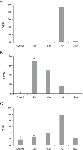
First, we used ELISA to look at the cytokine changes in the cornea. The analysis showed that the production of IL- 17 cytokines was the highest (46.95 pg/mL) at 1 week, and returned to almost the basal level (0.944 pg/mL) at 3 weeks after injury. Similarly, TNF-α levels increased at 1 week and then decreased at 3 weeks. In contrast, IL-6 showed the highest peak at 6 hours, and was subsequently reduced over time. However, IFN-γ was not detected throughout the entire post-injury time period (Fig. 1). We hypothesize that the IL-17-secreting cells maximally infiltrated into the cornea 1 week after the injury.
Time-dependent immunologic profiling of interferon gamma and interleukin-17-secreting cells in chemical injury
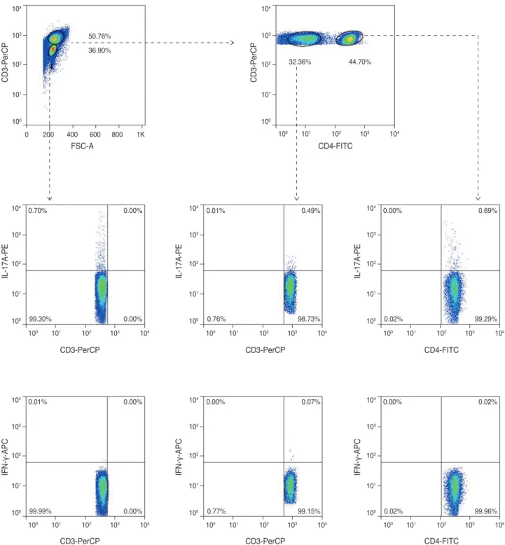
Next, we examined systemic changes of IFN-γ and IL-17-secreting cells in the chemically-injured mouse mod- el. We analyzed changes in sub-population secretion of IFN-γ and IL-17 in the cervical lymph nodes, based on the following outline (Fig. 2); CD3(-)CD4(-) cells, which in- cluded innate lymphoid cells and B cells; CD3(+)CD4(-) cells, which included gammadelta T-cells, NK cells, NK T-cells or CD8(+) T-cells; and CD3(+)CD4(+) cells, which represented CD4 T helper cells.
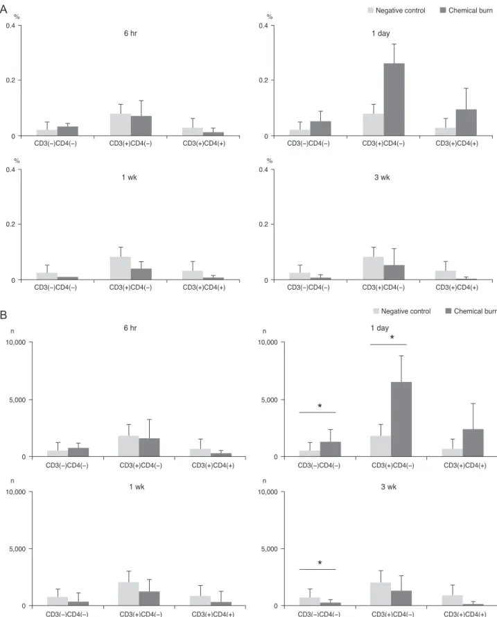
The results indicated that of the IFN-γ secreting cells, only CD3(+)CD4(-) cells showed high increases of IFN-γ at day 1 after the procedure, and the numbers abruptly de- creased from 1 to 3 week measurements (Fig. 3). A thor- ough analysis of the CD3(-)CD4(-) cells and the CD3(+) CD4(-) cells at day 1, and the CD3(-)CD4(-) cells at 3 weeks indicated that the cells numbers were significantly greater than the negative control group. This result suggests that the non-Th17 cells (gammadelta T-cells, NK cells, NK T-cells, and CD8(+) T-cells) were temporarily activated.
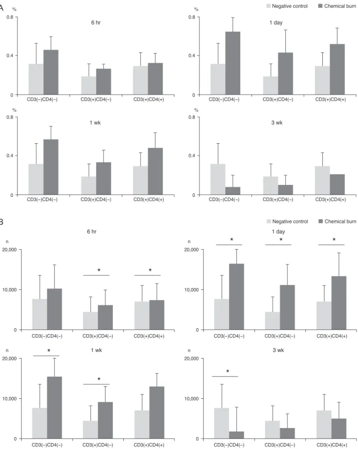
In comparison, the IL-17-secreting cells showed an early increase at 6 hours, and the elevated level of IL-17 was maintained through day 1 to 1week and returned to the basal level at 3 weeks (Fig. 4). A thorough analysis of the CD3(-)CD4(-) cells at day 1 and the 1 to 3 week time inter- val indicated that the CD3(+)CD4(+) cells at 6 to 24 hours, and the CD3(+)CD4(-) cells at 6 to 24 hours and 1 week had significantly increased numbers when compared with
Control 0
4 6 8 10 12
2
6 hr 1 day 1 wk 3 wk
pg/mL
Control 0
10 20 30 40 50
6 hr 1 day 1 wk 3 wk
pg/mL
Control 0
20 40 60 80
6 hr 1 day 1 wk 3 wk
pg/mL
Fig. 1. Enzyme-linked immunosorbent assay analysis of the corneal lysate after chemical injury. The bar charts show the mean concentrations of pro-inflammatory cytokines. (A) Interleukin (IL)-17. (B) IL-6. (C) Tumor necrosis factor alpha (TNF-α). Control represents the normal cornea. Note that the production of cytokines IL-17 and TNF-α peaked at 1 week post- injury, and in contrast, peak IL-6 levels increased immediately after injury. The experiments were performed in duplicate.
A
B
C
the negative control group. Specifically, the non-Th17 cells (CD3(-)CD4(-) cells and CD3(+)CD4(-) cells) secreted IL-17 earlier than CD3(+)CD4(+) Th17 cells; CD3(+)CD4(+) Th17 cells secreted IL-17 over 1 day.
Mesenchymal stem cells effect on interleukin-17- secreting cells in a chemical burn model
Although IL-17-secreting cells were systemically elevat- ed from 6 hours to 1 week, the cornea showed the highest
32.36%
50.76%
36.90%
1K 800 600 400 200 0 100 101 102 103 104
0.70% 0.00%
0.00%
0.01% 0.49%
0.76%
99.30% 98.73%
0.00% 0.69%
0.02% 99.29%
0.01% 0.00%
99.99% 0.00%
0.00% 0.07%
0.77% 99.15%
0.00% 0.02%
0.02% 99.96%
44.70%
CD3-PerCP
FSC-A CD4-FITC
CD3-PerCP CD3-PerCP CD4-FITC
CD3-PerCP CD3-PerCP CD4-FITC
CD3-PerCP
IL-17A-PE IL-17A-PE IL-17A-PE
IFN-γ-APC IFN-γ-APC IFN-γ-APC
100
100 101 102 103 104 101
102 103 104
100
100 101 102 103 104 101
102 103 104
100
100 101 102 103 104 101
102 103 104
100
100 101 102 103 104 101
102 103 104
100
100 101 102 103 104 101
102 103 104
100
100 101 102 103 104 101
102 103 104
100
100 101 102 103 104 101
102 103 104
Fig. 2. This photo represented a sort-out analyses of the interleukin (IL)-17/interferon gamma (IFN-γ) secreting cells in the cervical lymph nodes, following corneal chemical injury. The non-Th-cells (CD3(-)CD4(-) cells or CD3(+)CD4(-) cells) and CD3(+)CD4(+) T help- er cells were separated and were analyzed.
Negative control Chemical burn
CD3(−)CD4(−) 0
0.2 0.4%
CD3(+)CD4(−) CD3(+)CD4(+) 6 hr
CD3(−)CD4(−) 0
0.2 0.4%
CD3(+)CD4(−) CD3(+)CD4(+) 1 day
CD3(−)CD4(−) 0
0.2 0.4%
CD3(+)CD4(−) CD3(+)CD4(+) 1 wk
CD3(−)CD4(−) 0
0.2 0.4%
CD3(+)CD4(−) CD3(+)CD4(+) 3 wk
Fig. 3. The bar charts show the mean (A) percentages and (B) cell numbers of the interferon gamma (IFN-γ) secreting cells in the cervi- cal lymph nodes of the 4 groups (each group, n = 5), divided over the time course of 6 hours, 1 day, 1 week, and 3 weeks after the onset of chemical injury. Note that IFN-γ-secreting non-Th-cells (CD3(-)CD4(-) T-cells) reached the highest peak at day 1 after injury, and the Th- cells also increased at 1 day before the levels returned to baseline. *p < 0.05 (Moses extreem reactions test).
A
0 5,000 10,000
n
CD3(−)CD4(−) CD3(+)CD4(−) CD3(+)CD4(+) 6 hr
1 wk 3 wk
0 5,000
10,000
*
*
n
CD3(−)CD4(−) CD3(+)CD4(−) CD3(+)CD4(+) 1 day
0 5,000 10,000
n
CD3(−)CD4(−) CD3(+)CD4(−) CD3(+)CD4(+) 0
5,000 10,000
*
n
CD3(−)CD4(−) CD3(+)CD4(−) CD3(+)CD4(+) Negative control Chemical burn
B
CD3(−)CD4(−) 0
0.4 0.8
%
CD3(+)CD4(−) CD3(+)CD4(+) 6 hr
CD3(−)CD4(−) 0
0.4 0.8
%
CD3(+)CD4(−) CD3(+)CD4(+) 1 day
CD3(−)CD4(−) 0
0.4 0.8%
CD3(+)CD4(−) CD3(+)CD4(+) 1 wk
CD3(−)CD4(−) 0
0.4 0.8%
CD3(+)CD4(−) CD3(+)CD4(+) 3 wk
Negative control Chemical burn
0 10,000 20,000
n
CD3(−)CD4(−) CD3(+)CD4(−) CD3(+)CD4(+) 6 hr
* *
0 10,000 20,000
n
CD3(−)CD4(−) CD3(+)CD4(−) CD3(+)CD4(+) 1 day
* *
*
0 10,000 20,000
n
CD3(−)CD4(−) CD3(+)CD4(−) CD3(+)CD4(+) 1 wk
*
*
0 10,000 20,000
n
CD3(−)CD4(−) CD3(+)CD4(−) CD3(+)CD4(+) 3 wk
*
Negative control Chemical burn
Fig. 4. The bar charts show the mean (A) percentages and (B) cell numbers of the interleukin-17-secreting cells in the cervical lymph nodes in the 4 groups (each group, n = 5), divided over the time course of 6 hours, 1 day, 1 week, and 3 weeks after the onset of chemical injury. Note that both the non-T helper 17 (Th17) cells and Th17 cells increased 1 week after injury, and then gradually decreased. *p < 0.05 (Moses extreem reactions test).
A
B
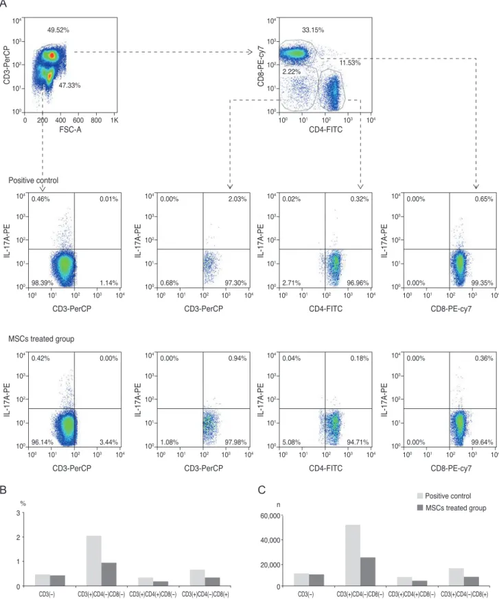
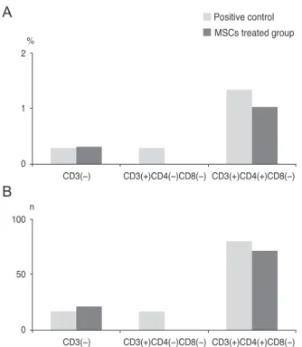
peak level of IL-17 at 1 week. Therefore, we chose the time point of 1 week to assess the anti-inflammatory effect of MSCs on the IL-17-secreting cells. The IL-17 secretion was reduced by 53.7%, 43.8%, and 50.8% in CD3(+)CD4(-) CD8(-) cells, CD3(+)CD4(+)CD8(-) cells (Th17), and CD3(+)CD4(-)CD8(+) cells, respectively, of the cervical lymph nodes when MSCs were applied (Fig. 5). Addition- ally, analysis of the cornea indicated that IL-17 secretion from CD3(+)CD4(-)CD8(-) cells was completely blocked, while the secretion of IL-17 in the CD3(+)CD4(+)CD8(-) cells (Th17 cells) was partially reduced by 10.5% (Fig. 6).
IL-17-secreting CD3(+)CD4(-)CD8(+) T-cells were not de- tected in the cornea. This result suggested that MSCs mainly modulate CD3(+)CD4(-)CD8(-) non-Th17 cells, and also partially suppress CD3(+)CD4(+)CD8(-) Th17 cells to inhibit IL-17 secretion in a chemical burn model.
Discussion
Our study revealed that both innate and adaptive IL-17-secreting cells were involved in chemically injured eye recovery, and that MSCs effectively suppressed IL-17 secretion in this model by blocking both CD3(+)CD4(-) CD8(-) non-Th17 cells and CD3(+)CD4(+)CD8(-) Th17 cells. To our knowledge, this is the first report to determine which cells are modulated by MSCs to attenuate IL-17 se- cretion in an ocular chemical burn model.
Systemically, in response to burn injury, IL-1b, TNF-a, and IL-6 are initially increased. However, after an initial pro-inflammatory phase, studies have determined that burn injury can result in a loss of Th1 cells, associated with depressed levels of IFN-g and a concomitant increase of Th2 cells. In regards to innate immunity, decreased macrophages and NK cells have been reported to increase the susceptibility of infection [23].
The initial responses of neutrophils and macrophages were time-dependent and the analysis in ocular chemical burn conditions was well dissected in order to understand innate immune reactions [24]. T cell involvement in ocular burns, as an adaptive immune response, was also reported, including the Th17 cells [17,25,26]; however, the details of T cell involvement have not been determined in sterile in- flammation.
Our chemical burn model showed that IL-17 was in- volved in the sterile inflammation process. ELISA data re-
vealed high IL-17 levels in the cornea at 1 week. However, contrary to IL-6, which was increased after 6 hours, the response of IL-17 appeared delayed; therefore we ques- tioned the main sources of IL-17. There are numerous sources that discuss IL-17-secreting cells, and the major cell sources of IL-17A and IL-17F are known to be Th17 cells, gammadelta T-cells, innate lymphoid cells, NK cells, NK T-cells, and CD8(+) T-cells. In addition, gammadelta T-cells are crucial players in the barrier function of muco- sal immunity and with the skin, including the eyes. Our results implied that non-Th17 cells (gammadelta T-cells, NK cells, NK T-cells, or CD8(+) T-cells) in addition Th17 cells seemed to be involved in the secretion of IL-17 in the early stage of the chemical burn model. The suppression effect of MSCs was more prominent on the non-Th17 cells (gammadelta T-cells, NK cells, and NK T-cells), which se- creted IL-17, instead of on the Th17 cells in the cornea.
This result provides indirect in vivo evidence in addition to recent in vitro recent evidence that MSCs can inhibit gammadelta T-cells and NK T-cells [16]. Although MSCs inhibit Th17 cells [11-14,27], MSCs seem to modulate the IL-17-secreting cells, Th17 cells, and also non-Th17 cells in sterile inflammation.
Although the results in this study cannot be directly ap- plied to human patients because of the application method (intraperitoneal injection), this study highlights the role of MSCs in sterile inflammation, such as chemical burns, and shows that they can reduce IL17-associated inflammation, in the early stages, that leads to ocular blindness. Addi- tional studies are needed pending the development of ap- propriate application methods for clinical usage. Our study was limited by the indirect evidence because we negatively sorted the non-Th17 cells, and these non-Th17 cells includ- ed mixed cell populations (gammadelta T-cells, NK cells, and NK T-cells). Nevertheless, it would be noteworthy to show the effect of MSCs on the non-Th17 cells in vivo model, which could help with understanding IL-17-secret- ing cell changes during an adaptive immune response in an ocular chemical burn. Because of the small number of experimental sets (all the cornea were pooled, lymph node data n = 5), we could not directly determine the statistical significance and relationships in the study. However, we demonstrated the immunological profile tendencies over time and the tendency of changes in the MSCs application, which is important for future studies. Another limitation is that we used rat MSCs in the mouse study. MSC immuno-
Fig. 5. Fluorescent-activated cell sorter analysis of cervical lymph nodes on day 7 (A), following corneal chemical injury in the group treated with mesenchymal stem cells (MSCs) and the control group (each group, n = 10). The bar charts show the (B) percentages and (C) cell numbers of interleukin (IL)-17=secreting cells in cervical lymph nodes. Both non-T helper 17 (Th17) cells (CD3(+)CD4(-)CD8(-) and CD3(+)CD4(-)CD8(+)) and Th17 cells were effectively reduced in the group treated with MSCs. All of the cervical lymph nodes for each group were pooled to perform the experiment.
0 200 400 600 800 47.33%
49.52%
100 1K 101 102 103 104
CD3-PerCP
MSCs treated group
IL-17A-PEIL-17A-PE IL-17A-PE IL-17A-PE IL-17A-PE
IL-17A-PE IL-17A-PE IL-17A-PE
CD8-PE-cy7
FSC-A CD4-FITC
CD4-FITC CD8-PE-cy7
CD3-PerCP
CD4-FITC CD8-PE-cy7
CD3-PerCP CD3-PerCP
100 101 102 103 104 2.22%
33.15%
11.53%
100 101 102 103 104
100 101 102 103 104
0.00% 2.03%
0.68% 97.30%
100 101 102 103 104
100 101 102 103 104
0.02% 0.32%
2.71% 96.96%
100 101 102 103 104
100 101 102 103 104
0.00% 0.65%
0.00% 99.35%
100 101 102 103 104
CD3-PerCP 100 101 102 103 104
0.46% 0.01%
98.39% 1.14%
100 101 102 103 104
100 101 102 103 104
0.00% 0.94%
1.08% 97.98%
100 101 102 103 104
100 101 102 103 104
0.04% 0.18%
5.08% 94.71%
100 101 102 103 104
100 101 102 103 104
0.00% 0.36%
0.00% 99.64%
100 101 102 103 104
100 101 102 103 104
0.42% 0.00%
96.14% 3.44%
100 101 102 103 104 Positive control
Positive control MSCs treated group
0 20,000 40,000 60,000 n
CD3(+)CD4(−)CD8(−)
CD3(−) CD3(+)CD4(+)CD8(−) CD3(+)CD4(−)CD8(+) 0
1 2 3%
CD3(+)CD4(−)CD8(−)
CD3(−) CD3(+)CD4(+)CD8(−) CD3(+)CD4(−)CD8(+)
A
B C
suppressive properties are believed to permit allogenic or even xenogenic transplantation. Spontaneous immortaliza- tion and malignant transformation of human adipose and mouse bone-marrow-derived MSCs after in vitro culture expansion have been reported [28-33]. In contrast to the behavior of mouse cells, MSC cultures derived from rat bone marrow mononuclear cells maintained contact inhi- bition in culture and showed no evidence of lung tumor formation after intravenous injection into syngeneic recip- ients [31]. Therefore, we preferred rat MSCs rather than mouse MSCs for this study based on this reason. However, further study is needed to determine the stability and xe- nogenic effects of cultured rat MSCs.
In conclusion, both IL-17-secreting, non-Th17 cells and Th17 cells were involved in the ocular sterile inflammation induced by a chemical burn, and MSCs effectively sup- pressed the non-Th17 cells in the cornea.
Conflict of Interest
No potential conflict of interest relevant to this article was reported.
Acknowledgements
This work was supported by the National Research Foundation of Korea Grant funded by the Korean Govern- ment (MEST) (NRF 2010-0010629 and NRF 2011- 0004128).
References
1. Bassi EJ, Aita CA, Camara NO. Immune regulatory prop- erties of multipotent mesenchymal stromal cells: where do we stand? World J Stem Cells 2011;3:1-8.
2. Bassi EJ, de Almeida DC, Moraes-Vieira PM, Camara NO.
Exploring the role of soluble factors associated with im- mune regulatory properties of mesenchymal stem cells.
Stem Cell Rev 2012;8:329-42.
3. Prockop DJ, Oh JY. Mesenchymal stem/stromal cells (MSCs): role as guardians of inflammation. Mol Ther 2012;
20:14-20.
4. Shi Y, Hu G, Su J, et al. Mesenchymal stem cells: a new strategy for immunosuppression and tissue repair. Cell Res 2010;20:510-8.
5. Chen SJ, Wang YL, Fan HC, et al. Current status of the immunomodulation and immunomediated therapeutic strategies for multiple sclerosis. Clin Dev Immunol 2012;2012:970789.
6. Raychaudhuri SP. Role of IL-17 in psoriasis and psoriatic arthritis. Clin Rev Allergy Immunol 2013;44:183-93.
7. Marwaha AK, Leung NJ, McMurchy AN, Levings MK.
TH17 cells in autoimmunity and immunodeficiency: pro- tective or pathogenic? Front Immunol 2012;3:129.
8. Maddur MS, Miossec P, Kaveri SV, Bayry J. Th17 cells: bi- ology, pathogenesis of autoimmune and inflammatory dis- eases, and therapeutic strategies. Am J Pathol 2012;181:8-18.
9. Hirota K, Ahlfors H, Duarte JH, Stockinger B. Regulation and function of innate and adaptive interleukin-17-produc- ing cells. EMBO Rep 2012;13:113-20.
10. Fossiez F, Banchereau J, Murray R, et al. Interleukin-17. Int Rev Immunol 1998;16:541-51.
11. Duffy MM, Pindjakova J, Hanley SA, et al. Mesenchymal
CD3(−) 0
1 2%
CD3(+)CD4(−)CD8(−) CD3(+)CD4(+)CD8(−) Positive control MSCs treated group
CD3(−) 0
50 100n
CD3(+)CD4(−)CD8(−) CD3(+)CD4(+)CD8(−)
Fig. 6. Fluorescent-activated cell sorter analysis of corneas on day 7, following corneal chemical injury in the group treated with mesenchymal stem cells (MSCs) and the control group.
The bar charts show the (A) percentages and (B) cell numbers of the interleukin (IL)-17-secreting cells in the corneas. Complete blockage of IL-17-secreting and the non-T helper 17 cells (CD3(+)/
CD4(-)/CD8(-) cells) was shown in the MSC treated group. All of the cornea and lymph nodes for each group were pooled to per- form the experiment.
A
B
stem cell inhibition of T-helper 17 cell- differentiation is triggered by cell-cell contact and mediated by prostaglandin E2 via the EP4 receptor. Eur J Immunol 2011;41:2840-51.
12. Duffy MM, Ritter T, Ceredig R, Griffin MD. Mesenchy- mal stem cell effects on T-cell effector pathways. Stem Cell Res Ther 2011;2:34.
13. Ghannam S, Pene J, Moquet-Torcy G, et al. Mesenchymal stem cells inhibit human Th17 cell differentiation and func- tion and induce a T regulatory cell phenotype. J Immunol 2010;185:302-12.
14. Ohshima M, Yamahara K, Ishikane S, et al. Systemic trans- plantation of allogenic fetal membrane-derived mesenchy- mal stem cells suppresses Th1 and Th17 T cell responses in experimental autoimmune myocarditis. J Mol Cell Cardiol 2012;53:420-8.
15. Qu X, Liu X, Cheng K, et al. Mesenchymal stem cells inhibit Th17 cell differentiation by IL-10 secretion. Exp Hematol 2012;40:761-70.
16. Prigione I, Benvenuto F, Bocca P, et al. Reciprocal interac- tions between human mesenchymal stem cells and gam- madelta T cells or invariant natural killer T cells. Stem Cells 2009;27:693-702.
17. Sasaki JR, Zhang Q, Schwacha MG. Burn induces a Th-17 inflammatory response at the injury site. Burns 2011;37:
646-51.
18. Ye J, Yao K, Kim JC. Mesenchymal stem cell transplanta- tion in a rabbit corneal alkali burn model: engraftment and involvement in wound healing. Eye (Lond) 2006;20:482-90.
19. Ye J, Lee SY, Kook KH, Yao K. Bone marrow-derived pro- genitor cells promote corneal wound healing following al- kali injury. Graefes Arch Clin Exp Ophthalmol 2008;246:
217-22.
20. Yao L, Li ZR, Su WR, et al. Role of mesenchymal stem cells on cornea wound healing induced by acute alkali burn.
PLoS One 2012;7:e30842.
21. Oh JY, Kim MK, Shin MS, et al. The anti-inflammatory and anti-angiogenic role of mesenchymal stem cells in cor- neal wound healing following chemical injury. Stem Cells
2008;26:1047-55.
22. Ma Y, Xu Y, Xiao Z, et al. Reconstruction of chemically burned rat corneal surface by bone marrow-derived human mesenchymal stem cells. Stem Cells 2006;24:315-21.
23. Church D, Elsayed S, Reid O, et al. Burn wound infections.
Clin Microbiol Rev 2006;19:403-34.
24. Oh JY, Choi H, Lee RH, et al. Identification of the HSPB4/
TLR2/NF-κB axis in macrophage as a therapeutic target for sterile inflammation of the cornea. EMBO Mol Med 2012;4:435-48.
25. Tazetdinova NR, Iurovskaia NN. Changes in the T-lym- phocyte subpopulations in patients with severe eye burns.
Oftalmol Zh 1990;(6):335-8.
26. Zhao M, Chen J, Yang P. Immunologic experimental stud- ies on the alkali burn of cornea in rats. Zhonghua Yan Ke Za Zhi 2000;36:40-2, 4.
27. Darlington PJ, Boivin MN, Renoux C, et al. Reciprocal Th1 and Th17 regulation by mesenchymal stem cells: Implica- tion for multiple sclerosis. Ann Neurol 2010;68:540-5.
28. Rubio D, Garcia-Castro J, Martin MC, et al. Spontaneous human adult stem cell transformation. Cancer Res 2005;65:
3035-9.
29. Wang Y, Huso DL, Harrington J, et al. Outgrowth of a transformed cell population derived from normal human BM mesenchymal stem cell culture. Cytotherapy 2005;7:
509-19.
30. Miura M, Miura Y, Padilla-Nash HM, et al. Accumulated chromosomal instability in murine bone marrow mesen- chymal stem cells leads to malignant transformation. Stem Cells 2006;24:1095-103.
31. Zhou YF, Bosch-Marce M, Okuyama H, et al. Spontaneous transformation of cultured mouse bone marrow-derived stromal cells. Cancer Res 2006;66:10849-54.
32. Aguilar S, Nye E, Chan J, et al. Murine but not human mesenchymal stem cells generate osteosarcoma-like lesions in the lung. Stem Cells 2007;25:1586-94.
33. Tolar J, Nauta AJ, Osborn MJ, et al. Sarcoma derived from cultured mesenchymal stem cells. Stem Cells 2007;25:371-9.