Postoperative maxillary cysts, also known as surgical ciliated cysts, are tardy complications after a radical sur- gery for maxillary sinus disease.1They are locally aggres- sive unilocular or multilocular and fully or partially sinus- occupying lesions.2,3As the size of the lesions increases, the cysts may displace or perforate adjacent sinus walls and invade adjacent tissues.2These lesions clinically pre- sent as expansive swelling of the dental alveolus and palate with pain, discharge, or fistula formation.4
Sugar et al5 first reported three cases of postoperative maxillary cysts that occurred after orthognathic surgery, and since then, several additional cases have been report- ed.6-8As the number of cases of orthognathic surgery has increased recently, the diagnosis of postoperative maxillary cysts has become more important. Although panoramic and Waters’ radiographs are the commonly used diagnos- tic imaging modalities for these lesions, the use of com- puted tomography (CT) has been proposed recently due
to its relatively high diagnostic accuracy.9
This report describes a case of bilateral postoperative maxillary cysts following orthognathic surgery performed approximately 21 years ago; these cysts were diagnosed using some imaging modalities including CT. To the best of our knowledge, this is the first report of postoperative maxillary cysts occurring in both maxillary sinuses.
Case Report
A 42-year-old woman came to Seoul National University Dental Hospital complaining of intermittent stinging pain in her right cheek. Previously, she had visited a local den- tal clinic due to intermittent pain in the past 6 months. The vitality test of upper-right teeth had been performed. Even though the teeth had been vital, she had received endo- dontic treatment on the upper-right second premolar due to worsening pain. Then, she was referred to the Depart- ment of Oral and Maxillofacial Radiology at our hospital when she complained of new sharpening pain on the right side of her face after two or three days.
There were no clinically abnormal findings in physical and oral examinations. The patient only had a history of
─ 321 ─ Received July 9, 2014; Revised August 10, 2014; Accepted August 22, 2014
*Correspondence to : Prof. Soon-Chul Choi
Department of Oral and Maxillofacial Radiology, School of Dentistry, Seoul National University, Daehak-ro 101, Jongno-gu, Seoul 110-749, Korea
Tel) 82-2-2072-2622, Fax) 82-2-744-3919, E-mail) [email protected]
Imaging Science in Dentistry 2014; 44: 321-4 http://dx.doi.org/10.5624/isd.2014.44.4.321
Copyright ⓒ 2014 by Korean Academy of Oral and Maxillofacial Radiology
This is an Open Access article distributed under the terms of the Creative Commons Attribution Non-Commercial License (http://creativecommons.org/licenses/by-nc/3.0) which permits unrestricted non-commercial use, distribution, and reproduction in any medium, provided the original work is properly cited.
Imaging Science in Dentistry∙pISSN 2233-7822 eISSN 2233-7830
Bilateral postoperative maxillary cysts after orthognathic surgery: A case report
Jung-Hye Lee1, Kyung-Hoe Huh1, Won-Jin Yi1, Min-Suk Heo1, Sam-Sun Lee1, Soon-Chul Choi1,*
1Department of Oral and Maxillofacial Radiology and Dental Research Institute, School of Dentistry, Seoul National University, Seoul, Korea
ABSTRACT
Postoperative maxillary cysts are locally aggressive lesions, usually developing as delayed complications many years after radical antral surgery. This report describes a case of bilateral postoperative maxillary cysts following orthognathic surgery performed approximately 21 years previously. The patient complained of stinging pain on her right cheek. Radiographic examination revealed low-attenuation lesions on both maxillary sinuses with discontinu- ously corticated margins without distinct expansion or bone destruction. The cysts were enucleated with the removal of metal plates and screws for pain relief. Histopathological examination confirmed the diagnosis of postoperative maxillary cysts lined by ciliated, pseudostratified columnar cells. The patient has remained asymptomatic thus far, and there was no evidence of local recurrence at 21 months of postoperative follow-up.
(Imaging Sci Dent 2014; 44: 321-4)
KEY WORDS: Maxillary Sinus, Orthognathic Surgery, Cysts, Radiography
orthognathic surgery on both the maxilla and the mandible performed about 21 years prior to this visit. Panoramic and Waters’ radiographs revealed increased haziness and dome-shaped homogeneous radiopacities in the lower regions of both maxillary sinuses (Fig. 1). Endodontic re-
treatment was performed on the upper-right second pre- molar because it was thought that radiologically, the ori- gins of the pain were not in the maxillary sinuses but in the teeth, resulting in only a temporary reduction of pain that later intensified.
─ 322 ─ Bilateral postoperative maxillary cysts after orthognathic surgery: A case report
Fig. 1.A. Panoramic radiograph shows dome-shaped homogeneous radiopacities at the lower regions in both maxillary sinuses. There are metal plates, screws in the maxilla, and wires on the mandible from the previous orthognathic surgery. B. Waters’ radiograph shows increas- ed haziness at the lower regions in both maxillary sinuses, but there is no distinct expansion or bone destruction.
A B
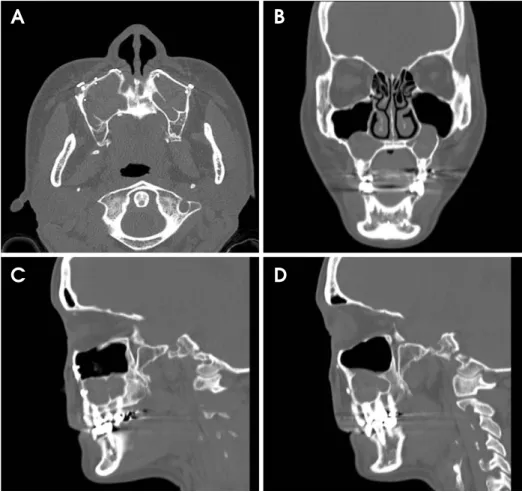
Fig. 2.Computed tomography images show low-attenuation lesions in both maxillary sinuses. A. Axial image reveals no distinct expansion and bone destruction of the lesions. Postopera- tive signs such as antral wall thicken- ing, sclerotic change, and loss of con- tinuity of the cortical antral wall lin- ing are not seen. B. Coronal image shows that the lesions extended slight- ly below the floor of both nasal sinus- es. C and D. Sagittal images of right (C) and left (D) maxillary sinuses reveal the undulated margins, which were discontinuously corticated.
A B
C D
Computed tomography (CT) was performed for finding other origins of the pain, and low-attenuation lesions were detected in both maxillary sinuses. The lesions measured approximately 36 mm×27 mm×20 mm in the right max- illary sinus, and 30 mm×21 mm×25 mm in the left max- illary sinus. The margins were undulated, while corticated discontinuously. The lesions extended slightly below the floor of both nasal sinuses. No internal calcifications were present. Postoperative signs including antral wall thicken- ing, sclerotic change, and loss of continuity of the cortical antral wall lining10,11were not observed on the CT images.
Furthermore, there were no characteristic features of dis- tinct expansion or bone destruction of soft tissue (Fig. 2).
The lesions were eventually diagnosed as retention pseu- docysts formed between septa.
The origins of the pain seemed to be not odontogenic lesions but the low-attenuation lesions in both maxillary sinuses. In order to resolve the patient’s severe pain, enu- cleation of the cysts with the removal of the plates and screws on both maxillary sinuses was performed, and biopsy specimens were obtained for histopathological examination.
Photomicrographs showed that inflammatory cells had infiltrated around the cystic cavity (Fig. 3A). The lesions had true cystic structures lined by ciliated, pseudostratified columnar epithelium (Fig. 3B). These findings of the micro- scopic examination were consistent with the postoperative maxillary cysts. Finally the lesions were diagnosed with them in both maxillary sinuses.
At 21 months of follow-up, the lesions healed unevent- fully and there was no evidence of local recurrence.
Discussion
Postoperative maxillary cysts are mainly complications that occur after radical surgery for sinus disease.3In Japan, they occur in up to 20% of patients who undergo radical maxillary sinus surgery.12
They may also occur after Le Fort I osteotomy. The maxillary sinuses, nasal sinuses, and mucosa of nasopala- tine duct can be damaged during Le Fort I osteotomy. At the time of such damage, the mucosal cells are inserted between bony edges of the osteotomies6and result in cys- tic degeneration.5The present case of postoperative max- illary cysts might have occurred due to this mechanism.
There have been a few reports of postoperative maxillary cysts after orthognathic surgery.5-8 These cysts occurred on various regions of the maxilla or the mandible, mostly 3-7 years after the surgery. In Korea, only two cases have been previously reported; one occurred between the left paramedian side of maxillary alveolar process and hard palate 3 years after the surgery,13and the other on the ante- rior region of maxilla 6 years after.14The patients com- plained of pain, tenderness, and swelling similar to other patients with postoperative maxillary cysts.
This report might be the first reported case of postoper- ative bilateral maxillary cysts following orthognathic sur- gery. Radiologically, none of the typical postoperative
─ 323 ─
Jung-Hye Lee et al
Fig. 3. A. Histopathological examination shows the inflammatory cells infiltrating around the cystic cavity (H&E stain, 40×). B. The cyst is lined by ciliated, pseudostratified columnar cells (H&E stain, 400×).
A B
symptoms, or distinct expansion or bone destruction, appe- ared on either maxillary sinus. The lesions occurred in both maxillary sinuses and had a similar appearance. The lesions extended slightly below the floor of both nasal sinuses; however, the sinuses were considered to be not pathologic but normal maxillary sinuses. In addition, they occurred as long as 21 years after the surgery and did not seem to be aggressive. Therefore, postoperative maxillary cysts could be excluded, and the initial diagnosis was retention pseudocysts formed between septa in both the maxillary sinuses.
However, the cystic cavities were histopathologically lined by ciliated, pseudostratified columnar cells with infiltrations of inflammatory cells, establishing the diag- nosis of bilateral postoperative maxillary cysts.
On the CT images, it was supposed that the origins of these lesions were the screws on the lower area of the anterior walls of both maxillary sinuses because the screws were the only surgical evidence around the lesions.
Postoperative maxillary cysts after orthognathic surgery have not been reported often; however, their prevalence might increase and the lesions might be located in various areas of the jaw because of the recent rapidly increasing number of the cases of orthognathic surgery for cosmetic purposes.13
In order to prevent this complication after orthognathic surgery, complete removal of respiratory mucosa, minimi- zation of bleeding, prevention of maxillary ostium obstruc- tions, and avoidance of irritation of the surrounding tissues are needed during the surgery.14However, it is difficult to prevent the formation of postoperative maxillary cysts in the maxillary sinus even when the surgeons pay attention to the abovementioned factors.
Panoramic and Waters’ radiographs are of value in the detection of maxillary sinus diseases;15-17however, CT is superior to plain films18and improves the ability to evalu- ate postoperative maxillary cysts.9 Therefore, although periodic plain radiographic follow-up is required for pati- ents who have undergone orthognathic surgery, these pati- ents may also need to undergo CT depending on the cir- cumstances, for finding the location, size, and extent of the lesions and making an accurate diagnosis.
References
1. Hasegawa M, Saito Y, Watanabe I, Kern EB. Postoperative mucoceles of the maxillary sinus. Rhinology 1979; 17: 253-6.
2. Pe MB, Sano K, Kitamura A, Inokuchi T. Computed tomo- graphy in the evaluation of postoperative maxillary cysts. J Oral Maxillofac Surg 1990; 48: 679-84.
3. Lee GI, Park TW. Clinical and radiological study of the post- operative maxillary cyst. J Korean Acad Oral Maxillofac Radiol 1994; 24: 47-55.
4. Shik CK. The post-operative maxillary cyst: report of 14 cases.
Taehan Chikkwa Uisa Hyophoe Chi 1989; 27: 1049-57.
5. Sugar AW, Walker DM, Bounds GA. Surgical ciliated (post- operative maxillary) cysts following mid-face osteotomies. Br J Oral Maxillofac Surg 1990; 28: 264-7.
6. Hayhurst DL, Moenning JE, Summerlin DJ, Bussard DA.
Surgical ciliated cyst: a delayed complication in a case of maxillary orthognathic surgery. J Oral Maxillofac Surg 1993;
51: 705-9.
7. Amin M, Witherow H, Lee R, Blenkinsopp P. Surgical ciliated cyst after maxillary orthognathic surgery: report of a case. J Oral Maxillofac Surg 2003; 61: 138-41.
8. Bourgeois SL Jr, Nelson BL. Surgical ciliated cyst of the mandible secondary to simultaneous Le Fort I osteotomy and genioplasty: report of case and review of the literature. Oral Surg Oral Med Oral Pathol Oral Radiol Endod 2005; 100: 36-9.
9. Heo MS, Song MY, Lee SS, Choi SC, Park TW. A compara- tive study of the radiological diagnosis of postoperative max- illary cyst. Dentomaxillofaci Radiol 2000; 29: 347-51.
10. Noyek AM. Radiology of the maxillary sinus after Caldwell- Luc surgery. Otolaryngol Clin North Am 1976; 9: 135-51.
11. Unger JM, Dennison BF, Duncavage JA, Toohill RJ. The radiological appearance of the post-Caldwell-Luc maxillary sinus. Clin Radiol 1986; 37: 77-81.
12. Kaneshiro S, Nakajima T, Yoshikawa Y, Iwasaki H, Tokiwa N. The postoperative maxillary cyst: report of 71 cases. J Oral Surg 1981; 39: 191-8.
13. Kim JM, Cho JH, Jung HV, Choi JM, Kim JS, Kim JK. Post- operative maxillary cyst after maxillary orthognathic surgery:
report of an unusual case. J Rhinol 2012; 19: 60-2.
14. Kim JK, Choi YS, Kim SY, Yi CK. Postoperative maxillary cyst after orthognathic surgery. J Korean Assoc Maxillofac Plast Reconstr Surg 1996; 18: 120-4.
15. Lyon HE. Reliability of panoramic radiography in the diagno- sis of maxillary sinus pathosis. Oral Surg Oral Med Oral Pathol 1973; 35: 124-8.
16. Ohba T, Katayama H. Comparison of panoramic radiography and Water’s projection in the diagnosis of maxillary sinus disease. Oral Surg Oral Med Oral Pathol 1976; 42: 534-8.
17. Choi SC, Ahn HK. Radiologic study of the maxillary sinusitis.
J Korean Acad Oral Maxillofac Radiol 1981; 11: 41- 50.
18. Cable HR, Jeans WD, Cullen RJ, Bull PD, Maw AR. Compu- terized tomography of the Caldwell-Luc cavity. J Laryngol Otol 1981; 95: 775-83.
─ 324 ─ Bilateral postoperative maxillary cysts after orthognathic surgery: A case report