http://dx.doi.org/10.4068/cmj.2012.48.1.57
ⒸChonnam Medical Journal, 2012 57 Chonnam Med J 2012;48:57-59
Images in Clinical Medicine
www.cmj.ac.kr
FIG. 2. Abdominal computed tomography confirmed perirenal and pelvic cavity fluid collection (white arrow). The black arrow in- dicates the double J stent.
FIG. 1. Renal ultrasound showing hydronephrosis and perirenal fluid collection (white arrow).
Perirenal Fluid Collection after Kidney Transplantation
Min Jee Kim, Chang Seong Kim, Joon Seok Choi, Eun Hui Bae, Seong Kwon Ma and Soo Wan Kim*
Department of Internal Medicine, Chonnam National University Medical School, Gwangju, Korea
A 30-year-old male presented with pitting edema. He had received a kidney trans- plantation 3 months previously. His serum creatinine level was increased, and a renal ultrasound showed hypoechoic fluid collection in the perirenal space and pelvic cavity.
We conducted sono-guided percutaneous drainage of the fluid collected in the pelvic cavity. The chemistry of the peritoneal fluid was more equivalent to serum chemistry values than to urinary values. Simple aspiration and treatment with antibiotics were performed. We have presented a case of lymphocele after kidney transplantation. This case suggests that physicians should remember how to differentiate the pelvic cavity fluid collection in patients who have received a kidney transplant.
Key Words: Lymphocele; Kidney; Transplantation
This is an Open Access article distributed under the terms of the Creative Commons Attribution Non-Commercial License (http://creativecommons.org/licenses/by-nc/3.0) which permits unrestricted non-commercial use, distribution, and reproduction in any medium, provided the original work is properly cited.
Article History:
received March 13, 2012 accepted March 20, 2012
Corresponding Author:
Soo Wan Kim
Department of Internal Medicine, Chonnam National University Medical School, 671 Jebong-ro, Gwangju 501-757, Korea
TEL: +82-62-220-6271 FAX: +82-62-220-8578 E-mail: skimw@chonnam.ac.kr
THE CASE: WHAT IS THE CAUSE OF IMPAIRED RENAL FUNCTION?
A 30-year-old male presented to Chonnam National University Hospital complaining of pitting edema of both lower extremities. He had received a kidney trans- plantation 1 month previously. A double J stent had been inserted to prevent urine leakage after kidney transplan-
tation. The patient’s medications consisted of tacrolimus, mycophenolate mofetil, and methylprednisolone. The physical examination showed a soft abdomen with right costovertebral angle tenderness. Laboratory studies were as follows: blood urea nitrogen, 24.7 mg/dl; serum crea- tinine, 1.4 mg/dl; and C-reactive protein, 3.5 mg/dl. One
58
Perirenal Fluid Collection after Kidney Transplantation
FIG. 4. (A) Percutaneous drainage from the intraabdominal fluid col- lection was conducted. (B) Abdomi- nal computed tomography con- firmed improved perirenal fluid collection. The black arrow in- dicates the double J stent.
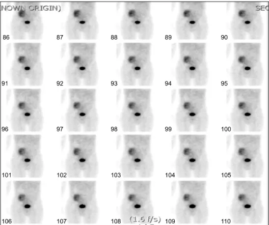
FIG. 3. Technetium99m diethylene- triamine penta-acetic acid renal scan showing focal tracer accumu- lation in the pelvic area with evi- dence of abnormal leakage from the transplanted kidney and ureter.
week later, the follow-up serum creatinine level had in- creased from 1.4 mg/dl to 3.0 mg/dl. The renal ultrasound showed a resistive index (RI index) of 0.53 and hypoechoic fluid collection in the pelvic cavity and around the trans- planted kidney. Follow-up renal ultrasound showed hydro- nephrosis of the transplanted kidney (Fig. 1) and pelvic cavity fluid collection, which was confirmed by abdominal computed tomography (Fig. 2).
We performed dynamic renal scintigraphy by use of tech- netium99m diethylenetriamine penta-acetic acid. The scan showed focal tracer accumulation in the pelvic area, and there was evidence of abnormal leakage from the trans- planted kidney and ureter (Fig. 3). To distinguish between lymphocele and urinoma, we conducted sono-guided percu- taneous drainage of the fluid collected in the pelvic cavity
(Fig. 4). Serum, urine, and aspirated peritoneal fluid chem- istries were analyzed. The chemistry of the peritoneal fluid was more equivalent to the serum values than to the uri- nary values (Table 1).
THE DIAGNOSIS: LYMPHOCELE AFTER KIDNEY TRANSPLANTATION
The common etiologies of perirenal fluid collections oc- curring after kidney transplantation include urinomas, hematomas, abscesses, and lymphoceles.1 Examination of the aspirate fluid is an essential method for specifying the kind of perirenal and pelvic cavity fluid collection. Analysis of fluid composition is also helpful in identifying urine leak- age, because higher creatinine and potassium concen-
59
Min Jee Kim, et al
TABLE 1. Chemical analysis of aspirated peritoneal fluid, serum, and urine
Serum Peritoneal fluid Urine BUN (mg/dl)
Creatinine (mg/dl) Na (mEq/L) K (mEq/L) Cl (mEq/L)
39.6 3.0 137 3.8 109
38.8 2.8 136 4.1 116
201.4 34.6
98 10.0
100
trations and lower sodium concentrations are detected in urine than in lymphocele fluid.2,3 Indeed, urine fistulae may be responsible for perirenal collection, particularly early after surgery, through ureteral or bladder leaks.
Perirenal hematomas occur during the postoperative peri- od or after traumatism and are easily diagnosed by examin- ing the fluid composition. Abscesses are also easily identi- fied by aspirate white blood cell composition and bacterial cultures.1
The cause of lymphocele formation is unclear, but it is believed to result from transection of the lymphatic vessels accompanying the external iliac vessels during trans- plantation surgery and subsequent lymph accumulation in a nonepithelialized cavity in the extra-peritoneal plane ad- jacent to the transplanted kidney.4 The therapeutic options for lymphoceles occurring after kidney transplantation in- clude simple aspiration under imaging control drainage with or without sclerotherapy and more invasive options of laparoscopic or open surgery to fenestrate the lympho- cele into the peritoneal cavity. However, treatment deci- sions seem to be center-dependent.5 Our patient under- went simple aspiration and antibiotic treatments. His se- rum creatinine level decreased to within the normal range,
and the hydronephrosis was resolved.
Because treatments of lymphocele and urinoma differ, it is important to clarify the cause of the perirenal fluid col- lection after kidney transplantation. In addition, early de- tection of transplantation-associated complications is ex- pected to preserve renal function in patients with kidney transplantation.
In summary, we have presented a case of perirenal and pelvic cavity fluid collection after kidney transplantation.
This case suggests that physicians should remember how to differentiate lymphocele and urinoma in postobs- tructive nephropathy in patients who have received a kid- ney transplant.
REFERENCES
1. Pollak R, Veremis SA, Maddux MS, Mozes MF. The natural history of and therapy for perirenal fluid collections following renal transplantation. J Urol 1988;140:716-20.
2. Lucewicz A, Wong G, Lam VW, Hawthorne WJ, Allen R, Craig JC, et al. Management of primary symptomatic lymphocele after kid- ney transplantation: a systematic review. Transplantation 2011;92:663-73.
3. Manahan KJ, Fanning J. Peritoneal fluid urea nitrogen and crea- tinine reference values. Obstet Gynecol 1999;93:780-2.
4. Zagdoun E, Ficheux M, Lobbedez T, Chatelet V, Thuillier-Lecouf A, Bensadoun H, et al. Complicated lymphoceles after kidney transplantation. Transplant Proc 2010;42:4322-5.
5. Iwan-Zietek I, Zietek Z, Sulikowski T, Nowacki M, Zair L, Romanowski M, et al. Minimally invasive methods for the treat- ment of lymphocele after kidney transplantation. Transplant Proc 2009;41:3073-6.