Postoperative Chylothorax: the Use of Dynamic Magnetic Resonance
Lymphangiography and Thoracic Duct Embolization
INTRODUCTION
Postoperative chylothorax resulting from thoracic duct injury is an uncommon complication. In order to visualize the leakage site of lymphatic channels, magnetic resonance (MR) lymphangiography has been considered to be a potential option (1).
After localizing the cisterna chyli and leakage site in MR lymphangiography, thoracic duct embolization could be performed. In this report, we present a case involving MR lymphangiography, in which we successfully performed thoracic duct embolization.
For the embolization of the thoracic duct, we directly entered the cisterna chyli and injected diluted histoacryl glue through the leakage site of the duct.
CASE REPORT
A 58-year-old male underwent left pleurectomy, decortication, and diaphragm and pericardial resection due to malignant epithelioid mesothelioma. Two days later, left- sided pleural effusion was persistent in the patient, and a turbid, milky pleural effusion was detected in a chest tube. Pleural fluid cytology showed a lymphocyte-rich effusion with a high triglyceride content (677 mg/dL), which was suggestive of chylothorax.
The left pleural effusion lasted for 16 days after the operation and increased in spite of management with a low-fat diet. One week prior to MR lymphangiography, the total serum protein was 4.6 g/dL and the albumin level was 2.3 g/dL.
This is an Open Access article distributed under the terms of the Creative Commons Attribution Non-Commercial License (http://creativecommons.org/licenses/
by-nc/3.0/) which permits unrestricted non-commercial use, distribution, and reproduction in any medium, provided the original work is properly cited.
Received: June 15, 2018 Revised: July 5, 2018 Accepted: July 16, 2018 Correspondence to:
Hyun Jung Koo, M.D., Ph.D.
Department of Radiology and Research Institute of Radiology, Asan Medical Center, University of Ulsan College of Medicine, Olympic-ro 43 gil 88, Songpa-gu, Seoul 05505, Korea.
Tel. +82-2-3010-0358 Fax. +82-2-476-0090 E-mail: [email protected]
Copyright © 2018 Korean Society of Magnetic Resonance in Medicine (KSMRM)
Case Report
Dynamic enhanced magnetic resonance lymphangiography can be used to provide anatomic and dynamic information for various lymphatic diseases, including thoracic duct injury, and can also help to guide the thoracic duct embolization procedure. We present a case of postoperative chylothorax demonstrated by dynamic enhanced MR lymphangiography. In this case, the chyle leakage site and location of cisterna chyli were clearly visualized by dynamic enhanced MR lymphangiography, thus allowing for management with thoracic duct embolization.
Keywords: Magnetic resonance lymphangiography; Thoracic duct injury; Chylothorax;
Thoracic duct embolization
Chae Woon Lee, Hyun Jung Koo, Ji Hoon Shin, Mi young Kim, Dong Hyun Yang
Department of Radiology and Research Institute of Radiology, Asan Medical Center, Seoul, Korea Magnetic resonance imaging
Prior to MR image acquisition, the patient was positioned in a supine fashion on a MR-feasible table, which was placed in an ultrasound room outside the MR room. While the patient was under local anesthesia with lidocaine injected subcutaneously in both inguinal areas, a cardiovascular radiologist inserted a 20-gauge spinal needle into the medulla of the inguinal lymph nodes on both sides using ultrasonographic (US) guidance (Fig. 1).
The needle was connected to a short connector with 2-3 cc of contrast material (Gadovist®, gadobutrol; Bayer Inc.).
The patient table was moved into the imaging room and carefully placed within the gantry. Then, using a 3-T MR image (Ingenia; Philips Healthcare, Best, the Netherlands), the T2-weighted (T2W) turbo spin-echo fat saturation sequence and breath-hold three-dimensional (3D) dynamic T1-weighted high-resolution imaging with volumetric excitation (THRIVE) enhancement, which covered the entire chest and abdomen, were performed (Table 1). The T2W turbo spin-echo fat saturation sequence with Cartesian k-space sampling had the following acquisition parameters:
repetition time ms/echo time ms, 1744.8/650.0; flip angle, 90°; Cartesian k-space acquisition; field of view, 340.0 mm.
The 3D dynamic THRIVE enhancement had the following acquisition parameters: repetition time ms/echo time ms, 3.0/1.5; flip angle, 15°; field of view, 340.0 mm. The acquisition duration for the 3D volume was 15-20 s. After obtaining T2W and unenhanced 3D THRIVE sequences, the
contrast material was gently injected into the bilateral inguinal lymph nodes while the 3D THRIVE sequence was obtained simultaneously. The 3D THRIVE sequence was acquired every 1-2 min in order to evaluate lymphatic flow into the cisterna chyli and thoracic duct leakage site.
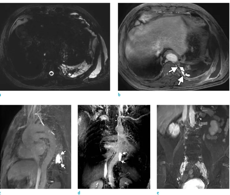
Postoperative multiloculated pleural effusion and subcutaneous fluid in the left hemithorax were noted on the T2W image (Fig. 2a). On the 3D enhanced THRIVE image, the contrast leakage site was detected on the left side of the T9-10 vertebral body level (Fig. 2b-d). The thoracic duct had a normal course and drained into the left subclavian vein. The leakage site was detected 5 min following
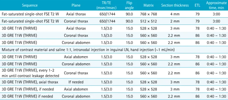
Table 1. Dynamic MR Lymphangiography Protocol
Sequence Plane TR/TE
(msec/msec) Flip
angle Matrix Section thickness ETL Approximate time, min Fat-saturated single-shot FSE T2 W Axial thorax 650/1744 90.0 768 × 768 4 mm 79 3:00 Fat-saturated single-shot FSE T2 W Coronal thorax 650/1744 90.0 512 × 512 2 mm 79 3:00
3D GRE T1W (THRIVE) Axial thorax 1.5/3.0 15.0 528 × 528 3 mm 78 0:40 - 1:30
3D GRE T1W (THRIVE) Coronal thorax 1.5/3.0 15.0 560 × 560 2.2 mm 86 0:40 - 1:30
3D GRE T1W (THRIVE) Coronal abdomen 1.5/3.0 15.0 560 × 560 2.2 mm 86 0:40 - 1:30
Mixture of contrast material and saline 1:1, intranodal injection in inguinal LN, hand injection (~1 mL/min)
3D GRE T1W (THRIVE) Axial abdomen 1.5/3.0 15.0 528 × 528 3 mm 78 0:40 - 1:30
3D GRE T1W (THRIVE) Coronal abdomen 1.5/3.0 15.0 560 × 560 2.2 mm 86 0:40 - 1:30
3D GRE T1W (THRIVE), every 1-2
min until contrast leakage detected Coronal thorax 1.5/3.0 15.0 560 × 560 2.2 mm 86 0:40 - 1:30 3D GRE T1W (THRIVE), axial thorax If needed 1.5/3.0 15.0 528 × 528 3 mm 78 0:40 - 1:30 3D GRE T1W (THRIVE), if needed Axial abdomen 1.5/3.0 15.0 528 × 528 3 mm 78 0:40 - 1:30 3D GRE T1W (THRIVE) if needed Coronal abdomen 1.5/3.0 15.0 560 × 560 2.2 mm 86 0:40 - 1:30 3D = three-dimensional; ETL = echo train length; FSE = fast spin-echo; GRE = gradient echo; TE = echo time; THRIVE = T1W high-resolution isotropic volume examination;
T1W = T1 weighted; TR = repetition time; T2W = T2 weighted
Fig. 1. Ultrasound-guided inguinal lymph node (arrowheads) selection.
contrast material injection. As we could not determine the exact time point at which to get cisterna chyli at the first abdominal acquisition 1 min following contrast injection, we repeated abdominal scanning after finding the leakage site. The cisterna chyli was noted on the left side of the T12 inferior endplate level (Fig. 2e).
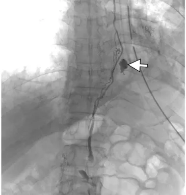
The patient underwent conventional lymphangiography for thoracic duct embolization. Lymphangiography was guided by both inguinal lymphangiography and using a 2.0 Fr Progreat catheter; the cisterna chyli was punctured
so as to enter the thoracic duct (Fig. 3). The contrast leakage site was confirmed at the same level noted on MR lymphangiography. Histoacryl glue (1 lipiodol:2 n-butyl-2 cyanoacrylate) was injected to fill the leakage site and distal thoracic duct. There were no procedure-related complications. Immediate chest radiography revealed an embolized thoracic duct. The chest drainage was resolved, and the patient was discharged three days later. The serum albumin level was normalized with follow-up laboratory results measured at the post-procedural 15th day.
Fig. 2. MR lymphangiography for visualizing the cisterna chyli and leakage site. (a) A T2W image showing postoperative multiloculated fluid collection in the left pleural space and chest wall. (b) An enhanced 3D THRIVE image and (c, d) volume- rendered image with the enhanced 3D THRIVE sequence (20 mm thickness) showing the leakage site (arrows) and that the thoracic duct drains into left subclavian vein (arrowheads). (e) Cisterna chyli (arrowhead) was noted on the left side of the T12 level.
a b
c d e
DISCUSSION
Lymphatic flow varies with patients’ conditions, as it will
be less than 1 ml/min during fasting but may return to more than 200 ml/min after eating a normal diet. An injury to the lymphatic system can result in the leakage of chyle into the
Fig. 3. Conventional lymphangiography with thoracic duct embolization. (a) The cisterna chyli (arrowhead) was selected using a 2.0 Fr Progreat catheter, and Lipiodol uptake was visualized along the thoracic duct course. (b) Thoracic duct embolization was performed at the leakage site (arrow) using histoacryl glue. (c) Immediate chest radiography revealed the embolized leakage site (arrow) and thoracic duct.
a
c
b
surrounding tissue or body cavity; chylothorax is a result of this condition. A dietary restriction of fat may reduce the lymphatic flow, and this is one of the management options for chylothorax. If dietary management fails, then it is important to identify the precise anatomic location of a lymphatic leakage, fistula, or obstruction for the effective management of this complication. Owing to the anatomic variation and small caliber of the thoracic duct and cisterna chyli, evaluating the central conducting lymphatic system is both technically challenging and time consuming. Typically, the gold standard imaging modality for lymphatic channels is bipedal lymphangiography, which is a fluoroscopic guided method used to visualize the cisterna chyli and thoracic duct. It is a useful imaging modality for the diagnosis of chyle leaks, localization of disrupted sites, and visualization of fine anatomic details (2). However, it only provides two-dimensional information, and the imaging quality can be degraded by overlapping structures. Moreover, it does not provide sufficient information on the central conducting lymphatics, including the thoracic duct and cisterna chyli, because of contrast material dilution and venous contamination (3). MR lymphangiography may be helpful in patients who are predicted to have high-flow lymphatic leakage or a large amount of lymphatic leakage, which can lead to contrast material dilution and venous contamination.
MR lymphangiography using heavily T2W images has demonstrated greater sensitivity in identifying the cisterna chyli compared to lymphangiography or CT (1).
This is a safe, noninvasive high-resolution technique for visualizing lymphatic abnormalities without ionizing radiation (4); however, this modality has several limitations, including difficulty in localizing a leakage site; a lack of dynamic information such as reflux or the adequacy of collateralization; and artifacts related to breathing, peristalsis, and cardiac pulsation. Dynamic MR lymphangiography with intra-nodal injection of gadolinium- based contrast material can provide useful information due to the dynamic monitoring of contrast transit within the lymphatic system. This minimally invasive modality is characterized by the rapid and selective opacification of the lymphatic system and provides pathologic information regarding obstruction, the degree of collateralization, and reflux (3).
For the management of chyle leakage, accurate identification of the leakage site is important regardless of
the management strategy decided on. Moreover, owing to the technical complexity of thoracic duct embolization, a structural assessment of the thoracic duct is necessary. In the present case, contrast material leakage was observed on the left side of the T9-10 vertebral body level, indicating chyle leakage. The visualization of the cisterna chyli is also important in MR lymphangiography. Lymphatic integrity disruption and anatomic variation are both common in patients who have had surgery (5). The cisterna chyli might be damaged following upper abdominal surgery, and this may interfere with interventional embolization via a transabdominal approach. A patient who has a prior history of abdominal surgery, which can cause lymphatic damage, may need MR lymphangiography before proceeding with conventional lymphangiography. For patients with nonvisualized cisterna chyli on MR lymphangiography images, surgical thoracic duct ligation may be considered.
In conclusion, we report a case of postoperative chylothorax managed by enhanced MR lymphangiography and following conventional thoracic duct embolization.
In this case, chyle leakage following thoracic surgery was effectively identified by dynamic enhanced MR lymphangiography, which accurately localized the cisterna chyli and thoracic duct, thus allowing for management with thoracic duct embolization.
REFERENCES
1. Erden A, Fitoz S, Yagmurlu B, Erden I. Abdominal confluence of lymph trunks: detectability and morphology on heavily T2-weighted images. AJR Am J Roentgenol 2005;184:35-40
2. Deso S, Ludwig B, Kabutey NK, Kim D, Guermazi A.
Lymphangiography in the diagnosis and localization of various chyle leaks. Cardiovasc Intervent Radiol 2012;35:117-126
3. Krishnamurthy R, Hernandez A, Kavuk S, Annam A, Pimpalwar S. Imaging the central conducting lymphatics:
initial experience with dynamic MR lymphangiography.
Radiology 2015;274:871-878
4. Lohrmann C, Foeldi E, Speck O, Langer M. High- resolution MR lymphangiography in patients with primary and secondary lymphedema. AJR Am J Roentgenol 2006;187:556-561
5. Pillay TG, Singh B. A review of traumatic chylothorax. Injury 2016;47:545-550