I. Introduction
Joint position sense (JPS) is derived from a complex array of information arriving at the brain from several different sources, including articular mechanoreceptors, cutane- ous afferents, and muscle receptors [1,2]. From the clinical point of view, an appropriate JPS is important to maintain the dynamic stability of the joint [3]. Joint position sense is important for the prevention of musculoskeletal injuries and diseases, such as ankle sprain and osteoarthritis, and for rehabilitation from central nervous system diseases and inju- ries, such as stroke and spinal cord injury. In particular, an- kle movements are important for a normal coordinated gait and smooth sinusoidal oscillation of the center of gravity [4].
Validation of Joint Position Sense of Dorsi-Plantar Flexion of Ankle Measurements Using
a Smartphone
Daehee Lee, PhD, PT, Seulki Han, PhD, PT
Department of Physical Therapy, U1 University, Yeongdong, Korea
Objectives: This study evaluated and validated the reliability of smartphones as measuring equipment for the dorsi-plantar flexion of ankle joint position sense (JPS) ability. Methods: The subjects were 20 healthy young students in their 20s. We con- firmed the concurrent validity by comparison with existing electrogoniometer data. The reliability of the smartphone was con- firmed using the test-retest method. Results: In the case of dorsiflexion, there was no significant difference between the smart- phone and electrogoniometer groups (p > 0.05). Regarding the correlation, it was significantly high (r = 0.65, p < 0.05), and ICC(3,1) was good (ICC(3,1) = 0.79). For the case of plantar flexion, there was no significant difference between the smart- phone and electrogoniometer groups (p > 0.05), the correlation was significantly high (r = 0.69, p < 0.05), and the ICC(3,1) was very good (ICC(3,1) = 0.82). In the case of dorsiflexion, there was no significant difference between test and retest (p > 0.05), the correlation was intermediate (r = 0.59, p < 0.05), and the ICC(3,1) value was good (ICC(3,1) = 0.74). For plantar flexion, there was no significant difference between test and retest (p > 0.05), the correlation was significantly high (r = 0.63, p < 0.05), and the ICC(3,1) was good (ICC(3,1) = 0.76). Conclusions: The results showed that smartphones provide high validity and reliability as measurement equipment for JPS of dorsi-plantar flexion of the ankle. Finally, the study also considers that smart- phone-based JPS measuring methods may replace the traditional and expensive methods that are currently being used for the same purpose.
Keywords: Proprioception, Smartphone, Ankle, Equipment, Validation Studies
Healthc Inform Res. 2017 July;23(3):183-188.
https://doi.org/10.4258/hir.2017.23.3.183 pISSN 2093-3681 • eISSN 2093-369X
Submitted: April 13, 2017
Revised: 1st, May 21, 2017; 2nd, June 17, 2017 Accepted: July 5, 2017
Corresponding Author Seulki Han, PhD, PT
Department of Physical Therapy, U1 University, 310, Daehak-ro, Yeongdong-eup, Yeongdong-gun, Chungcheongbuk-do 29131, Korea.
Tel: +82-43-740-1404, E-mail: [email protected]
This is an Open Access article distributed under the terms of the Creative Com- mons Attribution Non-Commercial License (http://creativecommons.org/licenses/by- nc/4.0/) which permits unrestricted non-commercial use, distribution, and reproduc- tion in any medium, provided the original work is properly cited.
ⓒ 2017 The Korean Society of Medical Informatics
Furthermore, the ankle is one of the most frequently injured joints of the human body [5,6]. Therefore, the measurement of ankle JPS is essential [7-9].
According to the work by Hwang et al. [10], isokinetic dy- namometers are reliable for the measurement of propriocep- tion. On the other hand, Bronner et al. [11] showed that the electrogoniometer has high reliability and validity, and it is therefore used for the movement of joints. Previous studies reported in the literature have suggested that both isokinetic dynamometers and electrogoniometers can be used as JPS measurement tools [12]. However, the penetration rate of the isokinetic dynamometer is low in clinical practice due to its cost, size, and weight. Therefore, it can be used for cases in which space is not a limitation. According to Kwon and Park [13], the weight of the lever arm of an isokinetic dyna- mometer may be discouraging in the assessment of proprio- ception. The electrogoniometer is smaller than the isokinetic dynamometer, but it also has a low penetration rate in clini- cal practice due to its high price. Although these types of measuring equipment are reliable and valid, they are not portable and their installation is time-consuming [14]. To overcome these issues and save time and cost, smartphones that include several sensors have been suggested as measure- ment and evaluation equipment [15-18]. Moreover, recent studies have reported on the reliability of smartphones for these purposes. For instance, the measurement of shoulder range of motion using a smartphone was reported by Lim et al. [19], and the measurement of heart rates using a smart- phone was investigated by Matsumura and Yamakoshi [20].
In addition, the measurement of balance using a smartphone was investigated by Han et al. [21]. However, the reliability and validity of smartphones to measure JPS of dorsi-plantar flexion of the ankle have not been studied so far.
Therefore, this study was conducted to contribute to the development of smartphone-based measurement of JPS of dorsi-plantar flexion of the ankle in an easy and economical fashion. Finally, the results obtained with this new method are compared to those obtained by the electrogoniometer, whose reliability and validity have already been proved.
II. Methods
1. Subjects
The sample size was calculated a priori using the software G*Power ver. 3.1.5. We considered a statistical power of 95% and a significance level (i.e., α-value) of 5%, and we referred to the results of Kim et al. [14]. The minimum number of subjects to be considered was 17 people, while
the number of subjects in order to account for the drop- outs was 20. Ethical approval for the study was granted by the Institutional Review Board of U1 University (Bioethics 2016-15). All subjects were fully informed of the objectives and methods of the study beforehand and gave informed consent to participate in the experiments. Only subjects with no musculoskeletal or neurological disorders affecting the upper or lower extremities, lesions, or history of surgery of the spine or upper or lower extremities were considered. A total of 20 healthy subjects were randomly selected from a group of people who responded to flyers that were strategi- cally placed throughout the university campus and also to word-of-mouth. All subjects received information about the procedures and signed an informed consent form before the testing session.
2. Measuring Equipment
The electrogoniometer used in this study was operated with a MP150 (BIOPAC System Inc., Santa Barbara, CA, USA) data acquisition system. This equipment is a basic device that digitizes the signal of the joint angle, and the extracted data is processed using a digital signal analysis program (AcqKnowledge 4.1, BIOPAC System Inc.) that ran on a per- sonal computer.
The smartphone used in this study was a Galaxy S4 (SHV- E330S; Samsung, Suwon, Korea), and the application was Sensor Kinetics Pro 2.1.2 by INNOVENTIONS Inc. (Hous- ton, TX, USA). We also used Mobizen (Rsupport, Seoul, Ko- rea) to display the output information from the smartphone and to synchronize and record it on the laptop.
3. Measurement Method
The test-retest method was used for sample 2, and all the measurements were performed at the same rate. The subjects in sample 2 were analyzed in two sessions separated by an interval of 7 days. We used an average value of 3 times the measurements in the test-retest analysis. The person who conducted the experiment was the same during the entire process, and he or she was experienced in the use of electro- goniometers and smartphones [22].
The JPS assessment was performed on the subjects posi- tioned in a sitting posture, and their maximum dorsiflexion and plantar flexion ranges of motion of the dominant side ankle were determined. The electrogoniometer was attached to sensors located at the front and back of the lateral mal- leolus of the ankle joint. The smartphone was attached to the facies plantares with Velcro (ankle support; Bioplus, Seoul, Korea) (Figure 1). During the JPS measurement, the subjects
wore shorts and used eye patches and ear plugs to minimize sensible compensation. The subjects were only allowed to move the ankle following given instructions, while the rest of the body remained at rest. Before the measurement process started, the subjects were given an explanation of the entire process. Then, a demonstration of the measurement process was shown, followed by a smooth start of the process itself.
The subjects were asked to maintain a position at a random target degree within the range of motion of their own ankle dorsiflexion, and they were asked to remain in that position for 10 seconds. After this, they were asked to return to the initial position and progressively move towards the target degree. JPS of dorsiflexion and plantar flexion of ankle joint retests were also processed, thus following the test-retest method.
4. Statistical Analysis
The software SPSS ver. 12.0 (SPSS Inc., Chicago, IL, USA) was used for the statistical analysis. A significance level of 5% (p < 0.05) was set. Each smartphone and electrogo- niometer used an average value for the middle 4 seconds within the total of 10 seconds of the manual setup and active reproduction. Subsequently, we calculated the absolute dif- ference between the values of the manual setup and active reproduction. Finally, we analyzed the absolute value. The data exhibited a normal distribution (p > 0.05), as verified by the Kolmogorov-Smirnov test, and it was presented in terms of its mean and standard deviation values. In this study, a matched-pairs t-test was carried out to determine the statistical differences between the electrogoniometer and smartphone data, namely, between each measurement of the
test-retest. In addition, the matching Pearson correlation was used to determine the correlation between the electrogoni- ometer and smartphone data, between each measurement of the test-retest. The reliability and validity was determined with the intraclass correlation coefficient (ICC(3,1)) for intra-raters [23,24]. ICC values were considered poor when below 0.20, fair from 0.21 to 0.40, moderate from 0.41 to 0.60, good from 0.61 to 0.80, and very good from 0.81 to 1.00 [23].
III. Results
1. General Characteristics of the Subjects
The general characteristics of the subjects are presented in Table 1.
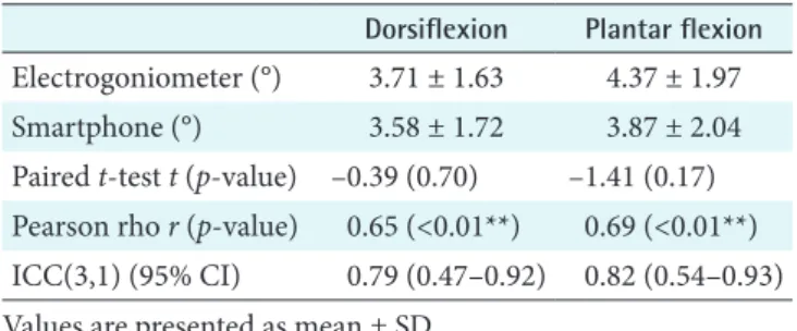
2. Validity of JPS of Dorsi-Plantar Flexion of Ankle Ob- tained Using a Smartphone
In the case of dorsiflexion, there was no significant difference between the smartphone and electrogoniometer groups (p >
0.05). Regarding the correlation, it was significantly high (r = 0.65, p < 0.05) and ICC(3,1) was good (ICC(3,1) = 0.79).
For the case of plantar flexion, there was no significant difference between the smartphone and electrogoniometer groups (p > 0.05). The correlation was significantly high (r = 0.69, p < 0.05), and the ICC(3,1) was very good (ICC(3,1) = 0.82) (Table 2).
Smartphone
Figure 1. Attached smartphone and electrogoniometer.
Table 1. General characteristics of the subjects Value
Gender (male:female) 10:10
Age (yr) 22.95 ± 4.05
Height (cm) 170.65 ± 8.29
Weight (kg) 72.50 ± 16.62
Table 2. Validity of smartphones for measuring JPS of dorsi- plantar flexion of the ankle
Dorsiflexion Plantar flexion Electrogoniometer (°) 3.71 ± 1.63 4.37 ± 1.97
Smartphone (°) 3.58 ± 1.72 3.87 ± 2.04
Paired t-test t (p-value) –0.39 (0.70) –1.41 (0.17) Pearson rho r (p-value) 0.65 (<0.01**) 0.69 (<0.01**) ICC(3,1) (95% CI) 0.79 (0.47–0.92) 0.82 (0.54–0.93) Values are presented as mean ± SD.
JPS: joint position sense, ICC: intraclass correlation coefficient, CI: confidence interval.
**p < 0.01.
3. Reliability JPS of Dorsi-Plantar Flexion of Ankle Ob- tained Using a Smartphone
In the case of dorsiflexion, there was no significant differ- ence between test and retest (p > 0.05). The correlation was intermediate (r = 0.59, p < 0.05), and the ICC(3,1) value was good (ICC(3,1) = 0.74).
For plantar flexion, there was no significant difference be- tween test and retest (p > 0.05). The correlation was signifi- cantly high (r = 0.63, p < 0.05), and the ICC(3,1) was good (ICC(3,1) = 0.76) (Table 3).
IV. Discussion
Losses in the proprioceptive system reduce the ability to continuously monitor motor sequences and interfere with coordination and balance. In particular, JPS of dorsi-plantar flexion of the ankle is important for maintaining balance during recovery [25,26].
The goal of this study was to evaluation the reliability and validity of smartphones as measuring equipment for JPS of dorsi-plantar flexion of ankle. The results of this study indi- cate that smartphones provide high validity and reliability as measurement equipment for JPS of dorsi-plantar flexion of the ankle.
Since home-based physical therapy is necessary [27], mea- surement equipment must also be easily portable. Thus, in the healthcare sector, including physical therapy, smart- phones can be broadly applied.
Some difficulties were faced in obtaining the data be- cause there are no applications for the measurement of JPS.
Therefore, efforts were directed towards the verification and consolidation of a huge amount of data. If an exclusive appli- cation to test JPS is to be developed, a thorough manual test should be made both in audio and video formats. Moreover,
the computed test results should be automatically linked to the cloud to enable users to compare them with previous re- sults. An intuitive interface should be provided so that users can easily use it in clinical tests. Moreover, the user interface should allow for the representation of graphs through which test results can be easily explained to patients.
In this study, the smartphones were attached to the facies plantares because it was more difficult to attach them to the instep or both sides of the feet. In contrast, smartphones attached to the facies plantares cannot be manipulated through their touch screens. For this reason, smartphones were remotely controlled with a laptop. The development of an exclusive JPS test application will therefore depend on the difficulty of touch screen operations. When the JPS of dorsi- plantar flexion of the ankle is measured using a smartphone, the method must be accurate. An electrogoniometer only measures the movement of the ankle to which the device is attached. However, when a smartphone is used, any joint except the ankle can be connected to the device since the movement is the same, in principle. On the other hand, the registered data was slightly influenced by the bending or straightening of the knee.
In this study, test-retest was performed in two sessions separated by an interval of 7 days. This interval was selected to represent the timeframe over which the measurement is likely to be used by clinicians to assess changes in JPS [28].
We confirmed the concurrent validity by comparing the results with those obtained by an electrogoniometer, whose validity has already been proved. According to Relph and Herrington [29], there were no significant correlations for the validity of knee JPS with image captures in the case of flexion; however, there were significant correlations in the case of extension. Hence, it can replace high-end equipment to a certain extent. In the present study, the electrogoniom- eter and smartphone concurrent validity was satisfactory.
In addition, Vuillerme et al. [30] reported that the knee JPS method showed a reliability of 0.75 points. In the present study, the reliability of measurements of JPS of dorsi-plantar flexion of the ankle obtained using smartphones was more than 0.74. Therefore, when applied to fixed joints, except for the ankle, a smartphone can be used to measure the JPS of dorsi-plantar flexion of the ankle. In this regard, this study is valuable because a protocol to process the measurement and an automatic data extraction scheme have been presented.
Moreover, it highlights the demand for the development of applications for JPS with save features and the main factors to be developed in this regard.
As the present study was conducted on young subjects only, Table 3. Reliability of smartphones for measuring JPS of dorsi-
plantar flexion of the ankle
Dorsiflexion Plantar flexion
Test (°) 3.58 ± 1.72 3.87 ± 2.04
Retest (°) 3.52 ± 1.46 3.97 ± 2.28
Paired t-test t (p-value) 0.21 (0.84) –0.53 (0.60) Pearson rho r (p-value) 0.59 (<0.01**) 0.63 (<0.01**) ICC(3,1) (95% CI) 0.74 (0.34–0.90) 0.76 (0.40–0.91) Values are presented as mean ± SD.
JPS: joint position sense, ICC: intraclass correlation coefficient, CI: confidence interval.
**p < 0.01.
our results cannot be generalized to older individuals or to those with shoulder conditions. There has been no previous study on the reliability and validity of measuring JPS of dor- si-plantar flexion of the ankle using smartphones. Therefore, the study was aimed at healthy young people. Based on the results obtained in this study, a further study will be planned with patients as subjects.
In conclusion, the evaluation of the reliability and validity of smartphones as measuring equipment for JPS of dorsi- plantar flexion of the ankle was the main goal of this study.
The results showed that smartphones provide high validity and reliability for this purpose. Finally, the results of this study also suggest that smartphone-based JPS measuring methods may replace the traditional and expensive methods that are currently being used for this purpose.
Conflict of Interest
No potential conflict of interest relevant to this article was reported.
Acknowledgments
This work was supported by the National Research Founda- tion of Korea (NRF) grant funded by the Korea government (Ministry of Science, ICT & Future Planning) (No. NRF- 2017R1C1B5014991).
References
1. Gurney, B., Milani, J., & Pedersen, M. E. (2000). Role of fatigue on proprioception of the ankle. J Exerc Physiol 2000;3(1):20-8.
2. Goble DJ, Noble BC, Brown SH. Where was my arm again? Memory-based matching of proprioceptive tar- gets is enhanced by increased target presentation time.
Neurosci Lett 2010;481(1):54-8.
3. Glencross D, Thornton E. Position sense following joint injury. J Sports Med Phys Fitness 1981;21(1):23-7.
4. Moriguchi CS, Sato TO, Gil Coury HJ. Ankle move- ments during normal gait evaluated by flexible electro- goniometer. Braz J Phys Ther 2007;11(3):205-11.
5. Garrick JG, Requa RK. The epidemiology of foot and ankle injuries in sports. Clin Sports Med 1988;7(1):29- 36.
6. Gehlson GM, Pearson D, Bahamonde R. Ankle joint strength, total work, ROM: comparison between pro- phylactic devices. J Athl Train 1991;26(1):62-5.
7. Callaghan MJ. What does proprioception testing tell us about patellofemoral pain? Man Ther 2011;16(1):46-7.
8. Lee JA, Kim DH, Shin HK. Difference of proprioceptive sense at elbow joint according to measurement meth- ods. Phys Ther Korea 2003;10(3):63-70.
9. Roberts D, Ageberg E, Andersson G, Friden T. Clinical measurements of proprioception, muscle strength and laxity in relation to function in the ACL-injured knee.
Knee Surg Sports Traumatol Arthrosc 2007;15(1):9-16.
10. Hwang JS, Lee DS, Cho YJ, Han NM, Kim HD. Mea- surement of proprioception of the knee in hemiplegic patients using an isokinetic dynamometer. J Korean Acad Rehabil Med 2010;34(1):27-33.
11. Bronner S, Agraharasamakulam S, Ojofeitimi S. Reli- ability and validity of electrogoniometry measurement of lower extremity movement. J Med Eng Technol 2010;
34(3):232-42.
12. Mohammadi F, Roozdar A. Effects of fatigue due to con- traction of evertor muscles on the ankle joint position sense in male soccer players. Am J Sports Med 2010;
38(4):824-8.
13. Kwon OY, Park DS. Effects of muscle fatigue on knee proprioception. J Korean Acad Rehabil Med 1998;22(4):
960-5.
14. Kim MC, Kim NJ, Lee MS, Moon SR. Validity reliability of the knee joint proprioceptive sensory measurements using a smartphone. J Korean Soc Phys Med 2015;
10(4):15-23.
15. Ferriero G, Vercelli S, Sartorio F, Munoz Lasa S, Ilieva E, Brigatti E, et al. Reliability of a smartphone-based goniometer for knee joint goniometry. Int J Rehabil Res 2013;36(2):146-51.
16. Jacquot F, Charpentier A, Khelifi S, Gastambide D, Rigal R, Sautet A. Measuring the Cobb angle with the iPhone in kyphoses: a reliability study. Int Orthop 2012;36(8):
1655-60.
17. Ozdalga E, Ozdalga A, Ahuja N. The smartphone in medicine: a review of current and potential use among physicians and students. J Med Internet Res 2012;14(5):
e128.
18. Boulos MN, Wheeler S, Tavares C, Jones R. How smart- phones are changing the face of mobile and participa- tory healthcare: an overview, with example from eCAA- LYX. Biomed Eng Online 2011;10:24.
19. Lim JY, Kim TH, Lee JS. Reliability of measuring the passive range of shoulder horizontal adduction using a smartphone in the supine versus the side-lying position.
J Phys Ther Sci 2015;27(10):3119-22.
20. Matsumura K, Yamakoshi T. iPhysioMeter: a new ap- proach for measuring heart rate and normalized pulse volume using only a smartphone. Behav Res Methods 2013;45(4):1272-8.
21. Han SK, Lee IH, Park NR. Reliability of static balance abilities measure using a smartphone's acceleration sen- sor. J Korea Acad Ind Cooperation Soc 2016;17(6):233- 8.
22. Perlau R, Frank C, Fick G. The effect of elastic bandages on human knee proprioception in the uninjured popu- lation. Am J Sports Med 1995;23(2):251-5.
23. Muraki T, Aoki M, Izumi T, Fujii M, Hidaka E, Miya- moto S. Lengthening of the pectoralis minor muscle during passive shoulder motions and stretching tech- niques: a cadaveric biomechanical study. Phys Ther 2009;89(4):333-41.
24. Borstad JD. Resting position variables at the shoulder:
evidence to support a posture-impairment association.
Phys Ther 2006;86(4):549-57.
25. Franco PG, Santos KB, Rodacki AL. Joint positioning sense, perceived force level and two-point discrimina- tion tests of young and active elderly adults. Braz J Phys Ther 2015;19(4):304-10.
26. Hoon AH, Stashinko EE, Nagae LM, Lin DD, Keller J, Bastian A, et al. Sensory and motor deficits in children with cerebral palsy born preterm correlate with diffu- sion tensor imaging abnormalities in thalamocortical pathways. Dev Med Child Neurol 2009;51(9):697-704.
27. Lee KJ, Roh JS. Research for the inclusion of home- based physical therapy in long-term care insurance system of physical therapists in elderly care facilities. J Korea Contents Assoc 2011;11(11):231-40.
28. Rosa DP, Borstad JD, Pires ED, Camargo PR. Reliability of measuring pectoralis minor muscle resting length in subjects with and without signs of shoulder impinge- ment. Braz J Phys Ther 2016;20(2):176-83.
29. Relph N, Herrington L. Criterion-related validity of knee joint-position-sense measurement using image capture and isokinetic dynamometry. J Sport Rehabil 2015 [Epub]. http://journals.humankinetics.com/pb- assets/hkj/JSR/Technical%20Reports/TR10_Relph%20 JSR_20130119.pdf.
30. Vuillerme N, Boisgontier M, Chenu O, Demongeot J, Payan Y. Tongue-placed tactile biofeedback suppresses the deleterious effects of muscle fatigue on joint position sense at the ankle. Exp Brain Res 2007;183(2):235-40.