bloodresearch.or.kr Blood Res 2017;52:218-33.
Letters to the Editor 219
Correspondence to: Pasquale Niscola Hematology Unit, S. Eugenio Hospital,
Rome 00144, Italy E-mail: [email protected]
Received on Nov. 6, 2016; Accepted on Jan. 17, 2017 https://doi.org/10.5045/br.2017.52.3.218
AuthorsÊ Disclosures of Potential Conflicts of Interest No potential conflicts of interest relevant to this article were reported.
REFERENCES
1. Niscola P, Vischini G, Tendas A, et al. Management of hemato- logical malignancies in patients affected by renal failure. Expert Rev Anticancer Ther 2011;11:415-32.
2. Niscola P, Tendas A, Luo XD, et al. The management of mem- branous glomerulopathy in allogeneic stem cells trans- plantation: updated literature. Cardiovasc Hematol Agents Med Chem 2013;11:67-76.
3. Launay-Vacher V, Oudard S, Janus N, et al. Prevalence of Renal Insufficiency in cancer patients and implications for anticancer drug management: the renal insufficiency and anticancer medi- cations (IRMA) study. Cancer 2007;110:1376-84.
4. Hong J, Lee S, Chun G, et al. Baseline renal function as a prog- nostic indicator in patients with newly diagnosed diffuse large B-cell lymphoma. Blood Res 2016;51:113-21.
5. Terpos E, Christoulas D, Kastritis E, et al. The Chronic Kidney Disease Epidemiology Collaboration cystatin C (CKD-EPI- CysC) equation has an independent prognostic value for overall survival in newly diagnosed patients with symptomatic multiple myeloma; is it time to change from MDRD to CKD-EPI-CysC equations? Eur J Haematol 2013;91:347-55.
6. Ofran Y, Tallman MS, Rowe JM. How I treat acute myeloid leuke- mia presenting with preexisting comorbidities. Blood 2016;
128:488-96.
7. Niscola P, Scaramucci L, Vischini G, et al. The use of major an- algesics in patients with renal dysfunction. Curr Drug Targets 2010;11:752-8.
8. Malik L, Mejia A, Weitman S. Eligibility of patients with renal impairment for Phase I trials: Time for a rethink? Eur J Cancer 2014;50:2893-6.
9. Salahudeen AK, Bonventre JV. Onconephrology: the latest fron- tier in the war against kidney disease. J Am Soc Nephrol 2013;24:26-30.
A case of synchronous multiple myeloma and chronic myeloid leukemia
TO THE EDITOR: Multiple myeloma (MM) is a hematologic malignancy caused by the proliferation of clonal plasma cells in the bone marrow, leading to uncontrolled production of monoclonal immunoglobulin. Chronic myeloid leukemia (CML) is a myeloproliferative neoplasm associated with the presence of the BCR-ABL1 fusion gene. These are un- common malignancies that account for approximately 1.6%
and 0.4% of all newly diagnosed cancers in the United States, respectively [1]. In Korea, these are also rare disease and the crude incidence rates of those are 2.5/100,000 and 0.8/100,000, respectively [2, 3]. The concurrent diagnosis of MM and CML in one patient is an extremely rare event.
In this report, we describe an additional case of synchronous MM and CML.
A 64-year-old man was referred to our hospital for evalua- tion of a right pleural soft-tissue mass and a sternal osteolytic lesion incidentally detected on chest computed tomography (CT). The patient had no specific symptoms and remarkable findings on physical examinations. A complete blood count showed a white blood cell counts of 13.1×10⁹/L, with a differential count of 65% neutrophils, 15% lymphocytes, 10% monocytes, 5.2% eosinophils, and 4% basophils; hemo- globin levels of 8.3 g/dL; and platelet counts of 234×109/L.
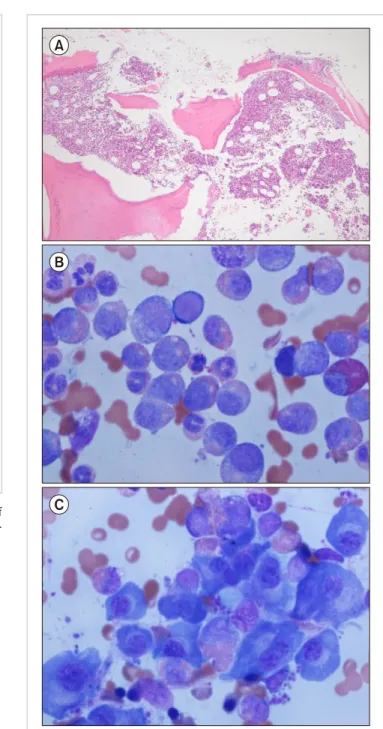
Blood chemistry showed reversal of the albumin-globulin ratio (total protein 11.8 g/dL, and albumin 2.3 g/dL), renal dysfunction (creatinine clearance 38.4 mL/min), and hyper- calcemia (calcium 11.8 mg/dL). A total of 7 g/dL of mono- clonal gammopathy (IgA lambda type) was detected with serum immunofixation electrophoresis. The ratio of serum kappa/lambda free light chains was 0.006 and serum β2 microglobulin level was 19.8 mg/L. Radiological inves- tigation revealed multiple osteolytic lesions in the skull and axial skeleton. The needle biopsy of pleural soft-tissue mass showed infiltrations of monotonous round cells with eccentrically located nuclei and different sizes, but no evi- dence of myeloid cell infiltration. Most of the infiltrated cells were positive for CD138 on immunohistochemical staining (Fig. 1). Bone marrow biopsy revealed increased cellularity (up to 90%), consisting of plasmacytoid round cell infiltrations and myeloid cells with varying maturation.
A patchy positive reaction for CD138 was compatible with myeloma involvement, and myeloid cells were positive for myeloperoxidase. The bone marrow aspirate demonstrated myeloid hyperplasia with increased eosinophils, basophils and plasma cells. Plasma cells showed mature forms with condensed nuclear chromatin, indistinct nucleoli, and abun- dant basophilic cytoplasm with a perinuclear halo (Fig. 2).
Chromosome analysis revealed 46,XY, t(2:3)(p15:q26), t(9:22)(q34:q11) in 20 of 21 cells, and fluorescent in situ
Blood Res2017;52:218-33. bloodresearch.or.kr
220 Letters to the Editor
Fig. 2. Bone marrow biopsy and aspiration at diagnosis. (A) Bone marrow biopsy shows increased cellularity up to 90% (H&E, ×100). (B) Bone marrow aspiration shows myeloid hyperplasia with increased eosinophils (Wright-Giemsa staining, ×1,000). (C) Bone marrow aspiration shows increased mature forms of plasma cells (Wright- Giemsa staining, ×1,000).
Fig. 1. Biopsy of the right pleural soft-tissue mass. (A) Infiltration of plasma cells (H&E staining, ×400). (B) Positive for immunohisto- chemical staining of CD138 (×100).
hybridization (FISH) indicated BCR/ABL translocations in 86.5% of cells. Molecular analysis revealed BCR/ABL re- arrangement based on results from reverse transcription polymerase chain reactions. Consequently, the patient was diagnosed with stage III MM and low-risk CML in accord- ance with the International Staging System [4] and Sokal scores [5], respectively. Considering the patient’s old age and toxicity of combination therapy for both CML and MM, the patient was first treated for high-stage MM.
Treatment was initiated with a regimen of thalidomide (100 mg daily) and dexamethasone (40 mg on days 1–4 and 15–18) every 4 weeks. However, the response to treatment was not evaluable, since the patient died of pneumonia caused by carbapenem-resistant Acinetobacter after 4 weeks.
Ide et al. [6] reviewed 12 cases in which MM and CML coexisted. In four of the cases, patients were diagnosed first with MM followed by CML, while in four other cases, MM developed after CML. The interval between the two types of cancer ranged from 14 months to 113 months.
In the remaining four cases, MM and CML were diagnosed simultaneously.
Although there are no established treatment regimens for simultaneously occurring MM and CML, there are two cases in which MM with CML was treated with a combina-
tion of bortezomib, dexamethasone, or lenalidomide, and imatinib or dasatinib [7, 8]. The combination treatment targeting both MM and CML resulted in successful outcomes in these cases. Our patient received only anti-MM therapy owing to old age and overall poor health status. Although data from a small study suggest that thalidomide treatment along with imatinib is efficacious for the treatment of CML [9], our patient did not respond to thalidomide treatment.
bloodresearch.or.kr Blood Res 2017;52:218-33.
Letters to the Editor 221
We presented our experience with a patient who was diagnosed with MM and CML simultaneously. Evaluation of other cases is required to shed light on clinical character- istics of the disease states, as well as to explore potential evaluable treatments.
Ji-Young Lee1, Sang-min Lee1, Hye-Kyoung Yoon2, Ki-Hyang Kim1, Moon-Young Choi1, Won-Sik Lee1
1Department of Internal Medicine, Hemato-Oncology,
2Department of Pathology, Inje University College of Medicine, Busan Paik Hospital, Busan, Korea
Correspondence to: Won-Sik Lee Department of Internal Medicine, Hemato-Oncology,
Inje University Busan Paik Hospital, 75, Bokji-ro, Busanjin-gu, Busan 47392, Korea
E-mail: [email protected]
Received on Oct. 14, 2016; Revised on Jan. 29, 2017; Accepted on Mar. 13, 2017 https://doi.org/10.5045/br.2017.52.3.219
AuthorsÊ Disclosures of Potential Conflicts of Interest No potential conflicts of interest relevant to this article were reported.
REFERENCES
1. Siegel RL, Miller KD, Jemal A. Cancer statistics, 2015. CA Cancer J Clin 2015;65:5-29.
2. Hong J, Lee JH. Recent advances in multiple myeloma: a Korean perspective. Korean J Intern Med 2016;31:820-34.
3. Au WY, Caguioa PB, Chuah C, et al. Chronic myeloid leukemia in Asia. Int J Hematol 2009;89:14-23.
4. Greipp PR, San Miguel J, Durie BG, et al. International staging system for multiple myeloma. J Clin Oncol 2005;23:3412-20.
5. Sokal JE, Cox EB, Baccarani M, et al. Prognostic discrimination in “good-risk” chronic granulocytic leukemia. Blood 1984;
63:789-99.
6. Ide M, Kuwahara N, Matsuishi E, Kimura S, Gondo H.
Uncommon case of chronic myeloid leukemia with multiple myeloma. Int J Hematol 2010;91:699-704.
7. Alsidawi S, Ghose A, Qualtieri J, Radhakrishnan N. A case of mul- tiple myeloma with metachronous chronic myeloid leukemia treated successfully with bortezomib, dexamethasone, and dasatinib. Case Rep Oncol Med 2014;2014:962526.
8. Offiah C, Quinn JP, Thornton P, Murphy PT. Co-existing chronic myeloid leukaemia and multiple myeloma: rapid response to le- nalidomide during imatinib treatment. Int J Hematol 2012;
95:451-2.
9. Monroy RH, Vargas-Viveros P, Ceballos EC, Velazquez JC, Munos SC. Imatinib (IM) plus thalidomide (Thali), a effective combination for the treatment of chronic myeloid leukemia (CML) Philadelphia chromosomepositive (Ph +) in IM-resistant disease. Report of 14 new cases from a single center in Mexico.
Blood 2013;122:5172.
Sequential heart and autologous stem cell transplantation for light-chain cardiac amyloidosis
TO THE EDITOR: Primary cardiac amyloidosis accompany- ing heart failure, angina, and/or arrhythmia is very serious and has a poor prognosis [1]. Sequential heart and autologous stem cell transplantation has resulted in some promising outcomes in a few series [2-4]. We present a case of primary amyloidosis with cardiac involvement that was successfully managed with these combined approaches.
Case
A 62-year-old woman was referred to our clinic with 3 months of dyspnea on exertion; she was categorized in New York Heart Association class III, and had abnormal echocardiographic findings. She had no other medical his- tory of note. On initial physical examination, her vital signs were as follows: blood pressure, 88/45 mmHg; pulse rate, 79 beats/min; and body temperature, 36.6oC. Neck vein en- gorgement and pretibial pitting edema were noted. Heart and lung sounds on auscultation were normal. Initial labo- ratory tests were as follows: white blood cell count, 5,600/μL;
hemoglobin, 12.4 g/dL; platelet count, 150,000/μL; protein, 6.0 g/dL; albumin, 3.6 g/dL, blood urea nitrogen, 21 mg/dL;
serum creatinine, 1.33 mg/dL; aspartate aminotransferase, 19 IU/L; alanine aminotransferase, 20 IU/L; alkaline phos- phatase, 128 IU/L; troponin I, 0.041 ng/mL; and brain natriu- retic peptide (BNP), 629 pg/mL. There were no abnormal findings on urinalysis.
A chest radiograph revealed cardiomegaly with a car- diothoracic ratio of 0.7 and increased interstitial markings suggesting pulmonary edema. Both costophrenic angles were blunted with bilateral pleural effusion. Electrocardiog- raphy displayed low voltage in leads I, II, and III and T-wave inversion in leads V5 and V6. Transthoracic echocardiog- raphy revealed thickened ventricle walls with minimal peri- cardial effusion and impaired diastolic function. Left ven- tricle (LV) filling pressure was high, with an E/E’ of 37.
No regional wall motion abnormality was observed and the LV ejection fraction was 59%. Cardiac magnetic reso- nance imaging indicated diffuse transmural or sub- endocardial enhancement at both ventricular walls on a delayed enhancement image, which were consistent with cardiac amyloidosis (Fig. 1A). Endomyocardial biopsy with a femoral venous approach was performed to confirm this diagnosis. On pathologic examination, amyloid deposits were confirmed by Congo-red staining. Immunohistochem- ical staining results were as follows: prealbumin (+); kappa chain (++); lambda chain (-); and amyloid A (-) (Fig. 1B, C). Although paraproteinemia or Bence-Jones proteinuria were not evident by electrophoresis and immunofixation, the patient’s serum free light-chain ratio was increased to 114 (kappa, 2,040.0 mg/L; lambda, 17.9 mg/L). A bone mar-