A Case of Primary Tracheal Lymphoma of a 65-year-old Female: Extremely Rare Primary Localization of
a Diffuse Large B-cell Lymphoma
You Sang Ko, M.D.1, Yu-Jin Lim, M.D.1, Jae-Hoon Yang, M.D.1, Chan-Soo So, M.D.1, Mi Jeong Kim, M.D.1, Su Hee Park, M.D.1, Sarah Park, M.D.1, Mi Na Huh, M.D.2,
Jung Han Kim, M.D.3, Chul Hong Kim, M.D.1, Jung Won Shim, M.D.4, Hee Sung Hwang, M.D.5, Young Iee Park, M.D.6 and Joo Young Jung, M.D.1
1Department of Internal Medicine, Hangang Sacred Heart Hospital, Hallym University College of Medicine,
2Department of Laboratory Medicine, Hangang Sacred Heart Hospital, Hallym University College of Medicine, 3Department of Internal Medicine, Kangnam Sacred Heart Hospital, Hallym University College of Medicine, 4Department of Pathology, Hangang Sacred Heart Hospital,
Hallym University College of Medicine, Seoul, 5Department of Diagnostic Radiology, Hallym Sacred Heart Hospital, Hallym University College of Medicine, Anyang,
6Department of Internal Medicine, National Cancer Center, Goyang, Korea
Primary extranodal non-Hodgkin’s lymphomas comprise approximately 10% of all non-Hodgkin’s lym- phomas. However, primary tracheal non-Hodgkin’s lymphoma is extremely rare, being mainly muco- sa-associated lymphoid tissue lymphoma. A 65-year-old female has dry cough for one year. She was diagnosed as diffuse large B-cell lymphoma via bronchoscopic-guided biopsy. She was treated with four cycles of the R-CHOP regimen and adjuvant radiotherapy. After completion of the combined treatment, the treatment response was complete remission, and the disease free survival was 26 months. (Korean J Hematol 2007;42:439-444.)
Key Words: Primary tracheal lymphoma, Diffuse large B-cell lymphoma, R-CHOP
439 접수:2007년 9월 14일, 수정:2007년 12월 14일
승인:2007년 12월 18일
교신저자:정주영, 서울시 영등포구 영등포동 94-200
150-719, 한림대학교 의과대학 한강성심병원 내과 Tel: 02-2639-5400, Fax: 02-2677-9736
E-mail: [email protected]
Correspondence to:Joo Young Jung, M.D.
Department of Internal Medicine, Hangang Sacred Heart Hospital, Hallym University College of Medicine
94-200, Yeongdeungpo-dong, Yeongdeungpo-gu, Seoul 150-719, Korea
Tel: +82-2-2639-5400, Fax: +82-2-2677-9736 E-mail: [email protected]
INTRODUCTION
Primary tumors of the trachea can be benign or malignant, and account for less than 0.1% of tumors. A majority of primary tracheal tumors are squamous cell or adenoid cystic carcinomas, while the rest are composed of adenoid cystic car- cinoma, mucoepidermoid carcinoma, adenoma, and others. However, squamous cell carcinoma, the most common type of primary tumor in the
trachea, is approximately 75 times more frequent in the larynx and 140∼180 times more frequent in the bronchi.1,2)
Extranodal lymphoma is defined as presented or developed disease outside the traditional lym- phoid tissue of the lymph nodes, spleen, thymus, tonsils, and Waldeyer’s ring. Extranodal non- Hodgkin’s lymphoma (NHL) is relatively com- mon; approximately 10% to 25% of NHL cases arise in extranodal sites, and their biological be-
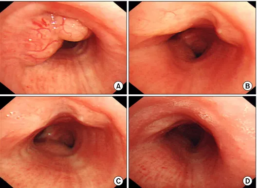
Fig. 1. Serial bronchoscopic findings. (A) At 20cm from upper incisor, multi-lobulated, multi-lobulated tumor with vasculariza- tion was seen. the tumor obstructed the tracheal lumen by about 50%. (B, C) Comparing previous bronchoscopic finding.
the size of tracheal lymphoma was marked decreased at about 5cm above carina. (B) After the patient received 2 cycles of combination chemotherapy with R-CHOP regimen. (C) After the patient received 4 cycles of combination chemotherapy with R-CHOP regimen. (D) Comparing previous bronchoscopic finding. Slight flat bilobulated nodule was more decreased at about 5cm above carina after additional RT was done.
havior and mode of dissemination may differ from those of nodal lymphoma.3) The sites of ana- tomical localization of extranodal NHL vary widely. Furthermore, there is wide variation in the geographic incidence of extranodal NHL, with reports ranging from 24 percent in the USA to 13 to 48 percent in Italy.4) The majority of ex- tranodal lymphomas arising from the muco- sa-associated lymphoid tissue are B-cell lympho- mas of follicle center cell origin.5) A few cases of extranodal NHL have been reported from Korea, but reports of primary malignant lymphoma of the trachea are extremely rare. Furthermore, no cases of primary tracheal diffuse large B-cell lym- phoma have been reported from Korea.
Here, we report a case of primary tracheal dif- fuse large B-cell lymphoma that was successfully managed with a combination of immuno-chemo- therapy and additional radiotherapy.
CASE REPORT
A 65-year-old female complained a persistent dry cough lasting for 1 year, which had been ag- gravated for one month. The patient also suffered from weight loss of 8kg, intermittent night sweats, and febrile sensation for 3 months. She had no other family history, nor prior medical history.
On admission, she was conscious and breath- less, vital signs were stable. Physical examination revealed no any other abnormalities of lymph node, liver, and spleen. Chest radiography find- ing was normal.
The initial laboratory fibdings were: WBC 8,600/mm3 (segmented neutrophils 53.7%, lym- phocytes 33%, monocytes 6.4%, eosinophils 4.4%, and basophils 0.9%), hemoglobin 12.8g/dL, hem- atocrit 37.0%, and platelets 327,000/mm3. Elec-
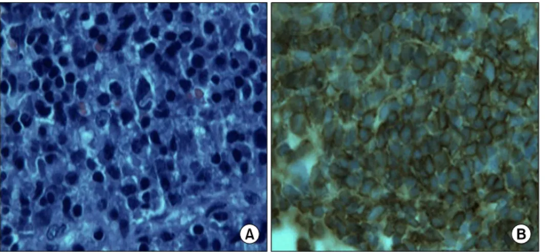
Fig. 3. Pathologic findings.
(A) Large, globular abnormal lymphocytes are observed (H&E stain, ×1,000). (B) Strongly stained tumor cell (CD20 stain, ×1,000).
Fig. 2. The chest CT shows a polypoid mass lesion in the Lt anterolateral aspect of the trachea.
trolyte, renal function, and liver function tests were within normal limits. The lactate de- hydrogenase level was 2,450U/L and the β2-mi- croglobulin level was 1.6mg/L.
Fiberoptic bronchoscopy showed an intratra- cheal multi-lobulated polypoid lesion with the tracheal lumen narrowing. Bronchoscopic biopsy was performed to establish a pathological diagno- sis (Fig. 1A).
The chest CT showed a 1.5×1.0cm sized poly- poid mass lesion in the Lt anterolateral aspect of the trachea (Fig. 2).
The histological finding showed diffuse in- filtration of large and globular abnormal lympho-
cytes (Fig. 3A). The malignant cells expressed CD20, indicating an increase of B-cells with a re- active response (Fig. 3B). The final diagnosis was a diffuse large B-cell lymphoma.
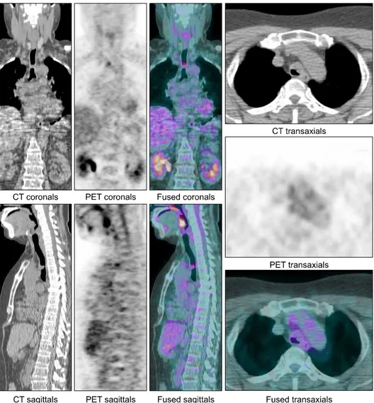
For the staging of the primary disease, there was no evidence of organ involvement of NHL via positron emission tomography - computed to- mography (PET-CT). A whole-body combined PET-CT scan demonstrated the high uptake of
18F-FDG within the 1.5×1.0cm sized polypoid mass in the middle area of the trachea. The PET-CT also revealed no increased 18F-FDG up- take at adjacent or distant lymph node sites, and showed no evidence of organ involvement (Fig.
4). Thus, we concluded that the diagnosis was primary tracheal diffuse large B-cell lymphoma, stage 1BE.
Based on recent reports of the synergistic bene- fits of immunotherapies in combination with standard chemotherapy for diffuse large B-cell lymphoma, treatment was initiated using CHOP (cyclophosphamide, adriamycin, vincristine, and prednisone) chemotherapy with rituximab anti- CD20 monoclonal antibody immunotherapy. Four courses of combination chemotherapy with the R-CHOP regimen (rituximab 375mg/m2 day 1, cyclophosphamide 750mg/m2 day 1, adriamycin 50mg/m2 day 1, vincristine 1.4mg/m2 day 1, and prednisone 100mg/day 1 to 5, q 3 weeks) were given. After two and four courses of chemo- therapy, the follow-up fiberoptic bronchoscopy
Fig. 4. PET-CT shows a 1.5×1.0cm sized polypoid mass lesion in trachea.
was performed. The follow-up bronchoscopic ex- am demonstrated that the size of the tracheal mass became reduced, thus indicating partial re- mission (Fig. 1B, C).
We performed the additional irradiation as ad- juvant therapy. The patient received a total of 3,960cGy in twenty 198cGy fractions. Complete re- mission was confirmed by bronchoscopic exam (Fig. 1D). Currently, the patient was found to be in complete remission and has been alive for 26 months after initial diagnosis.
DISCUSSION
Primary tumors of the trachea are rare, and are usually malignant in adults and benign in child- ren. Primary cancers of the upper respiratory
tract account for more than 1% of malignant dis- eases, but the frequency of tumors is not con- sistent throughout the upper respiratory tract.
Although the supraglottis is affected in 1.3 per 100,000 people and the glottis is affected in 2.3 per 100,000 people, fewer than 0.04 of 100,000 people have tumors in the subglottis or trachea.1) About 33% of NHL cases arise in tissues other than the lymph nodes, spleen, Waldeyer’s ring, and thymus, and these are referred to as primary extranodal NHL. Rosenberg et al. reported on 1,269 cases of primary extranodal non-Hodgkin’s lymphoma. This study showed that the most common site of anatomical localization was skin, followed by tonsil, bone, stomach, and others.
Furthermore, diffuse large B-cell lymphoma is a subtype of lymphoma that commonly has extra-
nodal presentation.6)
Primary tracheal tumors account for 0.1∼0.4%
of malignant diseases, with 2.6 new cases arising per million people every year.1) Fidias P et al. de- scribed primary tracheal lymphoma in only six cases in a review study published in 1999.4) Furthermore, from 1989 to 2005, only three cases of primary tracheal non-Hodgkin’s lymphoma were reported with literature review in Medline.7) In histological classification of primary trache- al lymphoma, the more common types are lym- phoblastic lymphoma and mucosa-associated lym- phoid tissue lymphoma.8) The most frequently presenting symptom/sign was cough (49%), fol- lowed by dyspnea (44%), hemoptysis (44%), whee- ziness (33%), stridor (20%), hoarseness (10%), and others.2) In Korea, the most common site of primary extranodal non-Hodgkin’s lymphoma was the stomach, followed by the intestine. Low incidence was found in the skin and bone. There were no reported cases for primary tracheal lymphoma.9) In this patient, the symptoms is dry cough, weight loss, intermittent night sweats, and febrile sensation.
Furthermore, a tendency for extranodal lym- phomas to remain localized for long periods of time is attributed to the migration of circulating mucosal B-lymphocytes back to their sites of ori- gin, thus rendering the lymphomas amenable to local therapy. In addition, the observation that extranodal lymphoma often disseminates to other mucosal sites rather than peripheral lymphoid tissue is attributed to the homing properties of the mucosal B-lymphocytes to other mucosal sites. These extranodal recurrences are frequently responsive to local treatment, even when the pa- tient is in Stage III or IV of the disease.3,5) Therefore, the combination R-CHOP regimen, consisting of rituximab plus CHOP (cyclopho- sphamide, doxorubicin, vincristine, and pre- dnisone), is now considered as the standard treat- ment for treating young and elderly patients with diffuse large B-cell lymphoma.10)
Although CT remains the gold standard for the
staging and follow-up of malignant lymphomas,
18F-FDG PET has a potential role in the accurate staging of disease and in predicting the response to therapy. This role has the potential to affect both the initial choice of chemotherapy and the decision to alter management based on the initial response to therapy.11)
As mentioned in reviews by Malik E. Juweid et al., IV contrast-enhanced PET/CT likely pro- vides at least equal information to that provided by the sum of the PET plus a separately per- formed contrast-enhanced CT, and therefore rep- resents an adequate alternative. In addition, if hepatic or splenic involvement was demonstrated at initial staging, PET-CT performed without IV contrast appears to be adequate for response as- sessment of lymphomatous involvement of nodes or other extralymphatic organs that may not be detectable by PET. It was recommended that in- vestigators should note that only PET or PET-CT systems, and not coincidence imaging, should be used for response assessment of lymphoma. In re- lation to bone marrow involvement, it was noted that bone marrow biopsy remains the standard procedure for assessment of bone marrow because of poor clinical correlation.12)
We diagnosed primary tracheal diffuse large B-cell lymphoma, that is actually extremely rare.
After R-CHOP based chemoradiotherpy, com- plete remission was obtained. We reported pri- mary tracheal diffuse large B-cell lymphoma with literature review.
요 약
비호지킨 림프종은 약 10% 정도는 림프절 외의 조직 에서도 일어난다. 기관에서 원발성으로 발생하는 경우 는 국제적으로도 극히 드문 것으로 보고되고 있으며, 대부분은 점막 관련 림프 조직 림프종으로 알려져 있 다. 본 저자들은 건성 기침을 주소로 내원한 65세 여자 환자에서 기관지 내시경을 이용하여 조직학적으로 미 만성 거대 B세 포 림프종을 확진하였다. 환자는 이후 4 cycle의 R-CHOP과 adjuvant RT 시행 이후 완전관해를 유도하였으며, 26개월간 완전 관해 상태가 유지되고 있
다.
REFERENCES
1) Macchiarini P. Primary tracheal tumours. Lancet Oncol 2006;7:83-91.
2) Gaissert HA, Grillo HC, Shadmehr MB, et al.
Uncommon primary tracheal tumors. Ann Thorac Surg 2006;82:268-73.
3) lsaacson P, Wright DH. Extranodal lymphoma aris- ing from mucosa-associated lymphoid tissue. Cancer 1984;53:2515-24.
4) Young GA. Lymphoma at uncommon sites. Hematol Oncol 1999;17:53-83.
5) Rudders RA, Ross ME, DeLellis RA. Primary ex- tra-nodal lymphoma: response to treatment and fac- tors influencing prognosis. Cancer 1978;42:405-16.
6) Rosenberg SA, Diamond HD, Jaslowitz B, Craver LF. Lymphosarcoma: a review of 1,269 cases. Medi- cine (Baltimore) 1961;40:31-84.
7) Zhang WD, Li SY, Ouyang M, Zhong NS. Primary endotracheal non-Hodgkin’s lymphoma in a Chinese
woman: a case report. Chin Med J (Engl) 2005;
118:702-4.
8) Kaplan MA, Pettit CL, Zukerberg LR, Harris NL.
Primary lymphoma of the trachea with morphologic and immunophenotypic characteristics of low grade B cell lymphoma of mucosa-associated lymphoid tissue. Am J Surg Pathol 1992;16:71-5.
9) Hahn JS, Ko YW, Min YH, et al. Statistical analysis of maliganat lymphoma in Korea. Korean J Hematol 1995;2:197-214.
10) Feugier P, Van Hoof A, Sebban C, et al. Long-term results of the R-CHOP study in the treatment of eld- erly patients with diffuse large b-cell lymphoma: a study by the Groupe d'Etude des Lymphomes de l'Adult. J Clin Oncol 2005;23:4117-26.
11) Jhanwar YS, Straus DJ. The role of PET in lym- phoma. J Nucl Med 2006;47:1326-34.
12) Juweid ME, Stroobants S, Hoekstra OS, et al. Use of positron emission tomography for response assess- ment of lymphoma: consensus of the Imaging Sub- committee of International Harmonization Project in Lymphoma. J Clin Oncol 2007;25:571-8.