ABSTRACT
Hyper-IgE syndrome (HIES) is a very rare primary immune deficiency characterized by elevated serum IgE levels, recurrent bacterial infections, chronic dermatitis, and connective tissue abnormalities. Autosomal dominant (AD) HIES involves a mutation in signal transducer and activator of transcription 3 (STAT3) that leads to an impaired TH17 response.
STAT3 signaling is also involved in the function of RORγt+ type 3 innate lymphoid cells (ILC3s) and RORγt+TH17 cells. The aim of this study was to investigate the role of innate immune cells such as innate lymphoid cells (ILCs), granulocytes, and monocytes in a patient with HIES. Peripheral blood mononuclear cells (PBMCs) from a patient with HIES and three age-matched healthy controls were obtained for the analysis of the innate and adaptive immune cells. The frequencies of ILCs in PBMCs were lower in the patient with HIES than in the controls. Moreover, granulocyte-macrophage colony-stimulating factor (GM-CSF) and IL-17A produced by ILC3s in PBMCs were lower in the patient with HIES than the controls. Compared with the controls, classical monocytes (CD14+CD16low), which have a high antimicrobial capability, were also lower in the patient with HIES, while non-classical monocytes (CD14lowCD16+) as well as intermediate monocytes (CD14+CD16intermediate) were higher. Taken together, these results indicate that the impaired immune defense against pathogenic microbes in the patient with HIES might be partially explained by functional defects in ILC3s and inflammatory monocytes.
Keywords: Hyper-IgE syndrome; STAT3 transcription factor; Innate immunity; Innate lymphoid cells; Monocytes; Cytokines
INTRODUCTION
Hyper-IgE syndrome (HIES) is a rare primary immunodeficiency disorder characterized by elevated serum IgE, eosinophilia, recurrent skin abscesses, and sinopulmonary tract infections, usually associated with Staphylococcus aureus (1). Most cases of HIES are sporadic,
Case Report
Received: Aug 8, 2017 Revised: Oct 4, 2017 Accepted: Oct 6, 2017
*Correspondence to Hye-Ryun Kang
Department of Internal Medicine, Seoul National University College of Medicine, 101 Daehak-ro, Jongno-gu, Seoul 03080, Korea.
E-mail: [email protected] Hye Young Kim
Laboratory of Mucosal Immunology in Department of Biomedical Sciences, Seoul National University College of Medicine, 103 Daehak-ro, Jongno-gu, Seoul 03080, Korea.
E-mail: [email protected]
†These authors contributed equally to this work.
Copyright © 2017. The Korean Association of Immunologists
This is an Open Access article distributed under the terms of the Creative Commons Attribution Non-Commercial License (https://
creativecommons.org/licenses/by-nc/4.0/) which permits unrestricted non-commercial use, distribution, and reproduction in any medium, provided the original work is properly cited.
Conflict of Interest
The authors declare no potential conflicts of interests.
Yuna Chang,1,2,† Sung-Yoon Kang,3,4,† Jihyun Kim,1,2 Hye-Ryun Kang,3,4,*
Hye Young Kim1,2,4,*
1Department of Biomedical Sciences, Seoul National University College of Medicine, Seoul 03080, Korea
2 Laboratory of Mucosal Immunology in Department of Biomedical Sciences, Seoul National University College of Medicine, Seoul 03080, Korea
3Department of Internal Medicine, Seoul National University College of Medicine, Seoul 03080, Korea
4 Institute of Allergy and Clinical Immunology, Seoul National University Medical Research Center, Seoul 03080, Korea
Functional Defects in Type 3 Innate Lymphoid Cells and Classical
Monocytes in a Patient with Hyper-IgE
Syndrome
Abbreviations
AD, autosomal dominant; CTRL, control; DC, dendritic cell; DOCK8, dedicator of cytokinesis 8; GM-CSF, granulocyte-macrophage colony- stimulating factor; HIES, hyper-IgE syndrome;
IFN, interferon; ILC, innate lymphoid cell; ILC1, type 1 innate lymphoid cell; ILC2, type 2 innate lymphoid cell; ILC3, type 3 innate lymphoid cell; PBMC, peripheral blood mononuclear cell; STAT3, signal transducer and activator of transcription 3; TNF, tumor necrosis factor;
Treg, regulatory T
but some familial cases of HIES have been reported, with either an autosomal dominant (AD) or autosomal recessive mode of inheritance. However, the pathogenesis of both the clinical manifestations as well as the immunologic abnormalities remains poorly defined.
Several genetic mutations, such as in signal transducer and activator of transcription 3 (STAT3), tyrosine kinase 2 (Tyk2), and dedicator of cytokinesis 8 (DOCK8), have been implicated in HIES (2-4). Among these genetic mutations, dominant-negative mutations in STAT3 were identified as a major molecular cause of HIES (2). STAT3 is a transcription factor that binds to the promoter regions of various genes, including those encoding acute-phase proteins. STAT3 is known to play a critical role in signal transduction for many cytokines, including those in the γc family (IL-2, IL-7, IL-9, IL-15, and IL-21), gp130 family (IL-6, IL-11, IL-27, and IL-31), IL-10 family (IL-10 and IL-22), and receptor-type tyrosine kinases (5-7). Therefore, STAT3 deficiency results in the upregulation of TH1 cytokines, but the downregulation of pro-inflammatory as well as anti-inflammatory responses (2).
Moreover, STAT3 has pleiotropic functions in both innate and adaptive immunity (8,9). A markedly decreased TH17 response is a hallmark of HIES, and STAT3 mutations have been demonstrated to result in the failure of TH17 CD4 cell differentiation (10,11). Since IL-17A signaling is involved in neutrophil chemotaxis and proliferation (12,13), impaired neutrophil responses due to impaired IL-17A signaling might account for the recurrent staphylococcal infections in patients with HIES (14,15).
Moreover, it has been reported that the production of antimicrobial molecules responsible for antifungal responses is tightly regulated by IL-17A (13). In patients with HIES with STAT3 mutations, elevated levels of tumor necrosis factor (TNF)-α, IL-6, and IL-12 in dendritic cells (DCs) were observed (16). Another study showed that tolerogenic DC generation failed in patients with STAT3-deficient HIES due to the disruption of the IL-10 signaling pathway (5). Moreover, increased TNF-α and IL-8 production was detected in HIES peripheral blood mononuclear cells (PBMCs) after stimulation with several concentrations of LPS and/or S.
Aureus (17). Finally, hyper-eosinophilia, which is a relatively consistent feature of HIES, has also been observed in mice with myeloid-specific STAT3 deficiency (18). Altogether, these results suggested that patients with STAT3-deficient HIES would have defects in the numbers and function of innate immune cells. Although few studies have been performed to date, the defects in innate immune cells might explain the increased susceptibility to infections in patients with HIES.
Human monocytes can be subdivided based on their expression of the Fcγ receptor III CD16 and lipopolysaccharide co-receptor CD14. “Classical” monocytes account for greater than 70% of total monocytes and are denoted as the CD14+CD16low population (19). Classical monocytes are highly phagocytic, have high myeloperoxidase activity, have high antibody- dependent cell-mediated toxicity, and produce high levels of IL-1, monocyte chemoattractant protein-1 (MCP-1), TNF-α, and IL-6. “Non-classical” monocytes express CD14lowCD16+ antigen and constitute about 10%–15% of blood monocytes. Non-classical monocytes express high levels of major histocompatibility class II receptor, migrate primarily to non-inflamed tissue, and produce high levels of TNF-α, IL-1, IL-6, and IL-12. These cells are more likely to become DCs involved in tissue surveillance. However, the recent identification of an intermediate population identified as CD14+CD16lntermediate supports a new idea that monocytes are a heterogeneous population. Intermediate monocytes are low cytokine producers but express high levels of angiogenic receptors (20). Understanding the different functions of the specific subpopulations of monocytes is necessary for better understanding human diseases.
Innate lymphoid cells (ILCs) have recently been identified as a unique group of innate immune cells (21). ILCs are characterized by a lack of T-cell and B-cell receptors (TCRs and BCRs, respectively), but they rapidly produce a variety of cytokines in response to innate signals, i.e., IL-25, IL-33, thymic stromal lymphopoietin (TSLP), and IL-1α. Traditionally, T-cells were thought as key players in the regulation of the immune response; however, recent emerging studies on ILCs have provided new paradigms for various immune responses, including host defense and tissue homeostasis (21-23). To date, the treatment for HIES has relied on prophylactic and therapeutic antibiotics and meticulous skin care, and a general role for ILCs in HIES is still unproven. Because both innate and adaptive responses are important in host defense, we evaluated the frequency and functional response of innate immune cells, including ILCs and monocytes, for the first time in patients with HIES who had recurrent infections.
MATERIALS AND METHODS
Case
PBMCs were taken from a 21-year-old woman who was diagnosed with HIES, and PBMCs from an age-matched healthy control were also used. This study was approved Institutional Review Board of Seoul National University Hospital, which waived the requirement of written informed consent (IRB No. 1707-130-871).
Cell isolation from human blood
PBMCs were diluted with a 3× volume of PBS. Then, 30 ml of diluted blood was carefully layered over 15 ml of Ficoll-Paque in a 50-ml conical tube, followed by centrifugation at 400 ×g for 30 min at 20°C in swinging bucket rotor without a brake. Mononuclear cells at the interface were collected with a transposable pipette. Collected cells were washed with PBS and centrifuged at 300 ×g for 10 min at 4°C. The cell pellets were resuspended in an appropriate volume of buffer and processed for flow cytometry.
Flow cytometry
Single cell suspensions from peripheral blood were stained with the following monoclonal antibodies: PerCP/Cy5.5-anti-CD45 (BD Biosciences, San Jose, CA, USA), fluorescein isothiocyanate (FITC)-anti-CD3 (BioLegend, San Diego, CA, USA), Alexa Flour 700-anti-CD4 (BioLegend, San Diego, CA, USA), Brilliant Violet (BV) 421-anti-CD8(BioLegend, San Diego, CA, USA), FITC-anti-Lineage Cocktail (anti-CD3, anti-CD14, anti-CD19, anti-CD11b, anti-CD11c, anti-CD49b, anti-FcεRIα (BioLegend, San Diego, CA, USA), PE/Cy7-anti-CD127(BioLegend, San Diego, CA, USA), PE-anti-CRTH2(BioLegend, San Diego, CA, USA), BV421-anti-c- kit(BioLegend, San Diego, CA, USA), allophycocyanin (APC)-anti-NKp44(BioLegend, San Diego, CA, USA), PE/Cy7-anti-CD15(BioLegend, San Diego, CA, USA), FITE-anti-
CD14(BioLegend, San Diego, CA, USA), and PE-anti-CD16 (BioLegend, San Diego, CA, USA). All samples were blocked with 1 μg Fc block (from 2.4G2 ATCC HB-197) for 15 min before antibody staining at 4°C for 30 min in PBS containing 2% FCS (2% FCS-PBS). Cells were washed twice in 2% FCS-PBS, and then, data were collected on a BD LSRII flow cytometer (BD Biosciences).
Analysis of the FACS plots was performed using FlowJo (Treestar, Ashland, OR, USA).
Intracellular cytokine staining
For intracellular cytokine staining, PBMCs were incubated in RPMI (WELGENE,
Gyeongsangbuk-do, Korea) containing 10% FBS with PMA (100 ng/ml), ionomycin (1 μg/ml), and BD GolgiStop (BD Biosciences) at 37°C for 4 h. After surface staining, the cells were fixed
and permeabilized with BD Cytofix/Cytoperm (BD Biosciences). Finally, the cells were stained with BV421-anti-interferon (IFN)-γ, BV421-anti-granulocyte-macrophage colony-stimulating factor (GM-CSF) (BD Biosciences), APC-anti-IL-5, PE-anti-IL-13, APC-anti-IL-17, and PE-anti- IL-22 (BioLegend). The respective isotype control antibody was also used for each experiment.
Statistical analysis
Statistical analysis was performed using Prism software (GraphPad, La Jolla, CA, USA). Data are displayed as individual data points with the mean±standard error of the mean (SEM).
RESULTS
Case presentation
A 21-year-old woman was referred to an allergy clinic for the evaluation of multiple skin axillary and buttock abscesses with eosinophilia. She was born full term without perinatal problems.
However, she had a history of repeated eczematous rashes on her body and skin abscesses not responding to symptomatic treatment. A diagnosis of HIES was made at 3 months of age based on recurrent skin infections and elevated total serum IgE (773.5 Ku/l) with increased eosinophil counts (38% of white cell blood count, 30,690/μL). She had septic arthritis of the hip joint, abscesses in the liver and lungs, and recurrent multiple cutaneous abscesses caused by methicillin-resistant S. aureus. There was no family history of immunodeficiency. On physical examination at the time of referral, her facial features were coarse with a broad nasal bridge and prominent nose. There was a significant presence of palpable skin lesions on the right axilla and buttock. Bronchial breath sounds were heard in all lung fields, and aeration was decreased in the left lung field. Pulmonary function tests showed severe restrictive patterns. Laboratory tests revealed a WBC count of 4,950/μL, with an elevated eosinophil count (1,100/μl) and total IgE level (158 kU/l). Other laboratory findings, such as liver and renal function tests, urinalysis, and C-reactive protein level, were within normal ranges. A computed tomography scan showed multifocal air cysts or bullae with right necrotic axillary lymph nodes and necrotic abscess in the right lower abdomen, buttock, and groin (Fig. 1).
Echocardiography and bone mineral density results were in normal ranges. The clinical and laboratory data were applied to the scoring system of the National Institute of Health, and the total score was 52 points, suggestive of AD-HIES (Table 1). She underwent surgical drainage and excisional biopsy that revealed abscess-forming inflammatory granulation tissue, and was then discharged with prophylactic oral antibiotics (Fig. 2).
Impaired type 3 immune responses in a patient with HIES
Several studies have measured the population of T cells in the PBMCs of patients with HIES;
however, the results varied according to the experimental conditions. Therefore, we first evaluated the T cell populations in the PBMCs of a patient with HIES (n=1) and age-matched controls (n=3) by flow cytometry. The percentages of CD3+ T cells in the PBMCs were similar between the patient with HIES and the controls (Fig. 3A and B). We further analyzed the T cell subpopulations and found that the percentages of CD4 and CD8 T cells were within normal ranges, but approximately 1.9 times more Foxp3+ regulatory T (Treg) cells were detected in the PBMCs of the patient with HIES than in those from the controls (Fig. 3A and B). Similar to our results, Heimall et al. (1) reported that normal numbers of CD4 and CD8 T cells existed in the PBMCs of patients with HIES. Additionally, a marked decrease in the TH17 response has been reported as a hallmark of HIES due to STAT3 mutations (24). Therefore, we stimulated PBMCs with PMA/ionomycin and then measured the production of various cytokines, including IL-17A
from T cells. Type 2 cytokine expression in T cells was similar between the patient with HIES and the control subjects. However, expression of type 1 and type 3 cytokines such as IFN-γ, IL-17 and IL-22, respectively, in T cells was lower in the patient with HIES than in the controls (Fig. 3C and D). These findings suggested that our patient with HIES may have a defective STAT3 signaling pathway, which resulted in reduced type 3 cytokine production from T cells.
A
B
Figure 1. Chest computed tomography scan (A, B). Images showing necrotic lesions in right axillary lymph nodes, subcutaneous tissue of the right buttock (A, white arrows), and multifocal air cysts or bullae of the both lungs (B).
Table 1. National Institutes of Health Score for the patient with HIES†
Clinical Finding Points
0 1 2 3 4 5 6 7 8 10
Highest serum-IgE level (IU/ml) <200 200–500 501–1,000* 1,001–2,000 >2,000
Skin abscesses None 1–2 3–4* >4
Pneumonia (episodes over lifetime) None 1 2 3 >3*
Parenchymal lung anomalies Absent Bronchiectasis Pneumatocoele*
Retained primary teeth None* 1 2 3 >3
Scoliosis, maximum curvature <10° 10°–14° 15°–20°* >20°
Fractures with minor trauma None* 1–2 >2
Highest eosinophil count (cells/μl) <700 700–800 >800*
Characteristic face Absent Mildly present Present*
Midline anomaly Absent* Present
Newborn rash Absent Present*
Eczema (worst stage) Absent* Mild Moderate Severe
Upper-respiratory infections per year 1–2 3* 4–6 >6
Candidiasis None* Oral Fingernails Systemic
Other serious infections None* Severe
Fatal infection Absent* Present
Hyperextensibility Absent* Present
Lymphoma Absent* Present
Increased nasal width (interalar distance) <1 SD 1–2 SD* >2 SD
High palate Absent* Present
Young-age correction (yr) >5 2–5 1–2 ≤1*
*Asterisks indicate the criteria that the patient satisfied; †Scores of at least 40 points were considered to indicate high likelihood of HIES, whereas scores below 20 indicated low likelihood.
Altered function of ILCs in a patient with HIES
There have been several reports regarding the roles of adaptive immune responses in the pathogenesis of HIES; however, the roles of innate immune responses in HIES remain unknown. For example, Gutierrez-Hincapié et al. (25) measured the number and frequency of innate immune cells in PBMCs from patients with HIES, but there were no differences in terms of the absolute number of leukocytes, neutrophils, monocytes, mDCs, pDCs, and NK cells in PBMCs from patients with HIES. However, they found that the number of invariant NK T (iNKT) cells was significantly reduced in the AD-HIES group; hence, these results indicated that innate immune responses were also impaired in the patients with HIES.
As an innate counterpart of TH cells, ILCs play important roles by producing types 1, 2, and 3 cytokines. However, the role of ILCs in HIES has not been reported. To determine the role of ILCs in our patient with HIES, we analyzed ILCs using flow cytometry. Compared to the control subjects, the percentage of total ILCs (Lineage−CD127+) was lower in the PBMCs from the patient with HIES (Fig. 4A). Next, we further analyzed the subset of ILCs by adding several surface markers such as CRTH2, c-kit, and NKp44 to distinguish type 2 innate lymphoid cells (ILC2s; Lineage−CD127+CRTH2+), type 3 innate lymphoid cells (ILC3s; Lineage−CD127+CRTH2−c-kit+), and type 1 innate lymphoid cells (ILC1s; Lineage− CD127+CRTH2−c-kit−). Interestingly, most of ILCs in the patient with HIES were ILC1s and not ILC2s or ILC3s (Fig. 4A and B). Intracellular staining of ILCs also revealed that IL-17A and IL- 22 (type 3) cytokine production in the PBMCs was lower in the patient with HIES than in the controls, but IFN-γ production was similar (Fig. 4C and D). These results indicated that ILCs, as well as T cells, had altered functions in the patient with HIES.
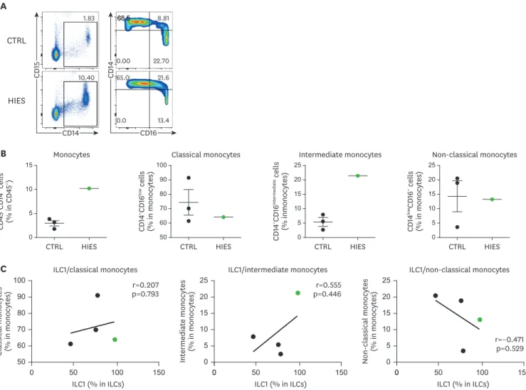
CD14+CD16intermediate monocytes are increased in a patient with HIES
To test the proportion of monocytes in the patient with HIES, we analyzed monocyte subsets using flow cytometry and antibodies to CD14 and CD16. The dot plot of the data indicates that was a large increase in monocytes with CD14 expression in the PBMCs of the patient with HIES compared with the normal controls (Fig. 5A and B). Interestingly, CD14+CD16lntermediate
monocytes were higher in the patient with HIES than in the controls, while there was no significant difference in the proportion of classical monocytes (CD14+CD16low) and non- classical monocytes (CD14lowCD16+).
Recently, Xiong et al. (26) reported on the crosstalk between inflammatory monocytes and ILCs. The authors suggested that IL-17A from ILCs could enhance the antimicrobial activities of inflammatory monocytes. To evaluate the correlation between monocytes and ILCs, we
A B
Figure 2. Excisional biopsy specimen revealing inflamed granulation tissue with abscess formation from the right axillary area (A, ×100; B, ×400).
CTRL T cells
HIES
B
CD45+CD3+ cells (% in lymphocytes) 60
70 85 75 80
65
CTRL CD4+ T cells
HIES
CD3+CD4+ cells (% in T cells)
40 50 65 55 60
45
CTRL CD8+ T cells
HIES
CD3+CD8+ cells (% in T cells)
30 40 50 45
35
CTRL Treg cells
HIES
CD3+CD4+Foxp3+ cells (% in CD4+ T cells)
0 15 10 5
0.18 0.75
18.80 80.30
0.27 0.92
8.76 90.00
0.29 1.37
0.96 97.40
0.21 2.11
0.86 96.80
0.45 0.40
9.15 90.00
0.15 0.48
0.70 98.70 0.086
0.082
2.770 97.100
0.013 0.200
0.180 99.600 IFN-γ
IL-5
IL-5
IL-13
GM-CSF
IL-17
IL-17
IL-22
CTRL
HIES C
68.4
77.6
10.20 4.57 46.5
44.2 35.5
50.2 CD3
CD45
CD4
CD8
CD4
Foxp3
CTRL
HIES A
CTRL IFN-γ
HIES
D
IFN-γ+ cells (% in lymphocytes)
0 10 25
15 20
5
CTRL IL-5
HIES
IL-5+ cells (% in T cells)
0 1.0 1.5
0.5
CTRL IL-13
HIES
IL-13+ cells (% in T cells)
0 2 3
1
CTRL GM-CSF
HIES
GM-CSF+ cells (% in T cells)
0 15 10 5
CTRL IL-17
HIES
IL-17+ cells (% in T cells)
0 0.4 1.0 0.6 0.8
0.2
CTRL IL-22
HIES
IL-22+ cells (% in T cells)
0 0.4 0.8 0.6
0.2
Figure 3. The distribution of T cells in PBMCs from healthy controls and a patient with HIES. (A, B) CD4+ T cells, CD8+ T cells, and Treg cells were analyzed by flow cytometry. (A) Representative flow cytometric profiles of the T cell population. (B) Graphs show the mean percentages of CD3+ T cells, CD4+ T cells, CD8+ T cells, and Treg cells±SEM (n=3). (C, D) Intracellular IFN-γ, IL-5, IL-13, GM-CSF, IL-17, and IL-22 expression in T cells was analyzed by flow cytometry. (C) Representative flow cytometric profiles of cytokine expression by T cells. (D) Graphs show the mean percentage of positive cells±SEM (n=3).
CTRL, control; GM-CSF, granulocyte-macrophage colony-stimulating factor; HIES, hyper-IgE syndrome; IFN, interferon; PBMC, peripheral blood mononuclear cell; SEM, standard error of the mean; Treg, regulatory T.
CTRL ILCs
HIES
B
CD45+Lin-CD127+ cells (% in lymphocytes) 0.40
0.50 0.65 0.55 0.60
0.45
CTRL ILC1
---CRTH2c-kitNKp44 cells (% in ILCs) HIES
0 100 150
50
CTRL ILC2
HIES
CRTH2+ cells (% in ILCs)
0 10 20 15
5
10 30 50 40
20
CTRL ILC3
HIES
CRTH2-c-kit+ cells (% in ILCs)
0.48 1.21
22.30 76.00
0.95 0.47
24.20 74.4
0.47 1.42
0.95 97.20
0.73 3.63
0.97 94.70
2.39 1.75
7.32 88.50
0.32 0.00
3.82 95.90 0.41
0.21
5.59 93.80
0.00 0.00
0.83 99.20 IFN-γ
IL-5
IL-5
IL-13
GM-CSF
IL-17
IL-17
IL-22
CTRL
HIES C
0.84
0.45
1.85 8.39
0.53
98.10 0.00
0.00 CD127
Lineage
CRTH2
c-kit
NKp44
c-kit
CTRL
HIES A
0.23 85.10
14.60 0.00
CTRL IFN-γ
HIES
D
IFN-γ+ cells (% in ILCs)
20 30 40 35
25
CTRL IL-5
HIES
IL-5+ cells (% in ILCs)
0 2 3
1
CTRL IL-13
HIES
IL-13+ cells (% in ILCs)
0 10 15
5
CTRL GM-CSF
HIES
GM-CSF+ cells (% in ILCs)
0 20
10 5 15
CTRL IL-17
HIES
IL-17+ cells (% in ILCs)
0 2 5
3 4
1
CTRL IL-22
HIES
IL-22+ cells (% in ILCs)
0 2 4 3
1
Figure 4. The distribution of ILCs in PBMCs from healthy controls and a patient with HIES. (A, B) ILC1s (CD45+Lin−CD127+CRTH2−c-kit−NKp44−), ILC2s (CD45+Lin− CD127+CRTH2+), and ILC3s (CD45+Lin−CD127+CRTH2−c-kit+) were analyzed by flow cytometry. (A) Representative flow cytometric profiles of ILC subsets. (B) Graphs show the mean percentage of total ILCs, ILC1s, ILC2s, and ILC3s±SEM (n=3). (C, D) Intracellular IFN-γ, IL-5, IL-13, GM-CSF, IL-17, and IL-22 expression in ILCs was analyzed by flow cytometry. (C) Representative flow cytometric profiles of cytokine expression in ILCs. (D) Graphs show the mean percentage of positive cells±SEM (n=3).
CTRL, control; GM-CSF, granulocyte-macrophage colony-stimulating factor; HIES, hyper-IgE syndrome; ILC, innate lymphoid cell; PBMC, peripheral blood mononuclear cell; SEM, standard error of the mean.
tested the association between subsets of monocytes and ILCs from healthy control and a patient with HIES (Fig. 5C). Although the number of samples was limited, the proportion of CD14+CD16lntermediate monocytes had a positive trend with an increase in ILC1s. In contrast to CD14+CD16lntermediate monocytes, non-classical monocytes (CD14lowCD16+) had a positive trend with an increase in ILC2s and ILC3s, but not ILC1s (Fig. 5C, Supplementary Fig. 1).
Taken together, these results indicated that innate immune cells are indeed altered in this patient with HIES; therefore, further studies to define the functional changes in innate immune cells during disease progression are needed.
DISCUSSION
HIES is a complex primary immunodeficiency disorder characterized by recurrent
staphylococcal skin abscesses and pneumonia, atopic dermatitis, and extremely high serum IgE levels. Recent studies have shown that dominant-negative mutations of the STAT3 gene CTRL
Monocytes
HIES
B
CD45+CD14+ cells (% in CD45+)
0 15 10 5
CTRL
Classical monocytes
HIES
CD14+CD16low cells (% in monocytes)
50 70 100 80 90
60
0 10 25 15 20
5
0 10 25 15 20
5
CTRL
Intermediate monocytes
intermediate+CD14CD16 cells (% inmonocytes) HIES
CTRL
Non-classical monocytes
HIES
CD14lowCD16+ cells (% in monocytes)
1.83
10.40
8.81
22.70 0.00
21.6
13.4 65.0
0.0 CD14
CD15
CD16
CD14
CTRL
HIES A
C
100
70 90 80
0 50
Classical monocytes (% in monocytes) 60
ILC1 (% in ILCs)
150
50 100
ILC1/classical monocytes r=0.207 p=0.793
0 50 100 150
25
10 20 15
0 0 5
Intermediate monocytes (% in monocytes)
ILC1 (% in ILCs) ILC1/intermediate monocytes
r=0.555 p=0.446
0 50 100 150
25
10 20 15
0 0 5
Non-
classical monocytes (% in monocytes)
ILC1 (% in ILCs) ILC1/non-classical monocytes
r=−0.471 p=0.529
Figure 5. The distribution of monocytes in PBMCs from healthy controls and a patient with HIES. (A, B) Monocytes (CD45+CD15−CD14+) were analyzed by flow cytometry. (A) Representative flow cytometric profiles of monocyte populations. (B) Graphs show the mean percentages of monocytes, classical monocytes (CD14+CD16low), intermediate monocytes (CD14+CD16intermediate), and non-classical monocytes (CD14lowCD16+)±SEM (n=3). (C) The correlations between ILC1s and classical monocytes, intermediate monocytes, and non-classical monocytes were analyzed in healthy controls (black) and a patient with HIES (red).
CTRL, control; HIES, hyper-IgE syndrome; ILC, innate lymphoid cell; SEM, standard error of the mean.
are a major molecular cause of HIES in most patients (2,27). However, our understanding of the immune alterations underlying HIES remains limited. In this report, we focused on the innate and adaptive immunological abnormalities in HIES.
The STAT3 signaling pathway was originally discovered in the context of components
downstream of the IFN-α, IFN-γ, and IL-6 signaling pathways (28,29). STAT3 is not only crucial for transducing signals, but it also functions as a critical transcription factor that regulates the expression of a wide range of genes. Therefore, STAT3 plays a crucial role in the function of various cell types, including stromal cells, tumor cells, epithelial cells, and immune cells. The susceptibility to infection observed in patients with HIES can be explained by the lack of IL-17A production from T cells due to impaired STAT3 signaling. In addition, impaired production of IFN-γ and TNF-α by T cells (30), diminished memory T-cell populations, and decreased delayed-type-hypersensitivity responses have variably been described in patients with HIES (15).
Although the functional alteration of adaptive immunity, such as changes in T cells and B cells, has been relatively well studied, innate immunological abnormalities that can explain the unique susceptibility to particular infections seen in HIES have not been identified. In this regard, we evaluated the innate immune cells observed in a HIES case. Of interest, the percentage of total ILCs in PBMCs was lower in the patient with HIES than in the controls, while the percentage of T cells remained relatively similar. Moreover, our data also indicated that ILC3s in PBMCs were significantly lower in the patient with HIES than in the controls. Although STAT3 is known for IL-17A production in adaptive arms, Guo et al. (31) reported that STAT3 expression in RORγt+ ILC3s, but not in T cells, was essential for protection against Citrobacter rodentium. They also suggested that transcriptional regulation of ILC3s, TH17, and TH22 cells might differ. Therefore, the functions of TH17 and ILC3s in HIES would be different, although they all produce type 3 cytokines and are regulated by STAT3. Thus, the mechanisms underlying the differential regulation by STAT3 in ILCs and T cells need to be further investigated.
In a current study, we also observed an increased number of intermediate monocytes in the PBMCs of a patient with HIES. In clinical settings, intermediate monocytes (CD14+CD16lntermediate) have been noted to increase in several diseases (32). For example, in patients with moderate and severe forms of asthma, a pronounced increase in intermediate monocytes was observed (19). In patients with rheumatoid arthritis, a strong increase in intermediate monocytes but a decrease in classical monocytes was observed (33). Finally, in patients with colorectal cancer, the percentage of intermediate monocytes was found to be increased, and this phenomenon was more pronounced in local than in metastatic disease (34). Altogether, these results show that intermediate monocytes are increased in several diseases; however, the roles of intermediate monocytes in the pathogenesis of disease still remain unclear. Moreover, there has been no report on the roles of monocytes in HIES except that IL-10 signal transduction was found to be defective in the monocyte derived DCs of patients with HIES (5). In terms of the effect of IL-10 on the function of monocyte, Skrzeczyńska-Moncznik et al. (35) reported that the highest IL-10 production was detected in intermediate monocytes in response to LPS and to zymosan. Extending this concept, Tsukamoto et al. (36) found that classical monocytes (CD14brightCD16−) became intermediate/non-classical monocytes (CD14brightCD16+) when they were stimulated with IL-10. Interestingly, we also observed that intermediate monocytes were increased in the PBMCs of a patient with HIES, and the increase in intermediate monocytes might have a suppressive effect of excessive immune responses. In our patient with HIES, we observed that intermediate monocytes negatively correlated with ILC3s. If we can demonstrate that
intermediate monocytes interact with a specific type of ILC and regulate inflammation in several disease conditions, it would support these novel and biologically meaningful concepts. However, further studies are required to fully elucidate this idea.
Taken together, our results suggest several important findings regarding HIES. First, our patient with HIES had few defects in peripheral T cells, but certain changes were observed in the innate immune cells, including ILCs and monocytes. Second, our data showed that type 3 and type 1 cytokine production from ILCs, which are essential for the response to acute bacterial infections, were significantly reduced in the PBMCs of the patient with HIES. Finally, the percentage of monocytes, especially intermediate monocytes, was significantly increased the PBMCs of the patient with HIES. Recent studies have suggested that intermediate monocytes play a regulatory role by producing IL-10. Therefore, a better understanding of the molecular mechanisms of intermediate monocytes is required.
Currently, the treatments for patients with HIES are primarily focused on controlling recurrent infections, such as bacterial and fungal infections. Moreover, previous studies have suggested the benefits of bone marrow transplantation, Ig replacement, and administration of IFN-γ and granulocyte colony-stimulating factor (G-CSF) for HIES treatment (37). However, our results indicate the possibility that the “innate arms” also play critical roles in patients with HIES. To improve the long-term quality of life of patients with HIES, it is necessary to develop a new treatment strategy based on both innate and adaptive immune mechanisms.
ACKNOWLEDGEMENTS
This study was supported by a grant of the Korea Healthcare Technology R&D Project, Ministry for Health & Welfare Affairs, Republic of Korea (HI15C3083) and the grants from the National Research Foundation of Korea (SRC 2017R1A5A1014560).
SUPPLEMENTARY MATERIAL
Supplementary Figure 1
The correlation between ILC2s, ILC3s with classical monocytes, intermediate monocytes, and non-classical monocytes were analyzed in healthy controls (black) and a patient with HIES (red).
Click here to view
REFERENCES
1. Heimall J, Freeman A, Holland SM. Pathogenesis of hyper IgE syndrome. Clin Rev Allergy Immunol 2010;38:32-38.
PUBMED | CROSSREF
2. Holland SM, DeLeo FR, Elloumi HZ, Hsu AP, Uzel G, Brodsky N, Freeman AF, Demidowich A, Davis J, Turner ML, et al. STAT3 mutations in the hyper-IgE syndrome. N Engl J Med 2007;357:1608-1619.
PUBMED | CROSSREF
3. Minegishi Y, Saito M, Morio T, Watanabe K, Agematsu K, Tsuchiya S, Takada H, Hara T, Kawamura N, Ariga T, et al. Human tyrosine kinase 2 deficiency reveals its requisite roles in multiple cytokine signals involved in innate and acquired immunity. Immunity 2006;25:745-755.
PUBMED | CROSSREF
4. Zhang Q, Davis JC, Lamborn IT, Freeman AF, Jing H, Favreau AJ, Matthews HF, Davis J, Turner ML, Uzel G, et al. Combined immunodeficiency associated with DOCK8 mutations. N Engl J Med 2009;361:2046-2055.
PUBMED | CROSSREF
5. Saito M, Nagasawa M, Takada H, Hara T, Tsuchiya S, Agematsu K, Yamada M, Kawamura N, Ariga T, Tsuge I, et al. Defective IL-10 signaling in hyper-IgE syndrome results in impaired generation of tolerogenic dendritic cells and induced regulatory T cells. J Exp Med 2011;208:235-249.
PUBMED | CROSSREF
6. Lin JX, Migone TS, Tsang M, Friedmann M, Weatherbee JA, Zhou L, Yamauchi A, Bloom ET, Mietz J, John S, et al. The role of shared receptor motifs and common Stat proteins in the generation of cytokine pleiotropy and redundancy by IL-2, IL-4, IL-7, IL-13, and IL-15. Immunity 1995;2:331-339.
PUBMED | CROSSREF
7. Wei L, Laurence A, Elias KM, O'Shea JJ. IL-21 is produced by Th17 cells and drives IL-17 production in a STAT3-dependent manner. J Biol Chem 2007;282:34605-34610.
PUBMED | CROSSREF
8. Hankey PA. Regulation of hematopoietic cell development and function by Stat3. Front Biosci (Landmark Ed) 2009;14:5273-5290.
PUBMED | CROSSREF
9. de Jong PR, Schadenberg AW, van den Broek T, Beekman JM, van Wijk F, Coffer PJ, Prakken BJ, Jansen NJ.
STAT3 regulates monocyte TNF-alpha production in systemic inflammation caused by cardiac surgery with cardiopulmonary bypass. PLoS One 2012;7:e35070.
PUBMED | CROSSREF
10. Milner JD, Brenchley JM, Laurence A, Freeman AF, Hill BJ, Elias KM, Kanno Y, Spalding C, Elloumi HZ, Paulson ML, et al. Impaired T(H)17 cell differentiation in subjects with autosomal dominant hyper-IgE syndrome. Nature 2008;452:773-776.
PUBMED | CROSSREF
11. Ma CS, Chew GY, Simpson N, Priyadarshi A, Wong M, Grimbacher B, Fulcher DA, Tangye SG, Cook MC.
Deficiency of Th17 cells in hyper IgE syndrome due to mutations in STAT3. J Exp Med 2008;205:1551-1557.
PUBMED | CROSSREF
12. Laan M, Cui ZH, Hoshino H, Lötvall J, Sjöstrand M, Gruenert DC, Skoogh BE, Lindén A. Neutrophil recruitment by human IL-17 via C-X-C chemokine release in the airways. J Immunol 1999;162:2347-2352.
PUBMED
13. Minegishi Y, Saito M, Nagasawa M, Takada H, Hara T, Tsuchiya S, Agematsu K, Yamada M, Kawamura N, Ariga T, et al. Molecular explanation for the contradiction between systemic Th17 defect and localized bacterial infection in hyper-IgE syndrome. J Exp Med 2009;206:1291-1301.
PUBMED | CROSSREF
14. Hill HR, Ochs HD, Quie PG, Clark RA, Pabst HF, Klebanoff SJ, Wedgwood RJ. Defect in neutrophil granulocyte chemotaxis in Job's syndrome of recurrent “cold” staphylococcal abscesses. Lancet 1974;2:617-619.
PUBMED | CROSSREF
15. Buckley RH, Becker WG. Abnormalities in the regulation of human IgE synthesis. Immunol Rev 1978;41:288-314.
PUBMED | CROSSREF
16. Giacomelli M, Tamassia N, Moratto D, Bertolini P, Ricci G, Bertulli C, Plebani A, Cassatella M, Bazzoni F, Badolato R. SH2-domain mutations in STAT3 in hyper-IgE syndrome patients result in impairment of IL-10 function. Eur J Immunol 2011;41:3075-3084.
PUBMED | CROSSREF
17. Yeganeh M, Henneke P, Rezaei N, Ehl S, Thiel D, Matamoros N, Pietrogrande C, Espanol T, Litzman J, Franco JL, et al. Toll-like receptor stimulation induces higher TNF-alpha secretion in peripheral blood mononuclear cells from patients with hyper IgE syndrome. Int Arch Allergy Immunol 2008;146:190-194.
PUBMED | CROSSREF
18. Panopoulos AD, Zhang L, Snow JW, Jones DM, Smith AM, El Kasmi KC, Liu F, Goldsmith MA, Link DC, Murray PJ, et al. STAT3 governs distinct pathways in emergency granulopoiesis and mature neutrophils.
Blood 2006;108:3682-3690.
PUBMED | CROSSREF
19. Moniuszko M, Bodzenta-Lukaszyk A, Kowal K, Lenczewska D, Dabrowska M. Enhanced frequencies of CD14++CD16+, but not CD14+CD16+, peripheral blood monocytes in severe asthmatic patients. Clin Immunol 2009;130:338-346.
PUBMED | CROSSREF
20. Wong KL, Yeap WH, Tai JJ, Ong SM, Dang TM, Wong SC. The three human monocyte subsets:
implications for health and disease. Immunol Res 2012;53:41-57.
PUBMED | CROSSREF
21. Artis D, Spits H. The biology of innate lymphoid cells. Nature 2015;517:293-301.
PUBMED | CROSSREF
22. Chang YJ, Kim HY, Albacker LA, Baumgarth N, McKenzie AN, Smith DE, Dekruyff RH, Umetsu DT.
Innate lymphoid cells mediate influenza-induced airway hyper-reactivity independently of adaptive immunity. Nat Immunol 2011;12:631-638.
PUBMED | CROSSREF
23. Kim HY, Lee HJ, Chang YJ, Pichavant M, Shore SA, Fitzgerald KA, Iwakura Y, Israel E, Bolger K, Faul J, et al. Interleukin-17-producing innate lymphoid cells and the NLRP3 inflammasome facilitate obesity- associated airway hyperreactivity. Nat Med 2014;20:54-61.
PUBMED | CROSSREF
24. Wang WB, Levy DE, Lee CK. STAT3 negatively regulates type I IFN-mediated antiviral response. J Immunol 2011;187:2578-2585.
PUBMED | CROSSREF
25. Gutierrez-Hincapié S, Muskus-López CE, Montoya CJ, Trujillo-Vargas CM. Quantitative defects in invariant NKT cells and TLR responses in patients with hyper-IgE syndrome. Allergol Immunopathol (Madr) 2015;43:553-561.
PUBMED | CROSSREF
26. Xiong H, Keith JW, Samilo DW, Carter RA, Leiner IM, Pamer EG. Innate Lymphocyte/Ly6C(hi) monocyte crosstalk promotes klebsiella pneumoniae clearance. Cell 2016;165:679-689.
PUBMED | CROSSREF
27. Minegishi Y, Saito M, Tsuchiya S, Tsuge I, Takada H, Hara T, Kawamura N, Ariga T, Pasic S, Stojkovic O, et al. Dominant-negative mutations in the DNA-binding domain of STAT3 cause hyper-IgE syndrome. Nature 2007;448:1058-1062.
PUBMED | CROSSREF
28. Taga T, Hibi M, Hirata Y, Yamasaki K, Yasukawa K, Matsuda T, Hirano T, Kishimoto T. Interleukin-6 triggers the association of its receptor with a possible signal transducer, gp130. Cell 1989;58:573-581.
PUBMED | CROSSREF
29. O'Shea JJ, Plenge R. JAK and STAT signaling molecules in immunoregulation and immune-mediated disease. Immunity 2012;36:542-550.
PUBMED | CROSSREF
30. Del Prete G, Tiri A, Maggi E, De Carli M, Macchia D, Parronchi P, Rossi ME, Pietrogrande MC, Ricci M, Romagnani S. Defective in vitro production of gamma-interferon and tumor necrosis factor-alpha by circulating T cells from patients with the hyper-immunoglobulin E syndrome. J Clin Invest 1989;84:1830-1835.
PUBMED | CROSSREF
31. Guo X, Qiu J, Tu T, Yang X, Deng L, Anders RA, Zhou L, Fu YX. Induction of innate lymphoid cell-derived interleukin-22 by the transcription factor STAT3 mediates protection against intestinal infection. Immunity 2014;40:25-39.
PUBMED | CROSSREF
32. Ziegler-Heitbrock L, Hofer TP. Toward a refined definition of monocyte subsets. Front Immunol 2013;4:23.
PUBMED | CROSSREF
33. Cooper DL, Martin SG, Robinson JI, Mackie SL, Charles CJ, Nam J, Isaacs JD, Emery P, Morgan AWYEAR Consortium. FcγRIIIa expression on monocytes in rheumatoid arthritis: role in immune-complex stimulated TNF production and non-response to methotrexate therapy. PLoS One 2012;7:e28918.
PUBMED | CROSSREF
34. Schauer D, Starlinger P, Reiter C, Jahn N, Zajc P, Buchberger E, Bachleitner-Hofmann T, Bergmann M, Stift A, Gruenberger T, et al. Intermediate monocytes but not TIE2-expressing monocytes are a sensitive diagnostic indicator for colorectal cancer. PLoS One 2012;7:e44450.
PUBMED | CROSSREF
35. Skrzeczyńska-Moncznik J, Bzowska M, Loseke S, Grage-Griebenow E, Zembala M, Pryjma J. Peripheral blood CD14high CD16+ monocytes are main producers of IL-10. Scand J Immunol 2008;67:152-159.
PUBMED | CROSSREF
36. Tsukamoto M, Seta N, Yoshimoto K, Suzuki K, Yamaoka K, Takeuchi T. CD14(bright)CD16+ intermediate monocytes are induced by interleukin-10 and positively correlate with disease activity in rheumatoid arthritis. Arthritis Res Ther 2017;19:28.
PUBMED | CROSSREF
37. Grimbacher B, Holland SM, Puck JM. Hyper-IgE syndromes. Immunol Rev 2005;203:244-250.
PUBMED | CROSSREF