Introduction
Fibrous dysplasia is an uncommon but important lesion affecting the maxillofacial region because it can cause severe deformity and asymmetry of face and jaw bones.
Fibrous dysplasia is a genetically-based sporadic disease of the bone; with mutations in the gene (GNAS I).1Fibrous dysplasia results from a localized change in normal bone metabolism that results in the replacement of all the com- ponents of cancellous bone by fibrous tissue containing varying amounts of abnormal appearing bone.2
Fibrous dysplasia is mainly divided into two forms, monostotic (single bone involvement) and polyostotic (multiple bone involvement) forms. Polyostotic form is further divided into Jaffe’s type and McCune Albright syndrome. In Jaffe-Litchenstein type, variable number of
bones plus pigmented lesion of skin ‘café au lait’ spots are seen. McCune-Albright syndrome is a severe form involving nearly all the bones with pigmented lesions, and endocrine disorder.3Another type identified by Daves and Yardley is craniofacial fibrous dysplasia which involves two or more facial and cranial bones.4Eversole et al report- ed that the monostotic type occurred in 74% polyostotic type in 13% and craniofacial type in 13%.5
Fibrous dysplasia can be investigated by conventional radiography, computed tomography (CT), scintigraphy, and histopathology. Fibrous dysplasia shows various radio- graphic findings according to the degree of maturation which determines the degree of opacification. Various appearances described on conventional radiographs are radiolucency, ground-glass, smoky, cloudy, peau d’orange, finger print, or diffuse sclerosis,2,6-8however CT features has not widely reported. Therefore, this study was under- taken to document and report the various CT appearances of fibrous dysplasia.
Computed tomographic features of fibrous dysplasia of maxillofacial region
Subodh Arun Sontakke, Freny R Karjodkar, Hemant R Umarji*
Department of Oral Medicine and Radiology, Nair Hospital Dental College, Mumbai, India
*Department of Oral Medicine and Radiology, Government Dental College and Hospital, Mumbai, India ABSTRACT
Purpose : This study was to find the computed tomographic features of fibrous dysplasia of the maxillofacial region.
Materials and Methods : All eight cases included in the study reported either to Government Dental College and Hospital or Nair Hospital Dental College, Mumbai between 2003 and 2009. The patients were prescribed computed tomogram in addition to conventional radiographs of maxillofacial region which were studied for characteristic fea- tures of fibrous dysplasia. The diagnosis of fibrous dysplasia was confirmed by histopathological report.
Results : All cases showed the ill-defined margins of lesions except in the region where the lesions were extending to cortex of the involved bone. Internal structure of all cases showed ground glass appearance. Four cases of maxil- lary lesion showed the displacement of maxillary sinus maintaining the shape of maxillary sinus. Two cases showed complete obliteration of maxillary sinus. Displacement of inferior alveolar canal did not follow any typical pattern in any of the cases but was displaced in different directions.
Conclusion : The craniofacial type of fibrous dysplasia is as common as fibrous dysplasia of jaw. The margins, extent, internal structure and effect on surrounding structure are well detected on computed tomographic images. (Imaging Sci Dent 2011; 41 : 23-8)
KEY WORDS : Fibrous Dysplasia of Bone; Tomography, X-Ray Computed; Jaw
Received January 7, 2011; Revised February 22, 2011; Accepted February 27, 2011 Correspondence to : Dr. Subodh Arun Sontakke
Department of Oral Medicine and Radiology, Nair Hospital Dental College, Opposite to Maratha Mandir, Mumbai Central-08, India
Tel) 91-98-33543328, Fax) 91-22-23080655, E-mail) [email protected]
Copyright ⓒ 2011 by Korean Academy of Oral and Maxillofacial Radiology
This is an Open Access article distributed under the terms of the Creative Commons Attribution Non-Commercial License (http://creativecommons.org/licenses/by-nc/3.0) which permits unrestricted non-commercial use, distribution, and reproduction in any medium, provided the original work is properly cited.
Imaging Science in Dentistry∙pISSN 2233-7822 eISSN 2233-7830
Materials and Methods
Eight cases of fibrous dysplasia which reported either to Government Dental College and Hospital or Nair Hospital Dental College, Mumbai from 2003-2009 were included in the study. The diagnosis of fibrous dysplasia was confirm- ed on the basis of radiologic and histological findings.
A thorough history of the cases was taken followed by radiographic examinations including initial conventional radiography and followed by CT. The lesions were studi- ed clinically with respect to the sex and age of the patient, the related complaint of the patients, and the jaw involve- ment with regards to maxilla, mandible (single or both), and sides (left or right or both).
The soft tissue and bone windows of the CT images were examined for the lesions. The CT images were studi- ed to evaluate the correlation between the clinical findings and the extent of involvement of the various facial bones.
The anatomical extension of the lesions was also assessed.
The lesions were studied on the axial, coronal, and sagit-
tal CT images, and the extent, borders, internal structure of the lesions, and its effect on the adjacent structures were noted.
Results
Out of 8 patients in our study male (4) and female (4) were equal in number. All the cases were unilaterally af- fected and right side (6) was more commonly affected than left side (2). The age ranged from 12 to 27 years, with ave- rage age 18.3 years (Table 1).
All the reported cases were either of monostotic form or craniofacial form (involving cranial bones and facial bones). The involved bones were maxilla, mandible, sphe- noid, temporal, zygomatic, and frontal bone (Table 2).
Only 3 cases showed involvement of cranial bones along with facial bones (Figs. 1A and B). Maxilla (4 cases) was most commonly involved followed by the mandible (2 cases). In two cases, both maxilla and mandible were involved. The anatomical extensions of the lesions are
Table 1.Clinical findings in patients with fibrous dysplasia
Case No. Sex Age Complaint Unilateral/ Bilateral Side Maxilla/Mandible
1 F 20 Swelling Unilateral Left Maxilla
2 F 27 Swelling Unilateral Right Maxilla
3 M 12 Swelling Unilateral Right Maxilla and Mandible
4 M 18 Swelling Unilateral Right Maxilla
5 M 15 Swelling Unilateral Left Mandible
6 M 17 Swelling Unilateral Right Maxilla and Mandible
7 F 19 Swelling Unilateral Right Mandible
8 F 19 Swelling Unilateral Right Maxilla
Table 2. CT Findings of patients with fibrous dysplasia
Case Facial/Cranial Bucco-lingual
Effect on Adjacent Structures
No. bones involved expansion Maxilla Mandible
Loss of with cortical
Orbital Nasal Lower Inferior Impacted lamina Root thinning Antrum floor cavity border alveolar tooth dura resorption
canal
1 Maxilla, sphenoid ++ ++ ++ ++ ++
2 Maxilla ++ ++ ++
3 Maxilla, frontal, ++ ++ ++ ++ Buccally ++
zygomatic, temporal, sphenoid, mandible
4 Maxilla ++ ++ ++
5 Mandible ++ ++ Inferiorly ++ ++
6 Maxilla, frontal, ++ ++ ++ ++ ++ Lingually ++ ++
zygomatic, sphenoid, sandible
7 Mandible ++ ++ Superiorly ++
8 Maxilla ++ ++ ++ ++
+
+present/involved
shown in Table 3.
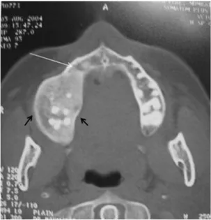
Two cases of the lesions involving the mandible showed ill-defined margin (Fig. 2) whereas other lesions were extending and merging into the cortex of the involved bone (Table 4).
The internal structure of the involved bone showed increased density of bone. Internal structure was found to be more homogenous in the maxilla than in the mandible.
All 8 cases showed the characteristic ground glass appear- ance (Fig. 3, Table 4).
Regarding the effect on surrounding structures (Table 2), all cases showed bucco-lingual expansion causing cortical
thinning of the involved bone. The lesions caused a dis- placement of adjacent structures such as inferior alveolar canal, maxillary sinus, and floor of orbit.
In 6 cases of maxillary involvement, the lesions displac- ed the maxillary sinus, however the shape of sinus was maintained (Fig. 4). Maxillary sinus in two cases and sphe- noid sinus in one case showed complete obliteration (Figs.
5A and B). The floor of orbit was raised in only two cases where the maxilla was involved. All 4 cases involving mandible showed a displacement of the inferior alveolar canal in all directions (Figs. 6A and B).
Loss of lamina dura was noted in all cases around the
Fig. 1. CT images of fibrous dyspla- sia. A. Coronal section shows cran- iofacial type of fibrous dysplasia with involvement 1. frontal, tempo- ral, 2. zygomatic, 3. maxilla, and 4.
mandible. B. Another coronal sec- tion shows maxillofacial type with involvement of 1. maxilla and 2.
zygomatic bone.
A B
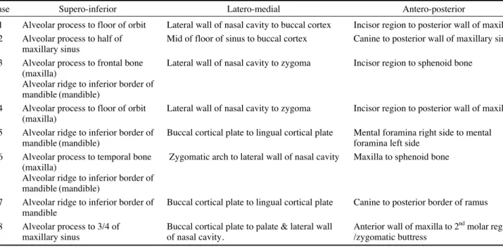
Table 3. CT findings of involved anatomical structures with extent of lesion
Case Supero-inferior Latero-medial Antero-posterior
1 Alveolar process to floor of orbit Lateral wall of nasal cavity to buccal cortex Incisor region to posterior wall of maxilla 2 Alveolar process to half of Mid of floor of sinus to buccal cortex Canine to posterior wall of maxillary sinus
maxillary sinus
3 Alveolar process to frontal bone Lateral wall of nasal cavity to zygoma Incisor region to sphenoid bone (maxilla)
Alveolar ridge to inferior border of mandible (mandible)
4 Alveolar process to floor of orbit Lateral wall of nasal cavity to zygoma Incisor region to posterior wall of maxilla (maxilla)
5 Alveolar ridge to inferior border of Buccal cortical plate to lingual cortical plate Mental foramina right side to mental
mandible (mandible) foramina left side
6 Alveolar process to temporal bone Zygomatic arch to lateral wall of nasal cavity Maxilla to sphenoid bone (maxilla)
Alveolar ridge to inferior border of mandible (mandible)
7 Alveolar ridge to inferior border of Buccal cortical plate to lingual cortical plate Canine to posterior border of ramus mandible
8 Alveolar process to 3/4 of Buccal cortical plate to palate & lateral wall Anterior wall of maxilla to 2ndmolar region
maxillary sinus of nasal cavity. /zygomatic buttress
teeth in the affected bone. Only 3 cases showed a displace- ment of the tooth and two cases were associated with im- pacted teeth, however none showed any evidence of root resorption.
Discussion
Fibrous dysplasia is encountered in younger age group with no specific sex predilection.5-7The craniofacial type of fibrous dysplasia is found to be as common as fibrous dysplasia of the jaw and was more commonly seen in
younger age. The unilateral nature of fibrous dysplasia was noted in all cases. The most commonly involved cra- nial bones were maxilla and frontal bones (Fig. 1). When the maxilla was affected, other adjacent bones separated by sutures such as zygomatic, sphenoid, frontal, and nasal bones might also get affected.9 This study showed that the right side was more commonly involved than the left, which was similar to the findings of previous studies.10
Eversole et al5suggested that the ill-defined margin of the lesion helped to differentiate it from other fibro-osse- ous lesions. In this study, the margins were ill-defined
Table 4. CT findings showing radiographic features
Internal Margins Cortex
structure
1 - - ++ - ++ ++ - - * - - ++ -
2 - - ++ - - ++ - - * - - ++ -
3 - - ++ ++ ++ ++ - - * - - ++ -
4 - - ++ - ++ ++ - - * - - ++ -
5 - - ++ - ++ ++ - - - ++ - ++ -
6 ++ - ++ - - - ++ - - ++ - ++ -
7 - - ++ - - ++ - - * - - ++ -
8 ++ - ++ - - - ++ - * - - ++ -
Total 2 0 8 1 4 6 2 0 6 2 0 8 0
*Well defined by the anatomic structures like cortex of involved bone. ++: present, - : negative.
Radiolucent
Case Peaud’orange Ground glass Proptosis Homogenous Heterogeneous Well defined Ill-defined Thick Thin AbsentResorption of toothDisplacement of tooth
Fig. 3.Coronal CT image shows lesion involving maxilla with increased density of maxilla giving ground glass appearance (black arrow).
Fig. 2. Axial CT image shows ill-defined margin (white arrow) except in region where lesion is extending to the cortex (black double arrow).
except in the region where they extended to the cortex of bone. Ground glass appearance was the most common radiographic appearance of internal structure of fibrous dysplasia in the present study, which was substantiated the diagnosis of fibrous dysplasia.10 Fibrous dysplasia shows bucco-lingual expansion causing thinning of the cortical plate. The expansion of the external surface of the affected bone assumed a grosser but still recognizable anatomical shape.6The lesion displaced the inferior alveo- lar canal in all four directions (buccal, lingual, superior, and inferior). The displacement was in contrast with the finding of Petrikowski11 who suggested that the upward displacement of inferior dental canal was a unique charac- teristic of fibrous dysplasia. However, the displacement of the canal in various directions might be explained by the location of epicenter in relation to the canal. For exam- ple if the epicenter was superior to the inferior alveolar
Fig. 4.Coronal CT image shows maxillary sinus occupied with lesion. Note: the size is reduced but the shape of maxillary sinus is maintained (black arrow).
Fig. 5.CT images. A. Coronal sec- tion shows obliteration of 1. maxil- lary sinus. B. Oblique section shows complete obliteration of 1. maxillary and 2. sphenoid sinus.
Fig. 6.CT images A. Oblique sec- tion shows displacement of inferior alveolar canal inferiorly (white ar- row). B. Axial section showing dis- placement of inferior alveolar canal lingually (black arrow).
A B
A B
canal, it would displace the canal inferiorly.
The lesion involving maxilla showed expansion on the external surface and on internal surface into maxillary sinus. The expansion of maxilla reduced the size of maxil- lary sinus cavity although the shape seemed unaltered.
This unique finding could help in differentiating fibrous dysplasia from other tumors such as ossifying fibroma.6 Fibrous dysplasia might also completely obliterate maxil- lary sinus and displace the floor of orbit.
Lamina dura was lost in all teeth which were involved in the lesion; this might be corroborated by the fact that the loss of lamina dura was unique to fibrous dysplasia and could be used as an ancillary diagnostic feature for fibrous dysplasia.11The impacted teeth which were noted in two cases may be caused by increased density of bone in the path of eruption. Although the lesion displaced the teeth without resorption, the displacement was found to be minimal. The minimal displacement of teeth such as inclination and rotation was noted but no bucco-lingual displacement similar to a study by Akintoye et al.12
The features of fibrous dysplasia such as the margins, internal structure, and effect on surrounding structure were well characterized on CT images. The margins of lesion, ground glass appearance, and displacement of maxillary sinus were characteristic and consistent with the findings of fibrous dysplasia. Although no single radiographic fea- ture is pathognomic of fibrous dysplasia, all the features together could be considered to be diagnostic of fibrous dysplasia.
References
1. Bastepe M. The GNAS Locus: quintessential complex gene
encoding Gsalpha, XLalphas, and other imprinted transcripts.
Curr Genomics 2007; 8 : 398-414.
2. White SC, Pharoah MJ. Oral radiology: principles and inter- pretation. 5th ed. St. Louis: Mosby-Year Book Inc; 2004. p.
485-91.
3. Waldron CA. Bone pathology. In: Neville BW, Damm DD, Allen CM, Bouquot JE. Oral and maxillofacial pathology.
Philadelphia: WB Saunders; 1995. p. 461-5.
4. Daves ML, Yardley JH. Fibrous dysplasia of bone. Am J Med Sci 1957; 234 : 590-606.
5. Eversole LR, Sabes WR, Rovin S. Fibrous dysplasia: a nosolo- gic problem in the diagnosis of fibro-osseous lesions of the jaws. J Oral Pathol 1972; 1 : 189-220.
6. MacDonald-Jankowski DS, Yeung R, Li TK, Lee KM. Com- puted tomography of fibrous dysplasia. Dentomaxillofac Radi- ol 2004; 33 : 114-8.
7. Ricalde P, Horswell BB. Craniofacial fibrous dysplasia of the fronto-orbital region: a case series and literature review. J Oral Maxillofac Surg 2001; 59 : 157-67.
8. Jacobsson S, Hallén O, Hollender L, Hansson CG, Lindström J. Fibro-osseous lesion of the mandible mimicking chronic osteomyelitis. Oral Surg Oral Med Oral Pathol 1975; 40 : 433- 44.
9. Posnick JC. Fibrous dysplasia of the craniomaxillofacial regi- on: current clinical perspectives. Br J Oral Maxillofac Surg 1998; 36 : 264-73.
10. MacDonald-Jankowski D. Fibrous dysplasia in the jaws of a Hong-Kong population: radiographic presentation and syste- matic review. Dentomaxillofac Radiol 1999; 28 : 195-202.
11. Petrikowski CG, Pharoah MJ, Lee L, Grace MG. Radiographic differentiation of osteogenic sarcoma, osteomyelitis, and fibr- ous dysplasia of the jaws. Oral Surg Oral Med Oral Pathol Oral Radiol Endod 1995; 80 : 744-50.
12. Akintoye SO, Lee JS, Feimster T, Booher S, Brahim J, King- man A, et al. Dental characteristics of fibrous dysplasia and McCune-Albright syndrome. Oral Surg Oral Med Oral Pathol Oral Radiol Endod 2003; 96 : 275-82.