Endocrinol Metab 2015;30:626-630
http://dx.doi.org/10.3803/EnM.2015.30.4.626 pISSN 2093-596X · eISSN 2093-5978
Case Report
A Rare Manifestation of Hypothyroid Myopathy:
Hoffmann’s Syndrome
Kang Won Lee1,*, Sun Hwa Kim1,*, Kyoung Jin Kim1, Sang Hyun Kim1, Hee Young Kim1, Byung-Jo Kim2, Sin Gon Kim1, Dong Seop Choi1
1Division of Endocrinology and Metabolism, Department of Internal Medicine, 2Department of Neurology, Korea University College of Medicine, Seoul, Korea
Hypothyroid myopathy is observed frequently and the resolution of the clinical manifestations of myopathy following thyroid hor- mone replacement is well known. However, a specific subtype of hypothyroid myopathy, Hoffmann’s syndrome, characterized by increased muscular mass (pseudohypertrophy), proximal muscle weakness, muscle stiffness and cramps, is rarely reported. Here- in, we describe a 34-year-old male who presented with proximal muscle weakness and non-pitting edema of the lower extremities.
He initially visited the neurology department where he was suspected of having polymyositis. Additional laboratory evaluation re- vealed profound autoimmune hypothyroidism and elevated muscle enzymes including creatine kinase. The patient was started on levothyroxine treatment and, subsequently, clinical symptoms and biochemical parameters resolved with the treatment. The pres- ent case highlights that hypothyroidism should be considered in the differential diagnosis of musculoskeletal symptoms even in the absence of overt manifestations of hypothyroidism. To our knowledge, this is the first case reported in Korea.
Keywords: Hypothyroidism; Muscular diseases; Creatine kinase
INTRODUCTION
Musculoskeletal symptoms are observed in 30% to 80% of pa- tients with hypothyroidism [1,2]. The symptoms consist of muscle weakness, pain, cramps, and stiffness and are usually mild but tend to become overt especially in untreated severe hypothyroidism [3]. There are four subtypes of myopathy asso- ciated with hypothyroidism: Hoffmann’s syndrome, myasthen- ic syndrome, atrophic form, and Kocher-Debre-Semelaigne syndrome [4].
Hoffmann’s syndrome, first reported by Johann Hoffmann in
1897, is characterized by increased muscular mass (pseudohy- pertrophy), muscle stiffness, proximal muscle weakness, and occasional muscle cramps [5]. The reported patient was an adult who developed peroneal muscular atrophy and myopathy associated with hypothyroidism after thyroidectomy. This syn- drome has a relatively favorable outcome if the thyroid hor- mone is replaced. Nevertheless, because of its rarity, this pre- sentation can be mistaken for primary muscular disorders such as polymyositis other than an initial manifestation of hypothy- roidism, especially when systemic signs of hypothyroidism are minimal or absent.
Received: 8 April 2015, Revised: 22 April 2015, Accepted: 11 May 2015 Corresponding author: Sun Hwa Kim
Division of Endocrinology and Metabolism, Department of Internal Medicine, Korea University College of Medicine, 73 Inchon-ro, Seongbuk-gu, Seoul 02841, Korea
Tel: +82-2-920-6270, Fax: +82-2-953-9355, E-mail: [email protected]
*These authors contributed equally to this work.
Copyright © 2015 Korean Endocrine Society
This is an Open Access article distributed under the terms of the Creative Com- mons Attribution Non-Commercial License (http://creativecommons.org/
licenses/by-nc/3.0/) which permits unrestricted non-commercial use, distribu- tion, and reproduction in any medium, provided the original work is properly cited.
Herein, we describe a 34-year-old male who presented main- ly with musculoskeletal symptoms and was diagnosed with Hoffmann’s syndrome associated with hypothyroidism. To our knowledge, this is the first case reported in Korea.
CASE REPORT
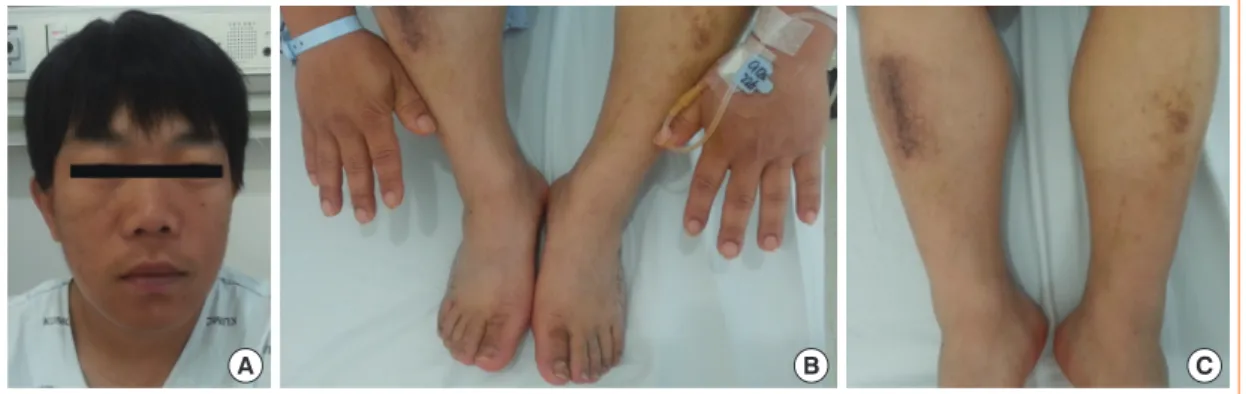
A 34-year-old male presented in July 2014 with progressive leg swelling and pain on exertion for approximately 6 months and was admitted to the neurology department. The patient had de- veloped considerable difficulty in ascending stairs for approxi- mately 1 month. He had become gradually lethargic and intol- erant to cold for 1 year. He had chronic constipation. He had no particular medical history including diabetes mellitus, prior thyroid disease, neuromuscular disorders, or autoimmune dis- eases and denied any history of medications. On physical ex- amination, his face was puffy with preorbital swelling (Fig.
1A) and his voice was hoarse. He had no goiter. His hands were edematous and had difficulty in grasping (Fig. 1B). His trapezius, biceps, triceps, and gastrocnemius muscles were hy- pertrophied although he denied that he exercised regularly. He also had non-pitting edema and induration of the skin on the lower extremities (Fig. 1C). On neurological examination, he had normal mentality. Upper and lower limb muscle weakness (grade 4/5) was observed, most markedly in the lower extremi- ties. The deep tendon reflexes were sluggish.
Laboratory findings revealed a hemoglobin level of 12.4 g/
dL (normal range, 13.2 to 17.3), elevated total cholesterol level of 327 mg/dL (normal range, 130 to 240), and elevated muscle enzymes as follows: aspartate aminotransferase (AST) 107 IU/
L (normal range, 3 to 45); alanine aminotransferase 106 IU/L (normal range, 3 to 45); creatine kinase (CK) 7,332 IU/L (nor-
mal range, 43 to 198); lactate dehydrogenase (LDH) 1,958 IU/
L (normal range, 238 to 422); serum myoglobin 235.4 ng/mL (normal range, 28 to 72); and aldolase 10.5 U/L (normal range, 0.0 to 7.6). Hepatitis markers, including hepatitis B surface an- tigen, hepatitis B surface antibody, hepatitis C virus antibody, and hepatitis A virus antibody immunoglobulin M were nega- tive. Urine myoglobin and protein were negative. Other bio- chemical blood tests revealed a blood urea nitrogen of 10.5 mg/
dL (normal range, 7 to 23), a creatinine of 1.58 mg/dL (normal range, 0.7 to 1.44), a protein of 7.9 mg/dL (normal range, 6.6 to 8.3), albumin of 4.7 mg/dL (normal range, 3.5 to 5.2), and total bilirubin of 0.45 mg/dL (normal range, 0.3 to 1.4). Electrolytes were in the normal range. The thyroid function test showed se- vere hypothyroidism with thyroid stimulating hormone (TSH) above 100 µIU/mL (normal range, 0.17 to 4.05), free thyroxine (FT4) of 0.03 ng/dL (normal range, 0.79 to 1.86), and total tri- iodothyronine of 75.8 ng/dL (normal range, 78 to 182). Anti- microsomal antibody was positive at 56.26 IU/mL (normal range, 0 to 5.61), and anti-thyroglobulin antibody was also ele- vated at 348.02 IU/mL (normal range, 0 to 4.11). The value of erythrocyte sedimentation rate was 9 mm/hr (normal range, 0 to 15) and C-reactive protein was 0.16 mg/L (normal range, 0 to 5). Fluorescent antinuclear antibody, anti-neutrophil cyto- plasmic antibody, and rheumatoid factor were negative.
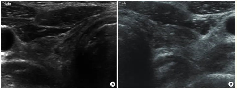
Ultrasonography of the neck revealed an atrophied thyroid (Fig. 2). Electrocardiogram showed sinus bradycardia (51 beats per minute) and a first-degree atrioventricular block; echocar- diography showed a small amount of pericardial effusion (post, 8 mm) with preserved left ventricular systolic function. Elec- tromyography showed mild spontaneous activity and short am- plitude with polyphasic myopathic motor unit potential (Fig. 3).
In addition, electroneurography revealed bilateral carpal tunnel
Fig. 1. (A) Puffy face and preorbital swelling. (B) Edematous hands and lower legs. (C) Non-pitting edema of the lower extremities and hypertrophy of the calf muscles, with skin changes (dry coarse skin and hyperpigmentation). These photos were taken after obtaining prior consent from the patient.
A B C
syndrome and reduced nerve conduction velocity on the super- ficial peroneal nerve. Magnetic resonance imaging of the lower extremities revealed symmetric multifocal patchy muscle en- hancements with diffuse muscle swelling in thighs and lower legs, especially in the superficial posterior compartment and gastrocnemius muscles that showed hyperintensity on T2- weighted images (Fig. 4). Muscle biopsy was recommended but was not performed due to the patient’s refusal.
Based on the above findings, the patient was diagnosed with autoimmune hypothyroidism with Hoffmann’s syndrome. He was treated with oral levothyroxine starting with 125 µg/day and gradually increased up to 200 µg/day. At the 1 month fol- low-up, his condition had improved significantly; the puffiness of his face and eyelids had regressed, the calf muscle size was decreased and muscle strength had returned to normal. After 2 months of thyroid hormone replacement, the muscle enzymes and creatinine were decreased as follows: CK, 103 IU/L; LDH, 409 IU/L; myoglobin, 55.8 ng/mL; and creatinine, 1.32 mg/dL.
At 3 months, the TSH value was reduced to 3.04 µIU/mL, FT4 increased to 1.26 ng/dL, and creatinine was decreased to 0.9
mg/dL. He is receiving levothyroxine (200 µg/day) at an out- patient clinic.
DISCUSSION
The manifestations of hypothyroid myopathy depend on the severity and duration of hypothyroidism [4]. Most patients with hypothyroidism have few apparent musculoskeletal symptoms at the time of diagnosis. The patient in our case suffered from proximal weakness of the lower extremities and initially visited Fig. 2. (A, B) Neck ultrasonography showed atrophied thyroid gland with no definite nodular lesions.
Right
A B
Left
100 μV Amp 1:20-10 k, 60 Hz 10 ms
Fig. 3. Electromyography showed mild spontaneous activity and short amplitude with polyphasic myopathic motor unit potential.
Fig. 4. Magnetic resonance imaging of the lower extremities shows symmetric multifocal patchy muscle enhancements with diffuse muscle swelling in both lower legs (superficial posterior compartment). Hypertrophy of the gastrocnemius; affected mus- cles show hyperintensity on T2-weighted images (arrows).
the neurology department where he was suspected of having polymyositis and was admitted for further evaluation of his muscular symptoms including progressive muscle weakness, hypertrophy and CK elevation. Polymyositis is an autoimmune inflammatory myopathy that presents with muscle weakness and elevated muscle enzymes, which requires long-term im- munosuppressive therapy [2] and can be confused with hypo- thyroid myopathy because of clinical and biochemical similari- ties. However, the laboratory findings on admission revealed highly elevated TSH and, therefore, he was referred to the en- docrinology department and was finally diagnosed as having autoimmune hypothyroidism with Hoffmann’s syndrome.
Hoffmann’s syndrome is a rare manifestation of hypothyroid myopathy characterized by pseudohypertrophy, proximal mus- cle weakness and stiffness [6]. The syndrome is usually ob- served in patients with severe and untreated hypothyroidism, as in our case. Pseudohypertrophy is a rare presentation of hypo- thyroidism and its etiology remains obscure [4]. However, its mechanism involves an increase in the deposition of glucos- aminoglycans and in the number and size of muscle fibers, as well as a change in muscle fiber type from fast twitch type II to slow twitch type I leading to delayed muscle contraction [7].
Previous reports on Hoffmann’s syndrome showed that calf muscles are usually affected, but any part of the muscles could be involved including thigh, arm, and forearm [7]. The pres- ence of calf muscle hypertrophy requires differential diagnosis from other diseases such as Duchenne and Becker muscle dys- trophy, focal myositis, sarcoid granulomas, and amyloid depos- its [4].
CK is the representative biochemical marker of myopathy [8]. However, CK is elevated even in patients with mild hypo- thyroidism regardless of the severity of myopathy [9,10]. Other enzymes such as aldolase, AST and LDH have a supportive role in the absence of hepatic dysfunction. The pathophysiolo- gy of elevated CK in hypothyroidism is not fully understood but, presumably, decreased CK clearance, a reversible defect in glycogenolysis and direct cell damage are the responsible fac- tors [11]. Due to the decreased CK clearance, the resolution time of elevated CK is usually longer in hypothyroidism com- pared with patients with myocardial infarction [12]. The CK level in our patient decreased to normal after 2 months of thy- roid hormone replacement.
Electrophysiological studies may reveal non-specific fea- tures suggestive of myopathic, neurogenic or mixed patterns in hypothyroid myopathy [6]. In Hoffmann’s syndrome, myo- pathic patterns are observed, especially in calf muscles, as in
our case [13]. Abnormal findings on electrophysiological study may persist in some patients despite the improvement of symp- toms and muscle enzymes [6]. On biopsy, the affected muscles may show type I fiber hypertrophy as a compensatory response to type II fiber atrophy with increased numbers of central nu- clei and increased connective tissue, which are the representa- tive findings of hypothyroid myopathy [14].
Hoffmann’s syndrome has a favorable prognosis after thy- roid hormone replacement has been initiated. Clinical symp- toms resolve slowly with time. It has been reported that the de- cline of CK to normal levels may not be a reliable indicator of the rate of recovery of hypothyroid myopathy. The decline of the muscle enzyme levels may occur slowly, varying from weeks, months, or even years [2].
If Hoffmann’s syndrome presents without other manifesta- tions of hypothyroidism, then a high degree of caution is neces- sary. Kaux et al. [15] reported a patient with Hoffmann’s syn- drome who presented emergently with severe asthenia and ar- thralgia without other prominent characteristics of hypothy- roidism; in that case, early diagnosis and hormone replacement brought rapid stabilization.
This case emphasizes that hypothyroidism should be consid- ered in the differential diagnosis of musculoskeletal symptoms in the absence of overt manifestations of hypothyroidism. Con- sideration of hypothyroidism and the routine measurement of serum TSH are important during the evaluation of patients with progressive muscle weakness and swelling.
CONFLICTS OF INTEREST
No potential conflict of interest relevant to this article was re- ported.
REFERENCES
1. Khaleeli AA, Griffith DG, Edwards RH. The clinical pre- sentation of hypothyroid myopathy and its relationship to abnormalities in structure and function of skeletal muscle.
Clin Endocrinol (Oxf) 1983;19:365-76.
2. Madariaga MG. Polymyositis-like syndrome in hypothy- roidism: review of cases reported over the past twenty-five years. Thyroid 2002;12:331-6.
3. Ciompi ML, Zuccotti M, Bazzichi L, Puccetti L. Polymyo- sitis-like syndrome in hypothyroidism: report of two cases.
Thyroidology 1994;6:33-6.
4. Senanayake HM, Dedigama AD, De Alwis RP, Thirumava-
lavan K. Hoffmann syndrome: a case report. Int Arch Med 2014;7:2.
5. Qureshi W, Hassan G, Khan GQ, Kadri SM, Kak M, Ah- mad M, et al. Hoffmann’s syndrome: a case report. Ger Med Sci 2005;3:Doc05.
6. Vasconcellos LF, Peixoto MC, de Oliveira TN, Penque G, Leite AC. Hoffman’s syndrome: pseudohypertrophic my- opathy as initial manifestation of hypothyroidism. Case re- port. Arq Neuropsiquiatr 2003;61:851-4.
7. Nalini A, Govindaraju C, Kalra P, Kadukar P. Hoffmann’s syndrome with unusually long duration: report on clinical, laboratory and muscle imaging findings in two cases. Ann Indian Acad Neurol 2014;17:217-21.
8. Giampietro O, Clerico A, Buzzigoli G, Del Chicca MG, Boni C, Carpi A. Detection of hypothyroid myopathy by measurement of various serum muscle markers: myoglo- bin, creatine kinase, lactate dehydrogenase and their isoen- zymes. Correlations with thyroid hormone levels (free and total) and clinical usefulness. Horm Res 1984;19:232-42.
9. Graig FA, Smith JC. Serum creatine phosphokinase activity
in altered thyroid states. J Clin Endocrinol Metab 1965;25:
723-31.
10. Mastropasqua M, Spagna G, Baldini V, Tedesco I, Paggi A.
Hoffman’s syndrome: muscle stiffness, pseudohypertrophy and hypothyroidism. Horm Res 2003;59:105-8.
11. Finsterer J, Stollberger C, Grossegger C, Kroiss A. Hypo- thyroid myopathy with unusually high serum creatine ki- nase values. Horm Res 1999;52:205-8.
12. Klein I, Mantell P, Parker M, Levey GS. Resolution of ab- normal muscle enzyme studies in hypothyroidism. Am J Med Sci 1980;279:159-62.
13. Tuncel D, Cetinkaya A, Kaya B, Gokce M. Hoffmann’s syndrome: a case report. Med Princ Pract 2008;17:346-8.
14. Khaleeli AA, Gohil K, McPhail G, Round JM, Edwards RH. Muscle morphology and metabolism in hypothyroid myopathy: effects of treatment. J Clin Pathol 1983;36:519- 26.
15. Kaux JF, Castermans C, Delmotte P, Bex M. Hoffmann syn- drome presenting to the emergency department. Ann Readapt Med Phys 2007;50:310-2.