https://doi.org/10.4174/astr.2020.99.6.352 Annals of Surgical Treatment and Research
The efficacy of the “no zone” approach for the assessment of traumatic neck injury: a case-control study
Ji Wool Ko1,2, Seong Chan Gong1,2, Myung Jun Kim1,2, Jae Sik Chung1,2, Young Un Choi1,2, Jun Hyuk Lee3, Pil Young Jung1,2
1Department of Surgery, Yonsei University Wonju College of Medicine, Wonju, Korea
2Regional Trauma Center, Wonju Severance Christian Hospital, Wonju, Korea
3Department of Biostatistics, Yonsei University Wonju College of Medicine, Wonju, Korea
INTRODUCTION
Traumatic neck injury comprises approximately 5%–10% of all traumatic injuries [1]. Despite its low incidence, the number of mortalities due to neck injury has still been high (10%–15%) [2-7]. Anatomical features like the trachea, esophagus, great vessels, and nerves are crowded in small spaces and are relatively unprotected [8]. Therefore, mandatory exploration is
generally performed in cases of zone II injuries, according to the classification of the anatomical zones of the neck: zone I spans from the clavicles to the cricoid; zone II spans from the cricoid to the angle of the mandible; and zone III ranges from the angle of the mandible to the base of the skull [9]. This approach has been referred to as the zone-based algorithm and has been used as a traditional assessment for traumatic neck injuries.
However, routine neck exploration in hemodynamically
Received January 29, 2020, Revised August 11, 2020, Accepted October 9, 2020
Corresponding Author: Pil Young Jung
Department of Surgery, Yonsei University Wonju College of Medicine, 20 Ilsan-ro, Wonju 26426, Korea
Tel: +82-33-741-0573, Fax: +82-33-741-0574 E-mail: [email protected]
ORCID: https://orcid.org/0000-0001-6460-8072
• This study was presented at the 71st Annual Congress of Korean Surgical Society 2019 (October 31, 2019) as an oral presentation.
Copyright ⓒ 2020, the Korean Surgical Society
cc Annals of Surgical Treatment and Research is an Open Access Journal. All articles are distributed under the terms of the Creative Commons Attribution Non- Commercial License (http://creativecommons.org/licenses/by-nc/4.0/) which permits unrestricted non-commercial use, distribution, and reproduction in any medium, provided the original work is properly cited.
Purpose: Recently, several studies have demonstrated symptom-based, non-zonal algorithms for approaching penetrating neck injuries. The purpose of this study was to confirm the effectiveness of the “no zone” approach in traumatic neck injuries.
Methods: Medical charts of patients with traumatic neck injuries who presented at the Regional Trauma Center in South Korea between January 2014 and December 2018 were retrospectively reviewed. Negative final neck findings (FNFs) were compared with positive FNFs (which include major vascular, aerodigestive, nerve, endocrine gland, cartilage, or hyoid bone injuries) using multivariate logistic regression analysis including values of the “zone” and/or no zone approach.
Results: Out of 168 trauma patients, 70 patients with a minor injury and 7 patients under the age of 18 years were excluded.
Of the remaining 91 patients, 74 (81.3%) had penetrating neck injuries and 17 (18.7%) had blunt neck injuries. Initial diagnosis most frequently revealed external wounds in zone II (84.6%). Twenty (22.0%) and 36 (39.5%) patients had hard and soft signs, respectively, using the no zone approach. Further, there was a significant difference between the negative and positive FNFs in patients with hard signs (11.6% vs. 54.5%; P < 0.01, respectively). According to the multivariate logistic regression analysis, the hard signs were associated with an odds ratio (OR) for FNFs (OR, 18.92; 95% confidence interval, 3.55–157.60).
Conclusion: Traumatic neck injuries classified as having hard signs based on the no zone approach may be correlated with internal organ injuries of the neck.
[Ann Surg Treat Res 2020;99(6):352-361]
Key Words: Neck injuries, “No zone” approach, Trauma centers
stable patients is reportedly known to result in a high rate of negative exploration, longer hospital stay, and an increased rate of complications, such as surgical site infections and sepsis [10]. Therefore, the management of neck injuries has changed from a mandatory exploration of all wounds that penetrate the platysma to a more selective approach based on patient symptoms. This selective operation is based on the “no zone”
approach in which one determines the treatment method based on the classification of the symptoms that may have resulted from damage to the major vascular, digestive, and respiratory systems. If the patients are hemodynamically unstable or indicate confirmed hard signs, such as active bleeding or severe emphysema, surgery or other therapeutic procedures like angioembolization should be considered without further examination. In contrast, the decision to perform a surgical treatment in hemodynamically stable patients is controversial.
The purpose of this study was to evaluate the efficacy of the no zone approach in traumatic neck injuries. We hypothesized that hard signs in symptomatic approaches may be useful for predicting internal organ injuries in the neck and may help make a more informed decision regarding an operation.
METHODS
Patients and data collection
Between January 2014 and December 2018, we prospectively enrolled patients with neck injuries who were treated at the Regional Trauma Center in South Korea and whose data were recorded in the Korean Trauma Data Bank (KTDB). The patients’ medical charts and data extracted from the KTDB
were analyzed retrospectively. This study was approved by the Institutional Review Board of Wonju Severance Christian Hospital (No. CR319118). Since the data were analyzed anonymously, informed consent was exempted. All patients who sustained a penetrating or blunt neck injury were included.
Patient characteristics including age, sex, psychiatric history, mechanism of trauma (penetrating or blunt), systolic blood pressure (BP), Glasgow Coma Scale (GCS) score, zone of injury and associated injuries, injury severity score (ISS), hemoglobin (Hb) level, lactate level, and PT-international normalized ratio (PT-INR) were measured on arrival at the emergency room (ER). The patients were classified by clinical presentation.
Operation findings of the neck injury were extracted from the medical charts and data from the CT scans were reviewed by a radiologist.
Negative or positive final neck findings: definitions and outcomes
The major study outcomes were final injury sites and final internal organ injuries, whereas other outcomes included primary repair rates (where primary repair is defined as a simple wound closure without internal organ injury under local anesthesia), conservative treatment rates (where conservative treatment is defined as surgical observation without any therapeutic procedures), hospital length of stay (LOS), length of intensive care unit (ICU) stay, complications (including surgical site infection, hepatic failure, acute kidney injury, acute respiratory failure, pneumonia, and sepsis), and mortality.
Positive final neck findings (FNFs) were defined as internal organ injuries of the neck that required surgical treatment as
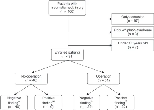
Patients with traumatic neck injury
(n = 168)
Enrolled patients (n = 91)
Negative findinga) (n = 40)
Positive findinga) (n = 0)
Negative findingb) (n = 29)
Positive findingb) (n = 22)
Only contusion (n = 67)
Only whiplash syndrome (n = 3)
Under 18 years old (n = 7)
No-operation (n = 40)
Operation (n = 51)
Fig. 1. Flowchart of study design. a)Internal organ injuries confirmed by CT scan. b)Internal organ injuries confirmed by an operation.
confirmed by CT scan or operation findings, such as major vascular, aerodigestive, nerve, endocrine gland, cartilage, or hyoid bone injuries. Absence of such internal organ injuries was defined as negative FNFs. In this study, we analyzed the differences between negative and positive FNFs.
The no zone approach with hard signs
Patients were classified as having “hard signs” or “soft signs,”
or being “asymptomatic,” according to the signs and symptoms they presented. As per several previous studies, hard signs were defined as the presence of either a shock, active bleeding, expanding/pulsatile hematoma, focal neurologic deficit, airway compromise, massive subcutaneous emphysema, air bubbling through wounds, or severe hematemesis; soft signs included stable hematomas, minor hemoptysis, hoarseness, dysphagia, and mild subcutaneous emphysema; and the asymptomatic group included all patients with no signs of neck injury and no symptoms related to neck injuries [11-13]. According to initial physical examination findings, patients who were hemodynamically unstable and displayed hard signs underwent surgery, whereas those who were stable were considered for imaging tests. Moreover, image inspection was considered for patients with soft signs and asymptomatic patients with conservative treatment.
Statistical analyses
Continuous variables are presented as mean ± standard deviation or median (range) and were compared using the Student t-test or the Wilcoxon rank-sum test. Categorical variables were presented as frequency (percentage) and were compared using the chi-squared test or Fisher exact test.
Multiple logistic regression analysis was used to identify values associated with final internal organ injuries of the neck, and results were presented as odds ratio (OR) with a 95% confidence interval (CI). A P-value of <0.05 was considered statistically significant for all analyses and all P-values were two-tailed. All statistical analyses were performed using SAS software, ver.
9.4 (SAS Institute, Cary, NC, USA) and R software, ver. 3.6 (R Foundation for statistical computing, Vienna, Austria).
RESULTS
Clinical characteristics
Of the 168 patients with neck injuries, 67 had only contusion of the neck, 3 had whiplash syndrome, and 7 were under the age of 18 years; hence, they were all excluded. Finally, 91 patients (mean age, 48.3 ± 14.7 years; male, 80.2%) were enrolled in the study (Fig. 1). Of these, 74 (81.3%) had penetrating injuries and 18 (19.8%) experienced trauma from self-inflicted injuries. The median ISS was 4 (0−75). There were 44 (48.4%) concomitant injuries including head, chest, abdomen, and extremities.
Table 1. Clinical characteristic and patients outcomes (n = 91)
Variable Data
Characteristic
Age (yr) 48.3 ± 14.7
Male sex 73 (80.2)
Penetrating injury 74 (81.3)
Blunt injury 17 (18.7)
Self-inflicted injury 18 (19.8)
Injury severity score 4 (0–75)
Accompanying injurya) 44 (48.4)
GCS score 13.2 ± 3.8
Systolic BP (mmHg) 125.2 ± 45.2
Hemoglobin (g/dL) 13.4 ± 2.4
Lactate (mmol/L) 3.8 ± 3.4
PT-INR 1.09 ± 0.27
“Zone” approach
Ib) 7 (7.7)
IIb) 64 (70.3)
IIIb) 7 (7.7)
Multiple zonesc) 13 (14.3)
“No zone” approach
Hard signs 20 (22.0)
Soft signs 36 (39.6)
Asymptomatic 35 (38.5)
Outcome
CT scand) 65 (71.4)
Operatione) 51 (56.0)
Primary repairf) 25 (27.5)
Conservative treatmentg) 15 (16.5)
Internal organ injuryh) 22 (24.2)
Injury sitesi)
Artery 6 (6.6)
Vein 13 (14.3)
Airway 3 (3.3)
Esophagus 1 (1.1)
Nerve 2 (2.2)
Endocrine glandj) 2 (2.2)
Thyroidal cartilage 3 (3.3)
Hyoid bone 1 (1.1)
Superficial injuryk) 34 (37.4)
Muscle 35 (38.5)
Multiple sites 7 (7.7)
Hospital LOS (day) 7 (1–1,107)
ICU LOS (day) 0 (0–40)
Complication 2 (2.2)
Mortality 5 (5.5)
Values are presented as number (%), mean ± standard deviation, or median (range).
GCS, Glasgow coma scale; BP, blood pressure; INR, international normalized ratio; LOS, length of stay; ICU, intensive care unit.
a)Injury of head, chest, abdomen, and extremities. b)Injury isolated to the zone. c)Zone II was included in all cases. d)Neck contrast CT or neck angiography CT. e)Surgery performed under general anesthesia.
f)Simple wound closure without internal organ injury under local anesthesia. g)Surgical observation without any therapeutic procedures. h)Injury to need surgical treatment confirmed by CT scan or operation. i)Variable duplicated. j)Thyroid gland or submandibular gland. k)Non-invasion of the platysma muscle.
Upon arrival at the ER, the mean values for the GCS score, systolic BP, Hb, lactate level, and PT-INR were 13.2 ± 3.8, 125.2
± 45.2 mmHg, 13.4 ± 2.4 g/dL, 3.8 ± 3.4 mmol/L, and 1.09
± 0.27, respectively. Based on the “zone” approach, the most common zone of injury was zone II (84.6%) including patients of multiple zones (in all multiple zone cases, zone II was included).
According to the no zone approach, 20 (22.0%), 36 (39.6%), and 35 patients (38.5%) were classified as hard signs, soft signs, and asymptomatic, respectively. CT scans were performed in 65 patients (71.4%), and emergency surgery was performed under general anesthesia in 51 patients (56.0) (Table 1).
Patient outcomes
Twenty-five patients (27.5%) underwent primary repair, 15 (16.5%) received conservative treatments, and 22 (24.2%) experienced internal organ injuries. The final injury sites were confirmed via the CT scans or surgical findings and classified as superficial (non-invasion of the platysma muscle), major vascular (artery or vein), aerodigestive (trachea or esophagus), nerve (cervical nerve), endocrine gland (thyroid gland or submandibular gland), cartilage or hyoid bone, muscle (muscle of neck including the platysma muscle without internal organ injury), or multiple injuries. The most common injury sites were muscle (38.5%) and superficial injuries (37.4%), whereas multiple injuries occurred in only 7 cases (7.7%). The median hospital LOS was 7 days (1−1,107 days) and the median ICU LOS was 0 days (0−40 days). Complications occurred in 2 patients (2.2%) and 5 patients (5.5%) died (Table 1).
Complications and mortalities
Two of the 91 patients had complications; both patients had penetrating injuries of zone II and cerebral infarctions as complications. One patient sustained an intraoperative left common carotid artery injury resulting in an infarction of the left anterior cerebral artery, whereas the other patient sustained an intraoperative left common carotid artery and jugular vein injury, which resulted in an infarction of the left middle cerebral artery received thrombectomy. Among the 20 patients
who had hard signs, 13 patients experienced a shock, 1 had a blunt injury, and all the other had a penetrating injury; in total, there were 5 mortalities. Dislocation of the cervical vertebrae was observed in the case of the patient with blunt injury; the patient died post cardiac arrest upon arrival at the ER. Out of the 4 patients, an emergency operation was performed in 2 patients, while another 2 patients died of an cardiac arrest that occurred during the visit to the hospital without operation.
Also, they died of massive bleeding caused by a carotid artery or jugular vein injury. Patients with an arrest at the hospital had major vessel injury highly suspected, although unable to perform the operation.
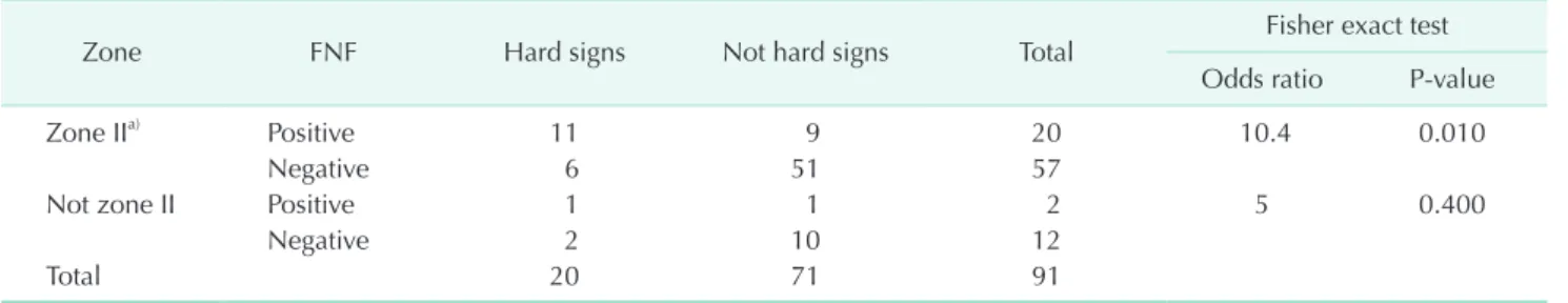
Zone I or III with hard signs and zone II without hard signs
For zone I or zone III cases, only 3 patients displayed hard signs, all of whom underwent operation; 1 patient had positive FNFs (subclavian artery injury). In the zone II cases, 51 patients had soft signs or were asymptomatic. In the multiple zones cases, 9 patients had soft signs or were asymptomatic. Of all the 60 patients in zone II (including 1 in multiple zones without hard sign), 31 underwent operation and 9 were diagnosed with positive FNFs. Eleven of the other 29 patients who did not undergo operation were confirmed with negative FNFs by CT scans and discharged without complications after conservative treatment.
When we performed the log-linear analysis with FNFs, hard sign, and zone, there was a statistically significant correlation between FNFs and hard sign compared to between FNFs and zone (χ2, 16.02; P < 0.001). Furthermore, Fisher exact test separately constructed in zone II showed that hard signs were highly correlated with the possibility of FNFs (OR, 10.4; P <
0.010) (Table 2).
Analysis of the final neck findings of total number of patients
When we compared the negative FNFs and positive FNFs, the median ISS of the positive FNFs was higher than that of
Table 2. Comparison between the “zone” approach and “no zone” approach
Zone FNF Hard signs Not hard signs Total Fisher exact test
Odds ratio P-value
Zone IIa) Positive 11 9 20 10.4 0.010
Negative 6 51 57
Not zone II Positive 1 1 2 5 0.400
Negative 2 10 12
Total 20 71 91
Values are presented as number.
FNF, final neck finding.
a)All multiple zone cases were included.
the negative FNFs group (2 vs. 9, P = 0.002). Further, patients with negative FNFs had a higher mean Hb level (13.8 ± 2.3 g/
dL vs. 12.3 ± 2.5 g/dL, P = 0.010). The positive FNFs group had a higher number of patients with hard signs (11.6% vs. 54.5%, P
< 0.001) than the negative FNFs group and no asymptomatic patients (35 vs. 0, P < 0.001). Furthermore, the rate of operation in the positive FNFs group was higher than that in the negative FNFs group (42.0% vs. 100.0%, P < 0.001), whereas the rate of conservative treatment in negative FNFs was higher than that in positive the FNFs group (21.7% vs. 0.0%, P < 0.018).
There were no significant differences in age, sex, mechanism of injury, accompanying injury, GCS score, systolic BP, lactate levels, PT-INR, zone of injury, soft signs, CT scan, hospital LOS, ICU LOS, complications, and mortality between the 2 groups (Table 3).
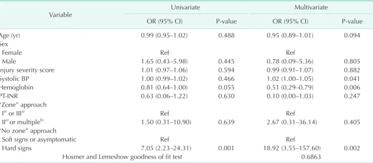
We constructed a multiple logistic regression model for FNFs for the total number of patients. Age, sex, systolic BP,
and PT-INR were the variables that played the least role as a compounding factor in the analysis. ISS and Hb were selected because there were significant differences in these variables between the negative and positive FNFs groups. The zone approach and no zone approach variables were included in the analysis because they were values to be confirmed. According to this analysis, the presence of hard signs was highly correlated with the possibility of the FNFs (OR, 18.92; 95% CI, 3.55–157.60;
P = 0.002) (Table 4). A receiver operating characteristic (ROC) curve of multivariate logistic regression analysis for the FNFs, with the use of the variables included in predicting FNFs, showed an area under the curve (AUC) of 0.846 (Fig. 2A).
Analysis of the final neck findings of patients who underwent neck exploration
We compared the negative and positive FNFs in the subgroup of patients receiving operation (defined as the exploration Table 3. Comparison between negative and positive FNFs in total patients
Variable Negative FNF (n = 69) Positive FNF (n = 22) P-value
Age (yr) 48.35 ± 15.2 48.2 ± 13.1 0.963
Male sex 56 (81.2) 17 (77.3) 0.761g)
Penetrating injury 53 (76.8) 21 (95.5) 0.062g)
Blunt injury 16 (23.2) 1 (4.5) 0.062g)
Self-inflicted injury 11 (15.9) 7 (38.9) 0.128
Injury severity score 2 (0−75) 9 (1−34) 0.002h)
Accompanying injurya) 32 (46.4) 12 (54.5) 0.626
GCS score 13.3 ± 3.8 13.0 ± 4.0 0.747
Systolic BP (mmHg) 124.1 ± 43.3 128.7 ± 51.9 0.679
Hemoglobin (g/dL) 13.8 ± 2.3 12.3 ± 2.5 0.010
Lactate (mmol/L) 3.8 ± 3.2 3.8 ± 3.9 0.962
PT-INR 1.09 ± 0.28 1.08 ± 0.24 0.893
“Zone” approach
Ib) 5 (7.2) 2 (9.1) 0.674g)
IIb) 49 (71.0) 15 (68.2) >0.999
IIIb) 7 (10.1) 0 (9.1) 0.189g)
Multiple zonesc) 8 (11.6) 5 (22.7) 0.291g)
“No zone” approach
Hard signs 8 (11.6) 12 (54.5) <0.001
Soft signs 26 (37.7) 10 (45.5) 0.618
Asymptomatic 35 (50.7) 0 (0) <0.001
CT scand) 50 (72.5) 15 (68.2) 0.788
Operatione) 29 (42.0) 22 (100.0) <0.001g)
Conservative treatmentf) 15 (21.7) 0 (0) <0.018g)
Hospital LOS (day) 5 (1–1,107) 11.5 (2–68) 0.790
ICU LOS (day) 0 (0 – 40) 3 (0–12) 0.219
Complication 0 (0) 2 (9.1) 0.056g)
Mortality 3 (4.3) 2 (9.1) 0.591g)
Values are presented as mean ± standard deviation, number (%), or median (range).
FNF, final neck finding; GCS, Glasgow coma scale; BP, blood pressure; INR, international normalized ratio; LOS, length of stay; ICU, intensive care unit.
a)Injury of head, chest, abdomen, and extremities. b)Injury isolated to the zone. c)Zone II was included in all cases. d)Neck contrast CT or neck angiography CT. e)Surgery performed under general anesthesia. f)Surgical observation without any therapeutic procedures.
g)Fisher exact test. h)Wilcoxon rank-sum test.
group) for removing a selection bias. In this exploration group, patients with negative FNFs had a higher Hb level than those with positive FNFs (13.8 ± 2.3 g/dL vs. 13.3 ± 2.4 g/dL, P = 0.027). Further, there was a significant difference between patients with hard signs and those who were asymptomatic (17.2% vs. 54.5%, P = 0.007; 27.6% vs. 0.0%, P = 0.007, respectively). However, there were no significant differences in age, sex, mechanism of injury, ISS, accompanying injury, GCS score, systolic BP, lactate, PT-INR, a zone of injury, soft signs, CT scan, hospital LOS, ICU LOS, complication, and mortality between the positive and negative FNFs patients in this
exploration group (Table 5). We constructed a multiple logistic regression model for FNFs in the exploration group. Age, sex, ISS, and systolic BP were the variables that played the least role as a compounding factor in the analysis. Hb was selected because there were significant differences in the Hb levels between the negative and positive FNFs in exploration group analysis. The zone approach and no zone approach variables were included in the analysis because they were values to be confirmed. According to analysis in the exploration group, the presence of the hard signs was highly correlated with the possibility of the FNFs in the exploration group (OR, 11.19; 95%
Table 4. Univariate and multivariate logistic regression analysis for negative and positive FNFs in total patient
Variable Univariate Multivariate
OR (95% CI) P-value OR (95% CI) P-value
Age (yr) 0.99 (0.95–1.02) 0.488 0.95 (0.89–1.01) 0.094
Sex
Female Ref Ref
Male 1.65 (0.43–5.98) 0.445 0.78 (0.09–5.36) 0.805
Injury severity score 1.01 (0.97–1.06) 0.594 0.99 (0.91–1.07) 0.882
Systolic BP 1.00 (0.99–1.02) 0.466 1.02 (1.00–1.05) 0.041
Hemoglobin 0.81 (0.64–1.00) 0.055 0.51 (0.29–0.79) 0.006
PT-INR 0.63 (0.06–1.22) 0.630 0.10 (0.00–1.03) 0.247
“Zone” approach
Ia) or IIIa) Ref Ref
IIa) or multipleb) 1.50 (0.31–10.90) 0.639 2.67 (0.31–36.14) 0.405
“No zone” approach
Soft signs or asymptomatic Ref Ref
Hard signs 7.05 (2.23–24.31) 0.001 18.92 (3.55–157.60) 0.002
Hosmer and Lemeshow goodness of fit test 0.6863
Compounding factor: age, sex, systolic BP, and PT-INR. Significant differences between the negative and positive FNFs: injury severity score and hemoglobin. Values to confirm: “zone” approach and “no zone” approach.
FNF, final neck finding; OR, odds ratio; CI, confidence interval; BP, blood pressure; INR, international normalized ratio.
a)Injury isolated to the zone. b)Zone II was included in all cases.
1.0
Sensitivity
1-Specificity 1.0
0.8
0.6
0.4
0.2
0
AUC: 0.846
0.2 0.4 0.6 0.8 1.0
Sensitivity
1-Specificity 1.0
0.8
0.6
0.4
0.2
0
AUC: 0.864
0.2 0.4 0.6 0.8
A B
Fig. 2. A receiver operating characteristic curve and area under the curve (AUC) of multivariate logistic regression analysis for the final neck findings in total patients (A) and exploration group (B).
Table 5. Comparison between negative and positive FNFs in the exploration group
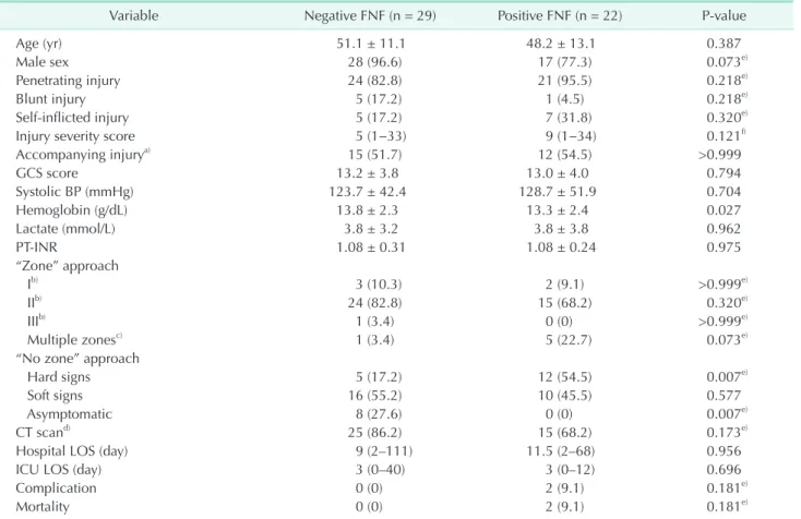
Variable Negative FNF (n = 29) Positive FNF (n = 22) P-value
Age (yr) 51.1 ± 11.1 48.2 ± 13.1 0.387
Male sex 28 (96.6) 17 (77.3) 0.073e)
Penetrating injury 24 (82.8) 21 (95.5) 0.218e)
Blunt injury 5 (17.2) 1 (4.5) 0.218e)
Self-inflicted injury 5 (17.2) 7 (31.8) 0.320e)
Injury severity score 5 (1−33) 9 (1−34) 0.121f)
Accompanying injurya) 15 (51.7) 12 (54.5) >0.999
GCS score 13.2 ± 3.8 13.0 ± 4.0 0.794
Systolic BP (mmHg) 123.7 ± 42.4 128.7 ± 51.9 0.704
Hemoglobin (g/dL) 13.8 ± 2.3 13.3 ± 2.4 0.027
Lactate (mmol/L) 3.8 ± 3.2 3.8 ± 3.8 0.962
PT-INR 1.08 ± 0.31 1.08 ± 0.24 0.975
“Zone” approach
Ib) 3 (10.3) 2 (9.1) >0.999e)
IIb) 24 (82.8) 15 (68.2) 0.320e)
IIIb) 1 (3.4) 0 (0) >0.999e)
Multiple zonesc) 1 (3.4) 5 (22.7) 0.073e)
“No zone” approach
Hard signs 5 (17.2) 12 (54.5) 0.007e)
Soft signs 16 (55.2) 10 (45.5) 0.577
Asymptomatic 8 (27.6) 0 (0) 0.007e)
CT scand) 25 (86.2) 15 (68.2) 0.173e)
Hospital LOS (day) 9 (2–111) 11.5 (2–68) 0.956
ICU LOS (day) 3 (0–40) 3 (0–12) 0.696
Complication 0 (0) 2 (9.1) 0.181e)
Mortality 0 (0) 2 (9.1) 0.181e)
Values are presented as mean ± standard deviation, number (%), or median (range).
FNF, final neck finding; GCS, Glasgow coma scale; BP, blood pressure; INR, international normalized ratio; LOS, length of stay; ICU, intensive care unit.
a)Injury of head, chest, abdomen, and extremities. b)Injury isolated to the zone. c)Zone II was included in all cases. d)Neck contrast CT or neck angiography CT. e)Fisher exact test. f)Wilcoxon rank-sum test.
Table 6. Univariate and multivariate logistic regression analysis for negative and positive FNFs in the exploration group
Variable Univariate Multivariate
OR (95% CI) P-value OR (95% CI) P-value
Age 0.98 (0.94–1.03) 0.518 3.95 (0.34–97.93) 0.301
Sex
Female Ref Ref
Male 7.50 (1.08–150.96) 0.078 0.99 (0.88–1.10) 0.878
Injury severity score 1.04 (0.96–1.12) 0.348 0.96 (0.89–1.02) 0.217
Systolic blood pressure 1.00 (0.99–1.02) 0.567 1.02 (1.00–1.05) 0.048
Hemoglobin 0.77 (0.58–0.98) 0.047 0.66 (0.42–0.96) 0.043
“Zone” approach
Ia) or IIIa) Ref Ref
IIa) or multipleb) 1.81 (0.32–14.14) 0.520 2.43 (0.23–34.80) 0.474
“No zone” approach
Soft signs or asymptotic Ref Ref
Hard signs 5.33 (1.51–21.32) 0.012 11.19 (2.12–90.55) 0.010
Hosmer and Lemeshow goodness of fit test 0.7188
Compounding factor: age, sex, injury severity score, and systolic blood pressure. Significant differences between the negative and positive FNFs: hemoglobin. Values to confirm: “zone” approach and “no zone” approach.
FNF, final neck finding; OR, odds ratio; CI, confidence interval.
a)Injury isolated to the zone. b)Zone II was included in all cases.
CI, 2.12–90.55; P = 0.010) (Table 6). A ROC curve of multivariate logistic regression analysis for the FNFs in the exploration group, with the use of the variables included in predicting FNFs, showed an AUC of 0.864 (Fig. 2B).
DISCUSSION
Historically, the traditional zone approach was applied for assessment of traumatic neck injuries [14]. According to this approach, mandatory exploration was performed and a surgical approach was found to be relatively easy for anatomical zone II injuries. However, zones I and III were relatively difficult to access surgically; therefore, if the patient’s vitality was stable, sufficient examinations were performed and an operation conducted upon injury confirmation [9].
Nevertheless, advances in the technology of CT scans have shown that these examinations can be completed and images obtained without any mandatory exploration to identify internal neck injuries [11,12,15]. Therefore, when patients arrive at the hospital, depending on their symptoms and vitality, they may be examined, undergo an operation immediately, or undergo conservative treatments. The clinical significance of the no zone approach, which is based on symptoms, has been reported recently [16,17]. According to Nowicki et al. [18], the no zone approach to penetrating neck injury evaluation and management is contemporary and goes against the grain of anatomical zones management. Furthermore, evidence is accumulating to suggest that the no zone approach is superior over traditional zone approaches to penetrating neck trauma, especially with respect to reduced negative neck explorations.
Our results further confirm the significance of the symptomatic approach in assessment and management of traumatic neck
injuries. When comparing negative FNFs to positive FNFs, there was no singularity in the zone approach in the total number of patients examined or in the exploration group, and there was a significant difference in the presence of hard signs with the no zone approach in both groups. In the multiple regression analysis, hard signs were an independent predictive factor for internal organ injuries of the neck while zone of injury was not.
We observed no differences in clinical characteristics including injury site, rate of hard sign, and mortality, as in previous studies. However, the number of operation and CT scan trials differed for each study. Hundersmarck et al. [17]
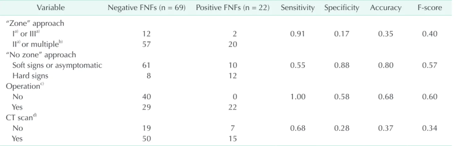
reported that operation was performed in 54% of total patients, CT scan was 72.1%, and positive FNFs was 53.5%. Ibraheem et al. [11] described different results wherein the operation rate was 36.2%, CT scan rate was 66.1%, and positive FNFs rate was 59.8%. In this study, 51 patients (56.0%) underwent an emergency surgical operation and 65 patients (71.4%) underwent CT scan, and internal organ injuries of the neck occurred in 22 patients (24.2%). Our results show that there were more unnecessary operations than in previous studies, despite similar CT scans rate. This result is thought to be due to lack of understanding of the no zone approach and absence of guidelines in our institution.
The results of FNFs were confirmed by operation findings and CT scans. However, the rate of precisely diagnosing neck injury using CT scan is relatively low, and additional imaging tests may be required for accurate diagnosis [11,12,18]. Moreover, the final diagnosis is often confirmed through operation. Our study showed that whereas in the zone approach, 5 patients of zone I, 39 patients of zone II, 1 patient of zone III, and 6 patients of multiple zones were operated on; in the no zone approach, 17 patients with hard signs, 26 with soft signs, and
Table 7. Diagnostic statistics of FNFs from several methods
Variable Negative FNFs (n = 69) Positive FNFs (n = 22) Sensitivity Specificity Accuracy F-score
“Zone” approach
Ia) or IIIa) 12 2 0.91 0.17 0.35 0.40
IIa) or multipleb) 57 20
“No zone” approach
Soft signs or asymptomatic 61 10 0.55 0.88 0.80 0.57
Hard signs 8 12
Operationc)
No 40 0 1.00 0.58 0.68 0.60
Yes 29 22
CT scand)
No 19 7 0.68 0.28 0.37 0.34
Yes 50 15
Values are presented as number.
FNFs, final neck findings.
a)Injury isolated to the zone. b)Zone II was included in all cases. c)Surgery performed under general anesthesia and 51 of total patients.
d)Neck contrast CT or neck angiography CT and 65 of total patients.
8 asymptomatic patients were operated on. CT scan can be helpful in patients with neck injury in previous studies, though this is still controversial [2,11,12,15]. In our study, upon analysis of the diagnostic statistics of the zone and no zone approach, we found that rather than CT scans for FNFs, operation had the highest sensitivity; in addition, the no zone approach had a higher specificity and accuracy (Table 7).
In addition to CT scans, there are many other imaging tests for accurate diagnosis of neck injuries, including laryngoscopy, bronchoscopy, endoscopy, esophagography, and angiography.
However, there is a lot of controversy over the cost and side effects compared to the accuracy of the diagnosis [3,11,12,18].
Our institution does not routinely conduct any additional imaging tests outside of CT scans and those conducted depend on the judgment of the standing trauma surgeon. In our study, an angiography was conducted on only one of the 91 patients and it revealed a zone II blunt injury. Angiography was conducted because the patient’s CT scan revealed a suspected intima injury of the right common carotid artery; and the angiography did reveal a common carotid artery dissection.
In addition, a laryngoscopy was performed in 4 patients, of whom 3 patients had blunt injury, 1 patient had a penetrating injury, and all the patients had a zone II injury. All the patients had oral bleeding findings. Although, when laryngoscopy was implemented for airway injury, no definite injury was found, and hence, conservative management was performed.
Bronchoscopy was performed in only one patient with multiple penetrating injuries in zone I and II; trachea injury was confirmed and the patient underwent operation.
Most of the existing studies of neck trauma are on penetrating injuries, and very few are on blunt injuries [3,4].
In our study, there were a total of 17 blunt injury patients.
Among them, 4 patients had a hard sign and 6 underwent operation. One of the 4 patients who had a hard sign visited the hospital with their neck bent and died due to cardiac arrest upon arrival at the ER. The remaining 3 were zone II injuries that were operated on because of airway obstruction findings resulting from a hematoma and neck swelling. Of these 3, only 1 had positive FNFs (hyoid bone fracture) and the other 2 had negative FNFs. Meanwhile, 3 of the remaining 13 patients without a hard sign had zone II injuries with negative FNFs and
underwent operation for a blunt mechanism accompanied by a neck laceration.
This study has some limitations. First, the study design was a retrospective analysis, and the number of patients enrolled was small. Most of the previous studies have analyzed only patients with penetrating injuries. However, in our study, we included patients with blunt injuries as well [19-21] due to the small number of patients, enrolled in our study. We do realize that patients with blunt injuries may need to be analyzed separately. However, their symptoms were not ambiguous, and 3 of the 17 patients were accompanied by a neck laceration. Six of the 17 patients underwent operation and simple contusion and whiplash syndrome due to blunt injury were excluded.
Thus, although our study was conducted including patients with blunt injury, further research and discussion regarding only blunt injury are needed. Future studies need to focus on multi-institutional studies with a large number of patients to accurately investigate the applicability of the no zone approach and compare it with the zone approach and consider blunt injury with the same approach.
In conclusion, traumatic neck injury with hard signs, according to the no zone approach, may correlate with internal organ injuries of the neck. Therefore, the no zone approach can make it easier to determine whether exploration of the neck should be considered.
ACKNOWLEDGEMENTS
Conflict of Interest
No potential conflict of interest relevant to this article was reported.
ORCID iD
Ji Wool Ko: https://orcid.org/0000-0001-8391-9941 Seong Chan Gong: https://orcid.org/0000-0001-9685-5924 Myung Jun Kim: https://orcid.org/0000-0002-6421-2112 Jae Sik Chung: https://orcid.org/0000-0001-7136-5128 Young Un Choi: https://orcid.org/0000-0003-2410-7788 Jun Hyuk Lee: https://orcid.org/0000-0002-7745-3599 Pil Young Jung: https://orcid.org/0000-0001-6460-8072
REFERENCES
1. Vishwanatha B, Sagayaraj A, Huddar SG, Kumar P, Datta RK. Penetrating neck injuries. Indian J Otolaryngol Head Neck Surg 2007;59:221-4.
2. Saito N, Hito R, Burke PA, Sakai O.
Imaging of penetrating injuries of the head and neck:current practice at a level I trauma center in the United States. Keio J
Med 2014;63:23-33.
3. Verdonck P, de Schoutheete JC, Monsieurs KG, Van Laer C, Vander Poorten V, Vanderveken O. Penetrating and blunt
trauma to the neck: clinical presentation, assessment and emergency management.
B-ENT 2016;Suppl 26:69-85.
4. Tessler RA, Nguyen H, Newton C, Betts J. Pediatric penetrating neck trauma:
hard signs of injury and selective neck exploration. J Trauma Acute Care Surg 2017;82:989-94.
5. Demetriades D, Theodorou D, Cornwell E, Berne TV, Asensio J, Belzberg H, et al.
Evaluation of penetrating injuries of the neck: prospective study of 223 patients.
World J Surg 1997;21:41-8.
6. Bell RB, Osborn T, Dierks EJ, Potter BE, Long WB. Management of penetrating neck injuries: a new paradigm for civilian trauma. J Oral Maxillofac Surg 2007;65:691-705.
7. Sim J, Lee J, Lee JC, Heo Y, Wang H, Jung K. Risk factors for mortality of severe trauma based on 3 years’ data at a single Korean institution. Ann Surg Treat Res 2015;89:215-9.
8. Mahmoodie M, Sanei B, Moazeni-Bistgani M, Namgar M. Penetrating neck trauma:
review of 192 cases. Arch Trauma Res 2012;1:14-8.
9. Monson DO, Saletta JD, Freeark RJ.
Carotid vertebral trauma. J Trauma 1969;9:987-99.
10. Shiroff A M, Gale SC, Martin ND, Marchalik D, Petrov D, Ahmed HM, et
al. Penetrating neck trauma: a review of management strategies and discussion of the ‘No Zone’ approach. Am Surg 2013;79:23-9.
11. Ibraheem K, Khan M, Rhee P, Azim A, O’Keeffe T, Tang A, et al. “No zone”
approach in penetrating neck trauma r e duc e s u n ne c e s s a r y c o mp ut e d tomography angiography and negative explorations. J Surg Res 2018;221:113-20.
12. Schroll R, Fontenot T, Lipcsey M, Heaney JB, Marr A, Meade P, et al. Role of computed tomography angiography in the management of Zone II penetrating neck trauma in patients with clinical hard signs. J Trauma Acute Care Surg 2015;79:943-50.
13. Sperry JL, Moore EE, Coimbra R, Croce M, Davis JW, Karmy-Jones R, et al. Western Trauma Association critical decisions in trauma: penetrating neck trauma. J Trauma Acute Care Surg 2013;75:936-40.
14. Low GM, Inaba K, Chouliaras K, Branco B, Lam L, Benjamin E, et al. The use of the anatomic ‘zones’ of the neck in the assessment of penetrating neck injury.
Am Surg 2014;80:970-4.
15. Inaba K, Branco BC, Menaker J, Scalea TM, Crane S, DuBose JJ, et al. Evaluation of multidetector computed tomography for penetrating neck injury: a prospective multicenter study. J Trauma Acute Care
Surg 2012;72:576-84.
16. Prichayudh S, Choadrachata-anun J, Sriussadaporn S, Pak-art R, Sriussadaporn S, Kritayakirana K, et al. Selective management of penetrating neck injuries using “no zone” approach. Injury 2015;46:1720-5.
17. Hundersmarck D, Reinders Folmer E, de Borst GJ, Leenen LP, Vriens PW, Hietbrink F. Penetrating neck injury in two Dutch level 1 trauma centres: the non-existent problem. Eur J Vasc Endovasc Surg 2019;58:455-62.
18. Nowicki JL, Stew B, Ooi E. Penetrating neck injuries: a guide to evaluation and management. Ann R Coll Surg Engl 2018;100:6-11.
19. Babu A, Garg H, Sagar S, Gupta A, Kumar S. Penetrating neck injury: collaterals for another life after ligation of common carotid artery and subclavian artery. Chin J Traumatol 2017;20:56-8.
20. Burgess CA, Dale OT, A lmeyda R, Corbridge RJ. An evidence based review of the assessment and management of penetrating neck trauma. Clin Otolaryngol 2012;37:44-52.
21. Siau RT, Moore A, Ahmed T, Lee MS, Tostevin P. Management of penetrating neck injuries at a London trauma centre.
Eur Arch Otorhinolaryngol 2013;270:2123- 8.