Original Article
Received: Mar 5, 2015; Revised: (1st) Mar 28, 2015, (2nd) May 6, 2015; Accepted: May 8, 2015 Correspondence to: Gilho Lee
Department of Urology, Dankook University Medical College, 119 Dandae-ro, Dongnam-gu, Cheonan 31116, Korea.
Tel: +82-41-550-3963, Fax: +82-41-551-6630, E-mail: [email protected] Copyright © 2015 Korean Society for Sexual Medicine and Andrology
This is an Open Access article distributed under the terms of the Creative Commons Attribution Non-Commercial License (http://creativecommons.
org/licenses/by-nc/4.0) which permits unrestricted non-commercial use, distribution, and reproduction in any medium, provided the original work is properly cited.
Chronic Prostatitis: A Possible Cause of Hematospermia
Gilho Lee
Department of Urology, Dankook University Medical College, Cheonan, Korea
Purpose: While hematospermia is mainly caused by genitourinary inflammatory disorders, very few studies have been published on prostatitis-associated hematospermia (PAH) diagnosed using robust prostatitis evaluation methods. Therefore, we have evaluated the incidence of PAH by using systematic methods for evaluating prostatitis.
Materials and Methods: We evaluated 37 hematospermia patients from a single hospital over the last five years. We classified the patients into PAH versus hematospermia without any evidence of prostatitis (HWP) by using a NIH-Chronic Prostatitis Symptom Index questionnaire and expressed prostatic secretion studies.
Results: The mean age was 55.89±14.87 years, and the patients were grouped into two groups: one group had 12 HWP patients and the other 25 PAH patients. PAH patients were further sub-classified: chronic bacterial prostatitis (3 patients), chronic nonbacterial prostatitis (10 patients), prostadynia (7 patients), and asymptomatic prostatitis (5 patients). We found Enterococcus faecalis in the three chronic bacterial prostatitis patients. We could not find any statistically significant difference between the PAH and the HWP groups in terms of the age interval, serum prostate-specific antigen level, and prostate volume. Even though there was no statistically significant difference in the items about urination between the two groups, we found a statistically significant difference in the quality of life (QoL) impact for the patients in this study.
Conclusions: Two-thirds of the hematospermia patients were associated with some evidence of prostatitis. Further, the patients with PAH revealed poor QoL compared with the patients with HWP. Therefore, we must evaluate the presence of prostatitis in hematospermia patients and alleviate the prostatitis-associated symptoms to improve their QoL.
Key Words: Hematospermia; Prostate; Prostatitis
INTRODUCTION
Hematospermia can be defined as the appearance of blood in ejaculate [1-3]. While it is usually self-limited in most patients, in some cases, persistent, recurrent symp- toms have been reported, causing serious concerns.
We could classify the many suggested etiologies for
hematospermia into inflammatory or infectious diseases and anatomical abnormalities or disorders [1-3]. Urethritis, prostatitis, and epididymitis are a prototype of in- flammatory hematospermia, while posterior urethral ob- struction, calculi in the ejaculatory duct, and prostate can- cer can be a prototype of anatomical hematospermia.
Tests for evaluating lower urinary tract infection such as
http://dx.doi.org/10.5534/wjmh.2015.33.2.103
urine analysis and Gram staining are routine tests for hem- atospermia [2,4,5]. Because transrectal ultrasonography (US) can show the anatomical structures of the prostate, seminal vesicles, and adjacent structures, many urologists have applied this modality for evaluating the anatomical causes of hematospermia [4-6].
Prostatitis, an inflammatory disorder of the prostate, has been considered an important disease causing bloody ejaculation [1-5]. It has been well known that the basic methods for evaluating prostatitis include the National Institutes of Health Chronic Prostatitis Symptom Index (NIH-CPSI) questionnaire, and the white blood cell (WBC) counts and bacterial culture of expressed prostatic secre- tion (EPS) [7-10]. Unfortunately, very few studies on the determination of prostatitis-related etiologies for the ori- gin of hematospermia have been performed using robust prostatitis evaluation methods. Therefore, in this study, we have aimed to evaluate the association between pros- tatitis and hematospermia by using systematic methods for evaluating prostatitis, and to clarify the role of prostate in- flammation in bloody ejaculate.
MATERIALS AND METHODS
1. Study design and participant characteristics Informed consent was obtained from the study partici- pants. The research protocol was also approved by the Institutional Review Board of Dankook University Hospital.
We collected data from 37 patients who visited a single university hospital complaining of hematospermia be- tween April 2009 and November 2014.
We evaluated prostatitis-associated symptoms by using a Korean version of the NIH-CPSI questionnaire, and pros- tate inflammation by using an EPS wet smear and culture.
On the basis of these evaluations, eligible participants were classified into patients without any evidence of pros- tatitis (negative prostatitis), with chronic bacterial prostati- tis (NIH-category II), with chronic nonbacterial prostatitis (NIH-category IIIA), with prostadynia (NIH-category IIIB), and with asymptomatic inflammatory prostatitis [8,10]. In particular, we defined the group of negative prostatitis pa- tients as hematospermia patients who did not have a spe- cific prostatitis symptom according to the NIH-CPSI ques- tionnaire or display any relevant signs in the EPS studies.
We defined chronic prostatitis as a condition of pelvic pain or discomfort that lasted for at least three months; we further divided the chronic prostatitis group into chronic bacterial prostatitis, prostadynia (<16 WBC per high- power field [hpf] in EPS), and chronic nonbacterial prosta- titis (≥16 WBC per hpf in EPS) groups [11]. Finally, we de- fined the asymptomatic prostatitis patients as people who had high WBC counts in EPS (≥16 WBC per hpf in EPS) without any specific symptom of chronic prostatitis.
The exclusion criteria were the presence of genito- urinary cancer, urinary stone disease, gonococcal or chla- mydial urethritis, acute epididymitis, acute cystitis, acute prostatitis, traumatic urethral stricture, or seminal vesicle or ejaculatory duct calculi, as well as experience of pros- tate surgery or biopsy within the past 6 months.
2. Laboratory tests
We strongly recommended serum prostate-specific an- tigen (PSA), which was determined with a PSA kit (PSA- RIACT; CIS Bio International, Gif-sur-Yvette, France). We also evaluated the structures of the seminal vesicles, pros- tate, and adjacent organs with transrectal US (HD7 Ultrasound System; Philips, Shenyang, China). The pros- tate size was automatically determined, and US-guided bi- opsy was mandatorily performed on the prostate nodule to differentiate prostate cancer. Patients with a serum PSA level of more than 4 ng/mL underwent transrectal biopsy to exclude prostate cancer.
Culture specimens for diagnosing prostatitis were ob- tained using the modified Meares-Stamey method [10-12].
In brief, after periurethral cleansing with an alcohol sponge, the patient provided a VB1 specimen, followed by a VB2 urine specimen. After the production of EPS by a digital prostatic massage, the patient provided 5 to 10 mL of voided urine for the VB3 specimen. We divided the VB1 urine sample for the VB1 culture, automatic urine analysis with Sysmex UF-1000i (TOA Medical Electronics, Kobe, Japan), and multiplex polymerase chain reaction (PCR) tests for sexually transmitted infections. The results of the WBC count in the first-voided urine were classified into two categories: WBC counts of 0 to 1 and ≥2. EPS was collected by a digital rectal massage into a sterile 1.5-mL tube. Using a micropipette, we placed 5 μL of EPS on a glass slide and covered it with a 22-mm2 No. 1 cover
www.wjmh.org Table 1. Clinical characteristics of patients
Variable Value
No. of patient 37
Age (yr) 55.89±14.87
Serum PSA (ng/mL) 2.35±1.84
Prostate size (mL) 34.35±15.25
NIH-CPSI
Sum of pain or discomfort (items 1∼4) 3.86±5.07 Sum of urination (item 5 and 6) 2.27±2.54 Sum of quality of life impact (items 7∼9) 3.64±3.04
Total (items 1∼9) 9.72±8.63 Classification of prostatitis
No evidence of prostatitis 12
Chronic bacterial prostatitis (NIH-cat II) 3 Chronic nonbacterial prostatitis
(NIH-cat IIIA) 10
Prostadynia (NIH-cat IIIB) 7
Asymptomatic prostatitis 5
Values are presented as number only or mean±standard deviation.
PSA: prostate-specific antigen, NIH-CPSI: NIH-Chronic Pros- tatitis Symptom Index, NIH: National Institutes of Health, USA.
glass. The slide was examined with Olympus BX40F (Olympus, Tokyo, Japan). At least 25 fields were examined. The results of the WBC count in EPS were clas- sified into two categories: <16 and ≥16 WBC per hpf [11]. Each VB1 specimen was cultured within 4 hours of collection by spreading 100 μL onto plates containing 5%
sheep blood agar, incubated aerobically, and examined for bacterial growth after 48 hours. Similarly, 100 μL of EPS was cultured onto the same culture plates for 2 days. The criterion for chronic bacterial prostatitis was that the bacte- rial colony concentration of the EPS specimens increased by at least 10-fold (one log) compared with the colony numbers of the VB1 specimen. Only one VB1 and one EPS specimen were recorded per person.
Using the stored VB1 and EPS samples, DNA extraction was performed with a genomic DNA purification kit (Qiagen, Hilden, Germany) according to the manufacturer’s instructions. Multiplex PCR was performed using KL1-KL2 primers for Chlamydia trachomatis infection and HO1-HO3 primers for Neisseria gonorrhoeae as pre- viously described [11].
3. Statistical methods
All data are expressed as mean±standard deviation (SD). A Mann-Whitney U test was performed to test for the differences in the mean ages, serum PSA level, prostate volume, and the items of the NIH-CPSI questionnaire be- tween the patients. Two-sided null hypotheses of no differ- ence were rejected if p values were less than 0.05. All analyses were performed using the SPSS software for Windows ver. 11 (SPSS Inc., Chicago, IL, USA).
RESULTS
The mean age±SD, mean serum PSA level, and mean prostate size of the patients were 55.89±14.87 years, 2.35±1.84 ng/mL, and 34.35±15.25 mL, respectively.
From the items of the NIH-CPSI questionnaire, we found that the sum scores of the pain or discomfort, urination, quality of life impact, and total items were 3.86±5.07, 2.27±2.54, 3.64±3.04, and 9.72±8.63, respectively.
Thirty-seven patients were classified into the following groups: no evidence of prostatitis (12 patients; 32.43%), chronic bacterial prostatitis (3 patients; 8.1%), chronic
nonbacterial prostatitis (10 patients; 27.02%), prostadynia (7 patients; 18.91%), and asymptomatic prostatitis (5 pa- tients; 13.51%) (Table 1).
We found Enterococcus faecalis in three chronic bacte- rial prostatitis patients.
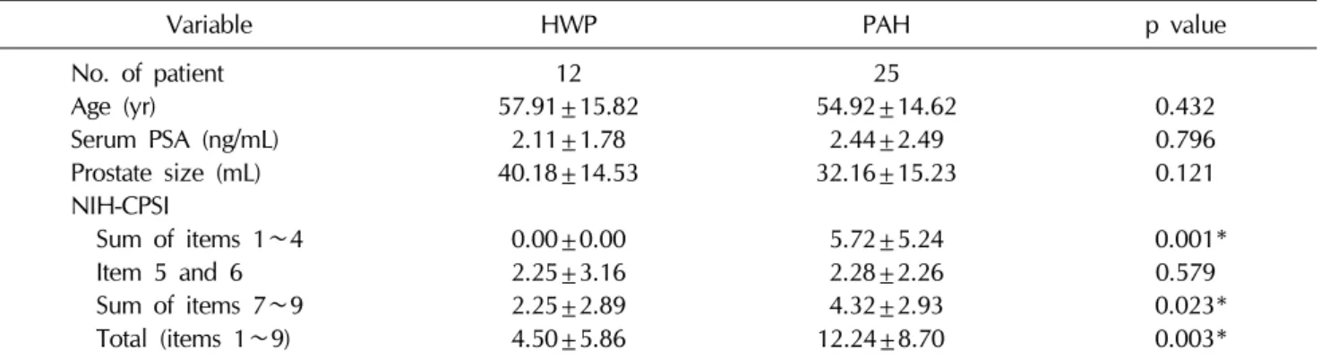
To differentiate patients with prostatitis-associated hem- atospermia (PAH) from those without any evidence of prostatitis (HWP), we divided the 37 hematospermia pa- tients into 12 patients for HWP and 25 patients for PAH.
We could not find any statistically significant differences between the two groups in terms of the age interval, serum PSA level, and prostate volume (Table 2). Even though no statistically significant difference was found in the sum items of urination (items 5 and 6) between the two groups, we were able to find a statistically significant difference in the sum of the quality of life impact (items 7∼9) (Table 2).
DISCUSSION
Even though the prevalence of hematospermia is not high in the general population and its clinical course is usually self-limited, it often causes considerable anxiety to
Table 2. Differences in clinical characteristics between patients of HWP and PAH
Variable HWP PAH p value
No. of patient 12 25
Age (yr) 57.91±15.82 54.92±14.62 0.432
Serum PSA (ng/mL) 2.11±1.78 2.44±2.49 0.796
Prostate size (mL) 40.18±14.53 32.16±15.23 0.121
NIH-CPSI
Sum of items 1∼4 0.00±0.00 5.72±5.24 0.001*
Item 5 and 6 2.25±3.16 2.28±2.26 0.579
Sum of items 7∼9 2.25±2.89 4.32±2.93 0.023*
Total (items 1∼9) 4.50±5.86 12.24±8.70 0.003*
Values are presented as number only or mean±standard deviation.
PAH: prostatitis-associated hematospermia, HWP: hematospermia patients without any evidence of prostatitis, PSA:
prostate-specific antigen, NIH-CPSI: National Institutes of Health Chronic Prostatitis Symptom Index.
*p<0.05.
patients [1-3,13,14]. Some patients presuppose hema- tospermia to be linked to sexual behaviors such as prolonged sexual abstinence or overindulgence, while others worry that genitourinary malignancies and sexually transmitted dis- eases are the key causes of hematospermia [1-6,15,16].
Theoretically, it may be derived from anatomical con- ditions along the sperm passageway, such as in the lesions of testis, epididymis, vas deferens, seminal vesicles, pros- tate, and posterior urethra. Urologists have also catego- rized hematospermia on the basis of pathophysiologic mechanisms such as inflammation and infections, ductal obstruction and cysts of accessory sexual glands, tumors, vascular abnormalities, and systemic and iatrogenic fac- tors [3].
Inflammatory or infectious conditions in the genito- urinary tract have appeared to be the most common causal factors of hematospermia [1-5]. An irritation of the muco- sa, mucosal edema, and hyperemia of the accessory sex- ual glands and their corresponding ducts may lead to bleeding and hematospermia [3]. The suggested infectious etiologies include viral, bacterial, mycobacterial, and par- asitic infections such as bilharziasis [3]. Bamberger et al [16] investigated sexually transmitted diseases from in- fected patients who demonstrated hematospermia and found infections of Herpes simplex virus (42%), C. tracho- matis (33%), E. faecalis (17%), and Ureaplasma ure- alyticum (8%). We also evaluated the C. trachomatis and gonococcal infection with a multiplex PCR method in this study. However, we could not find any trace of the two
pathogens in the specimens of urine or EPS. This discrep- ancy may be attributed to the difference in the age dis- tributions between the two studies. The age range of Bamberger et al’s [16] study was 17 to 66 years (median:
33 years), with 4 patients over the age of 40 years. In con- trast, the mean age of the patients in this study was 55.89±14.87 years. It has been well known that young age is an important factor associated with chlamydial in- fection [17].
Total ejaculate contains from 60% to 80% biological materials of prostate origin [18]. Therefore, we easily pre- suppose the association between hematospermia and prostatitis. We found E. faecalis infection in the prostate of the three hematospermia patients. E. faecalis is a gram-pos- itive, commensal bacterium inhabiting the human gastro- intestinal tract [19]. These bacteria frequently colonize the urinary tract and cause infection [11]. We have previously reported that only 41 samples from 1,021 patients with chronic prostatitis/chronic pelvic pain syndrome (CP/CPPS) revealed a significant E. faecalis manifestation in their prostate for defining chronic bacterial prostatitis [11].
When we compare the incidence of E. faecalis between the 8.1% (3 E. faecalis-infected patients/37 hematospermia pa- tients) found in this study with the 4% (41 E. faecalis-in- fected patients/1,021 CP/CPPS patients) in the previous study, we can infer that a bacterial prostatitis infection may lead to hematospermia in certain patients. Furthermore, the hematospermia symptoms of all E. faecalis patients im- proved with an appropriate antibiotic (fluoroquinolone)
www.wjmh.org
treatment.
Prostatitis, an inflammatory or infectious disorder of the prostate, can be classified into acute, chronic bacterial, chronic nonbacterial, prostadynia, and asymptomatic forms [8,20]. In this study, we could not find any acute prostatitis patients. Interestingly, 25 patients with hema- tospermia revealed prostatitis-like symptoms or signs.
Among them, three patients revealed an E. faecalis infection in the prostate, defined as chronic bacterial prostatitis.
With a strict criterion (≥16 WBC per hpf in the EPS specimen) for defining inflammatory prostatitis, we were able to diagnose 10 patients with chronic nonbacterial prostatitis.
We strongly recommend robust studies for CP/CPPS for patients with hematospermia for determining specific prostatitis etiologies because 25 of the 37 enrolled hema- tospermia patients were associated with CP/CPPS, as well as a higher incidence of chronic nonbacterial prostatitis than prostadynia.
From the meta-analysis studies, the mean age of hema- tospermia is 37 years, showing prevalence in young males [2,15]. However, the mean age in our study was 55.89 years. From the data of the mean serum PSA level (2.35±
1.84 ng/mL), prostate size (34.35±15.25 mL), and mean International Prostate Symptom Score (IPSS) score (9.64±8.27), our patients may include patients with be- nign prostatic hypertrophy. The mean age in our study was very similar to the mean age reported by Ng et al [1] (54 years; age range: 16∼82 years). Around this age, the ma- jor concern for many patients is to know whether they have prostate cancer. In general, the incidence of prostate cancer is not high in patients with hematospermia.
We could not find any statistically significant differences between the PAH patients and the patients without hema- tospermia in terms of the age interval, serum PSA level, prostate volume, and sum of items about urination (items 5 and 6 of the NIH-CPSI questionnaire). Interestingly, the PAH patients revealed a relatively poor quality of life com- pared with the patients without prostatitis. One of the treatment targets of prostatitis is to improve the quality of life in patients with chronic prostatitis [21]. Therefore, we must consider more specific treatments for hematospermia patients with prostatitis symptoms or signs.
We recognize two limitations to this study. First, even
though we ardently collected samples from hematospermia patients for five years, we found very few well-charac- terized samples for evaluating CP/CPPS. Second, this study can be classified as a correlation study. While our re- sults showed that the hematospermia patients were asso- ciated with a higher prevalence of prostatitis than the nor- mal population, as well as for inflammatory prostatitis than prostadynia, we could not determine whether this as- sociation is a true etiologic factor in the manifestation of hematospermia.
CONCLUSIONS
Two-thirds of the hematospermia patients were asso- ciated with evidence of prostatitis. We must carefully eval- uate the association of prostatitis in hematospermia patients. Further, hematospermia patients with prostatitis revealed a relatively poor quality of life compared with those without prostatitis. To improve the quality of life of the patients, we must control the symptoms of CP/CPPS in hematospermia patients with prostatitis.
ACKNOWLEDGEMENTS
This work was supported by the Research Fund of Dankook University in 2014.
CONFLICT OF INTEREST
No potential conflict of interest relevant to this article was reported.
REFERENCES
1. Ng YH, Seeley JP, Smith G. Haematospermia as a present- ing symptom: outcomes of investigation in 300 men.
Surgeon 2013;11:35-8.
2. Ahmad I, Krishna NS. Hemospermia. J Urol 2007;177:
1613-8.
3. Szlauer R, Jungwirth A. Haematospermia: diagnosis and treatment. Andrologia 2008;40:120-4.
4. Stefanovic KB, Gregg PC, Soung M. Evaluation and treat- ment of hematospermia. Am Fam Physician 2009;80:
1421-7.
5. Kumar P, Kapoor S, Nargund V. Haematospermia: a system- atic review. Ann R Coll Surg Engl 2006;88:339-42.
6. Raviv G, Laufer M, Miki H. Hematospermia: the added val-
ue of transrectal ultrasound to clinical evaluation: is trans- rectal ultrasound necessary for evaluation of hema- tospermia? Clin Imaging 2013;37:913-6.
7. Litwin MS, McNaughton-Collins M, Fowler FJ Jr, Nickel JC, Calhoun EA, Pontari MA, et al. The National Institutes of Health chronic prostatitis symptom index: development and validation of a new outcome measure. Chronic Prostatitis Collaborative Research Network. J Urol 1999;162:369-75.
8. Nguyen CT, Shoskes DA. Evaluation of the prostatitis patient. In: Shoskes DA, editor. Chronic prostatitis/chronic pelvic syndrome. Totowa, NJ: Humana Press; 2008;1-16.
9. Nickel JC, Shoskes D, Wang Y, Alexander RB, Fowler JE Jr, Zeitlin S, et al. How does the pre-massage and post-mas- sage 2-glass test compare to the Meares-Stamey 4-glass test in men with chronic prostatitis/chronic pelvic pain syn- drome? J Urol 2006;176:119-24.
10. Meares EM, Stamey TA. Bacteriologic localization patterns in bacterial prostatitis and urethritis. Invest Urol 1968;5:
492-518.
11. Park H, Sim SM, Lee G. The presence of Chlamydia is asso- ciated with increased leukocyte counts and pain severity in men with chronic pelvic pain syndrome. Urology 2015;85:
574-9.
12. Seo Y, Lee G. Antimicrobial resistance pattern in Enterococcus faecalis strains isolated from expressed prostatic secretions of patients with chronic bacterial prostatitis. Korean J Urol 2013;54:477-81.
13. Ganabathi K, Chadwick D, Feneley RC, Gingell JC. Haemo- spermia. Br J Urol 1992;69:225-30.
14. Jones DJ. Haemospermia: a prospective study. Br J Urol 1991;67:88-90.
15. Mulhall JP, Albertsen PC. Hemospermia: diagnosis and management. Urology 1995;46:463-7.
16. Bamberger E, Madeb R, Steinberg J, Paz A, Satinger I, Kra-Oz Z, et al. Detection of sexually transmitted pathogens in patients with hematospermia. Isr Med Assoc J 2005;7:
224-7.
17. Araújo RS, Guimarães EM, Alves MF, Sakurai E, Domingos LT, Fioravante FC, et al. Prevalence and risk factors for Chlamydia trachomatis infection in adolescent females and young women in central Brazil. Eur J Clin Microbiol Infect Dis 2006;25:397-400.
18. Mazzoli S. Conventional bacteriology in prostatitis patients:
microbiological bias, problems and epidemiology on 1686 microbial isolates. Arch Ital Urol Androl 2007;79:71-5.
19. Sood S, Malhotra M, Das BK, Kapil A. Enterococcal in- fections & antimicrobial resistance. Indian J Med Res 2008;128:111-21.
20. Schaeffer AJ. Epidemiology and demographics of prostatitis.
Andrologia 2003;35:252-7.
21. Calhoun EA, Duloy AMS, Quentin Clemens J. Quality of life and economic impact of chronic prostatitis. In: Shoskes DA, editor. Chronic prostatitis/chronic pelvic syndrome.
Totowa, NJ: Humana Press; 2008;59-75.