J Korean Soc Coloproctol Vol. 19, No. 3, 2003
177
Role of Protein Kinase C Signaling in In- testinal Ischemic Preconditioning
Jun Won Um, M.D.
Department of Surgery, Korea University College of Medicine, Seoul, Korea
Ischemic preconditioning (IPC) is a phenomenon that a brief episode of ischemia to a tissue renders the tissue resistance from a subsequent prolonged ischemia. It is generally accepted that this protection is a receptor-mediated proc- ess, and is realized via signal transduction pathways. Protein kinase C (PKC), known to play key regulatory roles in cellular processes, has been proposed as a primary cellular mediator of preconditioning. However, the role of PKC in eliciting cardioprotection remains controversial. The evi- dences for the 'PKC hypothesis' of preconditioning in various tissue and organs are summarized. Especially in intestine, a brief ischemia induced a reversible epithelial injury to the jejunum that is associated with activation of several PKC isoforms. Injury induced by an additional period of ischemia is reduced by the prior IPC, and this effect is abolished by non-selective PKC inhibition but not by a selective inhibitor of cPKC/or PKCδ. This result sug- gest that activation of nPKC isoform (especially PKCε) during and following ischemic insults may play an important role in protection against I/R injury in the intestine, and this mechanism is identical with previous study in heart tissue.
J Korean Soc Coloproctol 2003;19:177-191
Key Words: Protein kinase C, Ischemic preconditioning ꠏꠏꠏꠏꠏꠏꠏꠏꠏꠏꠏꠏꠏꠏꠏꠏꠏꠏꠏꠏꠏꠏꠏꠏꠏꠏꠏꠏꠏꠏꠏꠏꠏꠏꠏꠏꠏꠏꠏꠏꠏꠏꠏꠏꠏꠏꠏꠏꠏ
서 론
허혈을 받은 조직이나 기관에서 산소와 영양소의 결 핍으로 발생될 수 있는 조직의 손상을 감소시키거나
회복시키기 위해서는 허혈 받은 조직에 혈류를 조기 에 다시 공급해야 한다. 그러나 이러한 허혈 받은 조직 이나 기관으로의 재관류는 지속된 허혈로 발생할 수 있는 조직의 기능부전과 괴사 등 복잡한 병적 과정의 동일한 결과를 가져온다는 사실은 잘 알려져 있다.
선행의 일시적 허혈(brief episode of ischemia)을 받은 신체 일부 조직이나 기관은 후속의 지속적 허혈(sub- sequent subletal ischemia)로부터 보호된다는 “ischemic preconditioning (IPC)” 현상은 Murray 등1이 심근조직 에서 IPC하면 후속의 허혈로부터 심근 경색의 크기가 감소되는 것을 1986년 처음으로 보고하였다. 그 이후 여러 다양한 동물실험에서 심장1 이외에도 간,2 신장,3-5 근육,6,7 그리고 소장8 등에서도 선행의 일시적 허혈에 노출된 경우, 그 조직이나 기관은 후속의 허혈 손상으 로부터 보호되는 것으로 보고하고 있다.
이러한 IPC 현상이 어떠한 기전으로 발생하는지에 대해 알려지지 않고 있으나, 심근이나 심근세포에서 특히 Protein Kinase C (PKC)의 novel 동종효소(isoform) 의 하나인 epsilon9-14은 일시적 허혈에 의해 활성화되 며 IPC를 매개하는 중심적 역할이 보고되고 있어 이러 한 IPC 현상의 PKC의 활성과 관련이 깊다고 보고되고 있으며 심장 이외의 각각의 기관에 대해서도 연구가 진행 중이다.
본 론
1) Ischemic preconditioningIschemic preconditioning (IPC) 현상의 실험은 시간적 특성과 유사하거나 억제되는 현상에 관여되는 세포외 (extracellular)의 내재성(endogenous)과 외재성(exoge- nous)요인에 대해 중점을 두고 연구가 시작되었으며, 최근까지의 보고에 따르면 시간적 개념에서 일시적 허혈 후 후속의 허혈이 2시간 이내에 발생하였을 때 IPC 현상을 보이는 classic (acute) IPC와 일시적 허혈이
Role of Protein Kinase C Signaling in Intestinal Ischemic Preconditioning
고려대학교 의과대학 외과학교실
엄 준 원
책임저자: 엄준원, 경기도 안산시 단원구 고잔동 516 고려대학교의료원 안산병원 외과(우편번호: 425-707)
Tel: 031-412-5952, Fax: 031-413-4829 E-mail: [email protected]
발생하고 24시간 이상 경과한 후에 후속의 이차적 허 혈로 부터 조직을 보호하는 단백질에 의해 IPC 현상이 일어나는 delayed (late) IPC (ischemic tolerance)의 두 가 지로 나눌 수 있다.15 특히 classic ischemic precondition- ing에서의 조직보호현상은 심장조직이 지속되는 허혈 로부터 보호하는 내재성 보호(endogenous protection)현 상이며 이러한 현상은 수용체 의한 신호전달과정(sig- nal transduction)에 의해서 이루어진다고 알려져 있
다.14 일시적 허혈 후 심근세포 내 ATP의 감소를 보였
지만 후속의 허혈에서는 세포 내 ATP가 유지되어 있 었다.16 Adenosine은 ATP의 다양한 종류의 수용체의 분해산물로 허혈 조직에서 높은 농도를 보이지만 pre- conditioning을 가하기 전에 adenosine 수용체의 길항제 (antagonist)를 투여하면 preconditioning의 조직보호현 상이 소실되며, 5분간 adenosine이나 A1-수용체 작동제 (agonist)인 N6-1-(phenyl-2R-isopropyl) adenosine 투여 할 경우 5분간의 허혈에 의한 preconditioning과 유사하 게 심근경색의 병변 크기가 감소되었다.14,17 IPC 현상
의 중요 매개체로는 adenosine,3,4,14,18 PKC,2,4,7,9,19-22
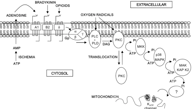
그리 고, heat shock proteins23이 거론되었지만 현재 그들의 정확한 역할과 기전은 아직 밝혀지고 있지 않다. 다만 토끼의 심장 조직이 보호되는 IPC 현상에서 adeno- sine,18 bradykinin,21 opioids,19 그리고 free oxygen radicals20 이 관여되었으며, 이들 각각은 PKC 억제제(inhibitor) 에 의해 IPC 현상이 소실되는 것으로 보아 IPC의 궁극 적 기전은 PKC 신호전달과정이 중요한 역할을 하는 것으로 알려져 있다(Fig. 1).14
따라서 심장 연구 초기의 classic (acute) ischemic pre- conditioning은 adenosine A1-receptor 혹은 K+ATP chan- nel opening의 활성과 관계가 깊다고 하였으나 최근에 는 ischemic preconditioning의 integral로 알려지는 신호 전달 과정 중 PKC의 역할이 연구의 대상이 되고 있다.
2) Protein kinase C (PKC)
Preconditioning을 유도하는 대부분의 작동제는 막 수용체와 결합하며, 이러한 수용체와 연관되는 세포내
Fig. 1. Schematic representation of the proposed mechanism of preconditioning in rabbit heart.14 Ischemia leads to release of adenosine, bradykinin, opioids, and free radicals that are seen attaching to their respective receptors, which in turn are coupled to a G protein (Gp). The latter activates either phospholipase C (PLC) or D (PLD), resulting in degradation of membrane phospholipids and generation of diacylglycerol (DAG), the required cofactor for activation and translocation of protein kinase C (PKC) from cytosol to cell membranes. Sufficient PKC is activated to translocate and initiate a complex kinase cascade that involves a down stream tyrosine kinase (possibly one of MKKs), p38 MAPK, and MAPKAPK-2. These kinases eventually open mitochondrial KATP channels that may be the end effectors. Many steps in this sequence remain unknown.
ATP Pi
ATP Pi ATP
Pi
A1 B2 δ
ADENOSINE
AMP
ATP
α γ
β PLC
PLD+ PKC
PKC
MKK
p38 MAPK
MAK KAP K2
ATP Pi TRANSLOCATION
?
KATP channel MITOCHONDION
ISCHEMIA
BRADYKININ OPIOIDS
OXYGEN RADICALS
EXTRACELLULAR
Gp
DAG
CYTOSOL
ATP Pi
ATP Pi ATP
Pi
A1 B2 δ
ADENOSINE
AMP
ATP
α γ
β PLC
PLD+ PKC
PKC
MKK
p38 MAPK
MAK KAP K2
ATP Pi TRANSLOCATION
?
KATP channel MITOCHONDION
ISCHEMIA
BRADYKININ OPIOIDS
OXYGEN RADICALS
EXTRACELLULAR
Gp
DAG
CYTOSOL
의 이차 전달체계는 심장의 preconditioning과의 관계 에 대해 많은 연구가 진행되고 있다. 작동제와 막 수용 체가 결합하게 되면, 수용체와 결합하는 G-단백질이 활성화되고 이는 막의 phospholipase를 활성화시키며, phospholipase는 phosphatidylinositol biphosphate를 ino- sitol triphosphate와 diacylglycerol (DAG)로 분리되고, 분리된 DAG가 ischemic preconditioning에 중요한 역할 을 하는 것으로 보고되었다.24 다양한 생화학적 기전을 가진 PKC는 많은 보고에서 preconditioning에 관여하는 작동제들의 수용체와 결합을 하는 것으로 잘 알려져 있다.14 하지만 PKC는 구조적이나 약물학적으로 매우 복잡하며, 활성과 효소 작용에 관여하는 4개의 con- stant region과 전위, 기질과의 결합, 그리고 동종효소 각각의 특이성을 지니는 5개의 variable region으로 갖 고 있으며,25 또한 적어도 11가지 이상의 동종효소가 있고 이들 동종효소들은 각각의 다른 고유의 기능을 지니고 있어 preconditioning의 정확한 기전을 밝히기 가 매우 어렵고 실험자 간의 결과가 서로 상이할 수 있다.25 일반적으로 PKC의 활성은 조직을 보호하고, PKC의 억제제를 사용하면 preconditioning 효과가 억 제된다고 보고하였지만,9 반면에 일부 실험들에서는 PKC의 억제제가 preconditioning을 억제하지 못한다는 보고도 있다.26
(1) 구조 및 분류: 일반적으로 PKC는 catalytic domain (carboxy-terminal)과 regulatory domain (amino-terminal) 의 두 부위로 구성되어 있는 phospholipid dependent serine/threonine kinase의 한 종류(family)로 PKC의 동종 효소들(isozymes)은 적어도 12가지 이상이 알려져 있 으며 이들은 regulatory domain의 구조와 활성화에 필 요한 보조인자들을 근거로 세 가지로 세분화된다.25 Conventional PKCs (PKCα, -βI, -βII, -γ)는 보조 인자로 지질(lipids), diacylglycerol (DAG)/phorbol ester, 그리고 칼슘(Ca+2)이 PKC의 활성화에 필요하다(Fig.
2A). 반면, novel PKCs (PKCδ, -ε, -η, -θ)는 지 질, DAG/phorbol ester가 필요하지만 칼슘은 필요하지 않 다(Fig. 2B). 한편 atypical PKCs (PKCζ, -λ, -ι, -μ) 는 칼슘과 DAG/phorbol ester가 모두 필요하지 않으며 단지 지질에 의해 활성화된다(Fig. 2C). 따라서 대부분 의 PKC 동종효소는 regulatory domain의 특이적 순서 에 의해 나누어진다.14,27,28
(2) PKC의 조직 특이적 분포와 역할: 생물체의 세포 나 조직에서 PKC의 존재와 그 역할은 세포 특이적 (cell-specific) 혹은 조직 특이적(organ-specific)의 역할 을 지니는 것으로 알려져 있다.29,30 조직이나 세포의
PKC 동종효소의 분포는 Northern/Western blot, 혹은 면 역조직검사에 의해 알려지게 되었으며,31,32 PKCα, - βI, -βII, -δ, -ε, 그리고 -ζ는 대부분의 모든 조 직에 존재하는 동종효소로 알려져 있다. PKCα는 거 의 모든 조직에 존재하고, PKCβI과 -βII는 뇌와 비 장에 대부분 존재한다. PCKδ는 뇌, 심장, 비장, 폐, 간, 난소, 비장, 그리고 부신에 존재한다. PKCε은 뇌, 신 장, 췌장에 존재하며 특히 뇌조직에 풍부하다. PKCζ 는 대부분의 조직에 존재하지만 특히 폐, 뇌, 간 조직 에 많다.33-35 PKCγ는 대부분 중추신경계 그리고 척수 에 제한되어 존재하고, PKCη는 피부와 폐 조직에 대 부분 존재하며, 또한 비장이나 뇌 조직에서도 일부 볼 수 있다.36 PKCθ는 대부분 근육조직에 존재하며 폐, 비장, 피부 뇌조직에서는 적은 양이 있다.37 PKCμ는 매우 다양하게 분포하며 특히 흉선과 폐에 강한 발현 을 보인다.38 비록 조직에서 PKC 존재에 대한 다양한 차이를 보고하고 있지만 대부분 다양한 각각의 조직 내에서 전사 정도(transcriptional level)의 차이에 대해 서는 정확히 알려진 바 없다. 따라서 조직 특이적 분포 에 대해서는 명확히 알고 있지 못한 실정이다. PKC는 세포의 성장(cell growth)과 분화(differentiation), 이온의 이동(ion transport), 세포 간의 상호작용(cell-cell com- munication), 세포골격의 구조(cytoskeletal architecture), 원형질막(plasma membrane) 등을 포함하는 다양한 생 물학적 기전(diverse biological process)을 조절하며, tumor promoter의 하나인 phorbol ester의 major cellular target이며, tumorigenesis뿐만 아니라 다양한 질환에서 점차 그 역할이 중요시되고 있다. 특히 PKC는 위장관 상피세포에서의 물질 이동(vectorial transport), 관문 작 용(barrier function), 세포의 형태(cell shape), 그리고 세 포의 이동(migration)에 중요 영향을 미치며, 세포의 분 화(differentiation)와 세포의 표현형(phenotype)의 적응 성(modulation)에도 중요한 작용을 한다고 알려져 있 다.29-33,39-42
(3) PKC 전위(translocation)의 개념 및 활성화(acti- vation): 일반적인 PKC 신호전달과정의 특징은 생물학 적 구획(biologic compartments)간의 전위(translocation) 되는 것으로, 활성화되지 않은 PKC는 대부분 세포질 (cytosol)에 위치하며, 동종효소가 활성화되면 대부분 세포막(plasma membrane)으로 전위되며 일부 세포나 조직에서는 그들 고유의 생물학적 목표(biological tar- gets)인 세포골격요소(cytoskeletal element) 핵(nucleus) 등의 세포 내(intracellular)의 위치로 전위된다고 알려 져 있다.43-45 PKC 동종효소는 세포 내의 분포가 일정하
지 않고, 특이적 생물학적 구획 혹은 구조물들(stru- ctures)과 관련이 많은 것으로 알려져 있고, PKC의 동 종효소들의 기능적 특이성은 그들의 초미세포성 위치 결정(subcellular localization)에 의해 결정된다. PKC 동 종효소들이 활성화되면, 각각의 동종효소는 각각 자신 고유의 subcellular site로 전위되어 RACKs (receptors for activated C kinases)이라고 명명된 특수 단백질과 결합하게 된다.46 즉 PKC 동종효소들의 고유의 특성은 활성화가 되었을 때 초미세포성 위치이동으로 Mochly- Rosen 등46은 면역형광법을 이용하여 비활성의 PKC 동종효소들이 활성화가 되면 심근의 핵과 핵 주변 지 역에서 cross-striated structure (contractile element)와 세 포-세포 접경지역으로 전위됨을 보였으며, 다양한 세
포내 구조물에 위치하는 RACKs은 활성화된 동종효소 와 결합을 이루게 된다고 하였다. 따라서 PKC의 활성 화의 연구에는 초미세포성 분획(subcellular fractiona- tion)이 필수적이며, 초미세포성 구획(subcellular com- partment) 사이의 상호 오염(contamination)으로 인한 실 수가 발생할 수 있으므로 면역조직형광 검사나 전자 현미경적 연구가 또한 필요하다. PKC 활성화와 그 이 후 세포막으로의 전위는 PKC 신호전달과정의 가장 중 요한 과정이다. 하지만 그 생물학적 의미에 대해서는 아직도 많은 연구가 진행되고 있다. PKC 활성을 측정 하는 대표적 방법으로는 PKC immunoblotting, 비방사 능기법(non-radioactive kit; ELISA 기법, 혹은 물리적/
화학적 방법으로 phosphorylated peptide를 찾는 방법), Fig. 2. Representation showing the sequence homology between the primary structures of protein kinase C (PKC) isoforms.50 The PKC structure can be divided into an N-terminal (N) regulatory domain and a C-terminal (C) catalytic domain. The conventional (cPKC) structure comprises four conserved regions (C1C4) and five variable regions (V1V5). The C1 region (C1A and C1B), which possesses the cysteine-rich zinc fingers, is involved in binding to diacylglycerol (DAG) and phorbol-ester activating compounds, whereas the C2 region is responsible for binding to Ca2+. Regions C3 and C4 are the binding sites for ATP and the PKC substrate, respectively. The novel PKC (nPKC) group and atypical (aPKC) group both possess a C2-like region within V1, and the aPKC group possesses only one zinc finger. Many isoform-selective inhibitors of PKC, such as the indolocarbazoles and bisindolylmaleimides, interact at the ATP-binding site (or C3), whereas many activating agents act at the DAG/phorbol-ester binding region (or C1). Peptide inhibitors and activators of PKC translocation target the C2 region of cPKC and the V1 region of nPKC.
N C
DAG, phobol Ester binding
Ca 2+
binding
ATP binding
Substrate binding Regulatory domain
C3 V4 C4 V5
DAG, phobol Ester binding C2-like
ATP binding
Substrate binding
C2-like
ATP binding
Substrate binding PKC inhibitors PKC inhibitors (Rottlerin)
PKC inhibitors (Go6976, LY333531) Peptide translocation
Inhibitors or activators PKC activators
Peptide translocation
Inhibitors or activators PKC activators
A. cPKC (α, βI , βII, γ)
B. nPKC ( δ, ε, η, θ)
C.aPKC ( ζ, ι/λ)
Regulatory domain
V1 C1 V2 C2 V3
N C
N C
N C
DAG, phobol Ester binding
Ca 2+
binding
ATP binding
Substrate binding Regulatory domain
C3 V4 C4 V5
DAG, phobol Ester binding C2-like
ATP binding
Substrate binding
C2-like
ATP binding
Substrate binding PKC inhibitors PKC inhibitors (Rottlerin)
PKC inhibitors (Go6976, LY333531) Peptide translocation
Inhibitors or activators PKC activators
Peptide translocation
Inhibitors or activators PKC activators
A. cPKC (α, βI , βII, γ)
B. nPKC ( δ, ε, η, θ)
C.aPKC ( ζ, ι/λ)
Regulatory domain
V1 C1 V2 C2 V3
N C
N C
그리고 방사능 동위원소 측정법(radioactive PKC assay;
PKC 특이 기질이 존재하는 상태에서 γ-32P-ATP를 측 정하는 방법) 등이 있다. 또한 알려진 많은 PKC 억제 제들을 이용하여 immunoblotting으로 측정하는 변형된 방법이 있다.26 저자들는 PKC 활성을 측정하기 위해 세포하 분획 후 Western blot, 면역조직형광법, 공초점 레이저현미경(Confocal microscopy), immunokinase as- say 등을 이용하였다.
(4) PKC에 대한 약물: PKC 억제제는 PKC 단백 구조 내에서 상호 작용을 하는 위치에 따라 분류할 수 있다.
먼저 regulatory domain의 억제제는 phospholipid 혹은 phorbol-ester와의 결합 부위에 작용하는 반면에 cata- lytic domain의 억제제는 substrate나 ATP와의 결합 부 위에 작용하게 된다. 사용 가능한 PKC 억제제는 다양 하지만 PKC 자체에만 혹은 개개의 PKC 동종효소 각 각에 대한 특이적인 억제제는 매우 적다.47 그러므로 앞으로 유망한 선택적 PKC 억제제는 ATP와의 결합부 위에서 작용하는 PKC 억제제 또는 약물이다. PKC 동 종효소에 대한 선택적 억제제(Isoform-selective inhibi- tors of PKC)의 약물로는 Rottlerin48 (selective PKC δ inhibitor), Indolocarbazole PKC inhibitors49 (Gö6976, UCN01, CGP41251), Bisindolylmaleimide PKC inhi- bitors49 (Gö6983, Gö6850, GF109203X, Ro317549, Ro318220) LY333531 (PKCβ-isoform-selective inhi-
bitor) 등이 있다(Table 1).50 그 외에 PKC gene inhibition 에는 antisense oligonucleotide inhibition of PKC, ribo- zyme inhibition of PKC가 있다.50 PKC 동종효소에 대한 선택적 activators에는 phorbol ester compounds Thymel- eatoxin (Thymelea hirsuta leaves), sapintoxin A (Sapium indicum), bistratene A, polyether toxin (Lissoclinum bis- tratum), polyphosphoinositides, phosphatidylinositol 3,4- bisphosphate [PtdIns(3,4)P2], phosphatidylinositol 3,4,5- trisphosphate [PtdIns(3,4,5)P3] 등이 알려져 있다.50 한편 PKC의 전위에 대한 억제제와 활성제(Inhibitors and activators of PKC translocation)에는 다음과 같은 특징 이 있다. 특이적 PKC 동종효소의 활성과 전위는 각각 의 PKC 동종효소의 독특한 생리적 작용을 부여하게 된다. 예를 들면 RACKs (Receptors for activated C kinases)와 같은 specific anchoring proteins과 활성화된 PKC 동종효소와 결합으로 PKC 동종효소의 활성과 전 위가 이루어진다고 알려져 왔다.28 Mochly-Rosen 등10 에 의해 개발된 PKC 전위의 peptide fragment의 억제제 와 활성제의 연구를 보면 rat neonatal cardiomyocytes에 서 PKCε에 의한 ischemic preconditioning의 효과가 PKCε의 peptide inhibitor인 εV1과 εV1-2에 의해 억 제되었고,10,28,51,52
adult rabbit cardiomyocytes에서도 PKCβ, -δ, -η들에 대한 각각의 peptide inhibitor에 의해서 ischemic preconditioning의 효과가 유지된 반면,
Table 1. IC 50 values (μM) for isoform-selective protein kinase C inhibitors50
ꠚꠚꠚꠚꠚꠚꠚꠚꠚꠚꠚꠚꠚꠚꠚꠚꠚꠚꠚꠚꠚꠚꠚꠚꠚꠚꠚꠚꠚꠚꠚꠚꠚꠚꠚꠚꠚꠚꠚꠚꠚꠚꠚꠚꠚꠚꠚꠚꠚꠚꠚꠚꠚꠚꠚꠚꠚꠚꠚꠚꠚꠚꠚꠚꠚꠚꠚꠚꠚꠚꠚꠚꠚꠚꠚꠚꠚꠚꠚꠚꠚꠚꠚꠚꠚꠚꠚꠚꠚꠚꠚꠚꠚꠚꠚꠚꠚꠚꠚꠚꠚꠚꠚꠚ Protein kinase C isoform
Agent ꠏꠏꠏꠏꠏꠏꠏꠏꠏꠏꠏꠏꠏꠏꠏꠏꠏꠏꠏꠏꠏꠏꠏꠏꠏꠏꠏꠏꠏꠏꠏꠏꠏꠏꠏꠏꠏꠏꠏꠏꠏꠏꠏꠏꠏꠏꠏꠏꠏꠏꠏꠏꠏꠏꠏꠏꠏꠏꠏꠏꠏꠏꠏꠏꠏꠏꠏꠏꠏꠏꠏꠏꠏꠏꠏꠏꠏꠏ
α β βI βII γ δ ε ζ η
ꠏꠏꠏꠏꠏꠏꠏꠏꠏꠏꠏꠏꠏꠏꠏꠏꠏꠏꠏꠏꠏꠏꠏꠏꠏꠏꠏꠏꠏꠏꠏꠏꠏꠏꠏꠏꠏꠏꠏꠏꠏꠏꠏꠏꠏꠏꠏꠏꠏꠏꠏꠏꠏꠏꠏꠏꠏꠏꠏꠏꠏꠏꠏꠏꠏꠏꠏꠏꠏꠏꠏꠏꠏꠏꠏꠏꠏꠏꠏꠏꠏꠏꠏꠏꠏꠏꠏꠏꠏꠏꠏꠏꠏꠏꠏꠏꠏꠏꠏꠏꠏꠏꠏꠏ Naturally derived inhibitors
Rottlerin 30 42 ND ND 40 3∼6 100 100 82
Indolocarbazoles
UCN01 0.029 - 0.034 ND 0.030 0.590 0.53 - ND
CGP41251 0.024 - 0.017 0.032 0.018 0.360 4.500 >103 0.060
Gö6976 0.0023 - 0.006 ND ND - - - ND
Bisindolylmaleimides
Gö6983 0.007 0.007 ND ND 0.006 0.010 ND 0.060 ND
Gö6850 0.0084 ND 0.0180 ND ND 0.210 0.132 5.8 ND
Ro317208 0.160 - 0.310 0.28 0.377 ND 0.330 ND ND
Ro317549 0.053 - 0.195 0.163 0.213 ND 0.175 ND ND
Ro318220 0.005 - 0.024 0.014 0.027 ND 0.024 ND ND
R0318420 0.008 - 0.008 0.041 0.031 ND 0.039 ND ND
Ro320432 0.009 - 0.028 0.031 0.037 ND 0.108 ND ND
LY333531 0.360 - 0.0047 0.0059 0.400 0.250 0.600 >105 0.052
ꠏꠏꠏꠏꠏꠏꠏꠏꠏꠏꠏꠏꠏꠏꠏꠏꠏꠏꠏꠏꠏꠏꠏꠏꠏꠏꠏꠏꠏꠏꠏꠏꠏꠏꠏꠏꠏꠏꠏꠏꠏꠏꠏꠏꠏꠏꠏꠏꠏꠏꠏꠏꠏꠏꠏꠏꠏꠏꠏꠏꠏꠏꠏꠏꠏꠏꠏꠏꠏꠏꠏꠏꠏꠏꠏꠏꠏꠏꠏꠏꠏꠏꠏꠏꠏꠏꠏꠏꠏꠏꠏꠏꠏꠏꠏꠏꠏꠏꠏꠏꠏꠏꠏꠏ Abbreviation: ND = not determined in reference article, -, no effect.
εV1-2에 의해서는 preconditioning의 효과가 억제되는 유사한 결과를 보였다.53 심장보호기능에 PKC의 관여 하며, PKCε의 전위를 활성화하면 허혈성 심질환에서 치료제로 사용될 수 있다고 하였으며, neonate와 adult cardiomyocytes에서 선택적으로 PKCε의 전위의 활성화 를 유도할 수 있는 PKC의 V1 region에서 8-amino acid 위치에서 pseudo εRACK peptide를 개발하였다.54 Pseudo εRACK의 투여하거나 mouse heart에 도입된 유 전자 동물의 출생 후 발현은 지속된 저산소증으로부터 심장세포의 생육력(viability)과 심장 기능의 개선을 보 였다.55,56
3) Role of protein kinase C signaling in ischemic preconditioning of the various tissue and organs (1) 심장: 심근조직에서 처음으로 보고된 ischemic preconditioning은 가장 활발한 연구가 진행되고 있는 기관이며 대부분의 결과는 심장에 대한 연구이다.
Ischemic preconditioning은 내재적 조직보호 현상이며, 이것은 수용체에 의한 신호전달과정에 의해서 이루어 진다고 알려져 있다.14 심근조직에서 다양한 수용체의 작동제와 길항제들을 사용한 실험 결과 adenosine,18 bradykinin,21 opioids19 등의 수용체 의존형의 endoge- nous trigger와 free oxygen radicals,20 nitrate,57 calcium 등 의 수용체 비의존형의 exogenous trigger 등에 의한다고 알려져 있으며 이와 관련하여 Tyrosine kinase, PKC, Mitogen-activated protein kinase (MAPK), ERK, JNK, p38 MAPK 등이 신호전달과정의 매개체로 알려져 있 으며, 특히 PKC와의 관련에 대한 연구가 그 중심이 되 고 있다.14,58
심장근육세포에서 적어도 6가지 이상의 PKC 동종 효소가 발현되었으며,59 그들 중 preconditioning과 관련 되어 PKCδ와 PKCε만이 활성화를 보였다.10 PKCε의 peptide 억제제인 εV1-2은 preconditioning에 의한 심장 근육세포의 보호효과를 억제하였고, 그 이외의 peptide 억제제는 심장보호효과를 억제하지 못하여 PKCε의 활성이 심장보호효과에 필요하다고 하였다.10 한편 PKCε의 활성 작동제인 ψεRACK은 neonate와 adult cardiomyocytes에서 허혈성 세포 괴사의 70% 가량을 감 소시키는 효과를 보였고, PKC 억제제와 PKCε의 선택 적 억제제인 εV1-2에 의해 ψεRACK의 심장보호효 과는 소실되었다.54 ψεRACK을 이용한 transgenic mice는 허혈에 따른 심근의 손상이 non transgenic mice 에 비해 60% 가량의 감소를 보여 세포나 transgenic mice의 허혈성 심장손상을 보호하기 위해서는 PKCε
의 활성이 필요하다고 하였다.54 그리고 ψεRACK 혹 은 유사 약물에 의한 인간의 허혈성 심장 질환의 치료 에 사용될 수 있는 연구가 진행 중에 있다.54
(2) 간: 간에 대한 ischemic preconditioning에 대한 연 구는 심장 연구에서 비하여 아직 초기 단계이다. Heat shock (즉 체온을 42oC까지 올려 15분간 유지시킴) 혹 은 ischemic preconditioning은 간의 warm ischemia로부 터 효과적으로 보호하며,60,61 또한 coldstorage injury로 부터 보호한다고 하였다.62 인체의 간조직의 ischemic preconditioning63에 대한 효과는 adenosine이 가장 중요 한 매개체로 알려져 있고,64 Adenosine은 NO64,65를 생 성하는 초기 단계의 adenosine A2 receptor를 자극하 여,65-67 PKC, adenosine monophosphate-activated protein kinase, 그리고 p38 MAPK를 활성화시킨다고 하였
다.68-71 이러한 세포 내 신호전달과정의 활성화는 허혈
로부터 간세포와 내피세포들의 내성을 증가시킬 뿐만 아니라 세포 대사를 정지시키고 다시 재생할 수 있는 조건을 유발시킨다.71 Recombinant IL-6의 투여로 허혈 (warm ischemia) 후 발생하는 재관류 손상을 감소시키 는 것으로 볼 수 있으며,72 heat shock proteins60 (특히 HSP70)과 heme oxygenase-1 (HSP32)73,74의 발현으로 preconditioning의 기전을 암시하였다. HSP의 발현은 proinflammatory transcription factors와 핵의 결합을 감 소시키며,75 세포의 항산화능(antioxidant capacity)을 증 가시킬 수 있다.76 이러한 두 가지 효과는 precondi- tioned liver에서 TNF-α의 생성을 감소시키고 염증반 응을 저하시키는 작용을 하는 것으로 생각되고,77,78 허 혈/재관류 손상에서 발생하는 heme oxygenase-1의 부 산물인 carbon monoxide는 p38 MAPK를 활성화 시키 는 것으로 보인다.79 따라서 ischemic preconditioning된 간에서는 허혈 손상으로부터 이상과 같은 여러 요인 에 의해 보호될 수 있다고 생각되며, 최근 간이식 동물 실험에서 먼저 preconditioning을 한 경우 일반적인 간 이식편에 비해 담즙의 분비의 증가, 간 이식편 혈류의 증가, 그리고 간손상의 표지자인 LDH의 혈중 농도가 감소되었으나, PKC 억제제로 전처치한 preconditioned 간이식편은 preconditioning 효과가 억제됨을 보여 간 의 ischemic preconditioning은 PKC의 활성과 관계가 깊 다고 보고하였다.2
(3) 골격근계: 골격 근육에서의 latissimus dorsi flap에 서 preconditioning으로 허혈에 대한 내성의 증가됨이 처음 보고되었으며,80 또한 canine gracilis 근육을 이용 한 IPC 실험에서 KATP channels의 활성화의 기전으로 모세혈관의 혈류가 증가되어 근육조직으로부터 손상
을 줄인다고 하였다.81 Intravital microscopy를 이용한 murine 고환거근의 허혈/재관류에서 백혈구의 adhe- sion과 emigration은 preconditioning으로 감소시킬 수 있었으며, 이 기전에는 adenosine이 관여한다고 보고하 였다.82 심장 근육(contractile muscle)의 preconditioning 에 많은 관련이 있다고 보고된 adenosine은 일반 근육 (striated muscle)에서도 유사한 작용을 하는 것으로 생 각된다. 최근에는 latissimus dorsi 근육의 in vivo 실험 에서 PKC 억제제와 K+ATP channel 길항제로 전처치하 여 preconditioning을 하면 근육 경색의 보호 효과가 감 소한 반면에 PKC activators로 전처치한 경우 근육 경 색의 보호 효과는 preconditioning을 한 경우와 유사한 근육 보호효과를 보였으며, Western blot에서 PKC (α, βI, βII, δ, ε, θ, ζ, ι)의 동종효소가 발현되었고, 발 현된 동종효소 중 IPC 동안 PKCε만이 유일하게 세포 질 분획에서 세포막 분획으로 전위됨이 관찰되었다.83 이는 심장근육에서와 유사한 결과를 보여, 일반 근육 계에서의 preconditioning 기전도 .PKC와 K+ATP channel 의 신호전달 체계가 그 일부를 차지하며, 특히 PKC가 중요 작용을 하는 것으로 보고하였다.83
(4) 신장: 1984년 Zager 등84에 의해 처음 precon- ditioning 개념의 실험은 시작되었으나 첫 허혈(15분간 의 허혈) 후 이차 허혈은 오히려 허혈에 대한 신장의 내성을 낮춘다고 하였다.85 그러나 최근의 보고에 따르 면 8분간의 허혈(각각 5분간 재관류) 4회의 전처치 후 45분간 이차 허혈에 대한 내성을 보였으며,86 또한 5, 10, 15, 20분간의 허혈(각각 10분간 재관류) 후 40분간 의 이차 허혈 후 1, 2, 3, 그리고 7일 후의 혈청 크레아 티닌이 감소된 것으로 보고하였다.87 다른 실험에서는 2분간 허혈 그리고 5분간 재관류를 연속으로 2회하고 24시간 경과하여 측정한 혈청 크레아티닌은 감소를 보였으나 3회의 허혈과 재관류를 한 경우는 효과가 없 었다.88 또한 돼지에서 10분씩 3회의 허혈(각각 10분간 재관류)은 60분간의 이차 허혈에 대해 내성이 없다고 보고하였다.89 Adenosine 전처치에 따른 신장의 precon- ditioning은 심장의 preconditioning과 달리 bradykinin, muscarinic, opioid 수용체 혹은 ATP-sensitive K+ATP
channels에 의한 기전과는 관계가 없었으나, pertussis toxin-sensitive G-proteins과 PKC와는 관계가 깊었다.4 그리고 cultured human proximal tubule (HK-2) cells에서 ATP depletion preconditioning (antimycin A/ 2-deoxy- glucose) 또는 adenosine (adenosine A1 or A2a receptor selective agonist)으로 전처치하고 후속의 ATP de- pletion으로 손상을 받게 되면 ATP depletion으로 인한
신장세포의 손상이 감소되었지만, adenosine 수용체 길 항제로 전처치한 경우 신장세포의 손상 보호효과가 억제되었으며, ATP depletion preconditioning과 adeno- sine에 의해 매개된 조직보호 현상은 mitogen-activated kinases, PKC, 그리고 tyrosine kinase의 억제제에 의해 그 보호 효과가 소실되었다.5
(5) 위장관계: 위장관계, 특히 소장에서의 허혈에 관 계된 논문은 너무나 다양하고 많다. 그러나 위장관의 IPC에 대한 기전에 대한 연구에서는 heat shock pro- tein,23 nitric oxide,90-92 adenosine22,93과 관계된다는 초기 단계의 연구가 보고되고 있다. 체온의 상승을 유발시 킨 후 허혈/재관류 손상 모델에서 소장점막의 손상은 보호되었으며 이는 leukotriene B4의 생성의 감소, 백혈 구와 chemotaxis의 활성화 감소, heat shock protein의 발 현의 증가가 IPC와 유사한 효과를 가져온다고 하였 다.23 상장간 동맥의 IPC(각각 10분간의 허혈/재관류) 하고 30분간 후속의 허혈 손상을 주고 24시간 경과하 여 검사한 소장 조직에서 점막의 손상은 감소하였으 며 장간막, 비장, 간, 혈액에서 bacterial translocation이 감소되었다. 이는 IPC에 의한 소장에서 inducible nitric oxide (iNOS)의 발현이 증가한 것으로 설명하였다.92 허혈/재관류손상에서 흔히 볼 수 있는 소장점막의 hy- perpermeability는 IPC하여 permeability를 감소시켰으 며, adenosine receptor blocker로 전처치하고 IPC하면 permeability가 다시 증가하는 결과로 소장에서의 IPC 와 adenosine과 관련에 대해 보고된 바 있으며,93 adeno- sine 차단제와 PKC 억제제를 이용한 허혈 후 발생하는 백혈구의 활성화의 과정, 특히 leukocyte rolling에 관여 하는 P-selectin의 발현이 감소된 점이 adenosine과 PKC 와의 관계된다고 하였다.22
수없이 많은 논문에서 위장관의 허혈/재관류 손상에 관한 실험을 볼 수 있다. 하지만 그 허혈과 재관류의 시간설정은 매우 다양하여 천차만별이다, 또한 알아보 고 검증하고자 하는 내용도 다르다. Heat stress에 의한 heat-shock protein (HSP72)의 유발은 소장의 허혈/재관 류 손상으로부터 점막의 손상을 줄일 수 있었으며 이 는 소장점막의 leukotriene B4의 감소로 인한 백혈구의 활성과 chemotaxis가 억제된 것으로 그 이유를 설명하 였다.23 또한 상장간막동맥의 일시적 허혈 후 24시간 경과한 delayed ischemic preconditioning rat 모델에서 대조군에 비해 선행의 precondition을 받은 실험군의 소장점막이 leukotriene B4와 백혈구의 활성에도 불구 하고 조직학적으로 보호되었으며, 이는 goblet cell의 증가에 따른 mucus 분비에 의해 점막이 보호된 것으로
보고하였다.8 Rat 소장의 classic ischemic preconditioning 모델에서 염증반응의 leukocytes rolling에 관여하는 P-selectin의 발현은 preconditioning의 실험군에서 감소 하였으며, preconditioning의 전처치로 adenosine A1 수 용체 길항제 또는 PKC의 억제제들로 전처치한 경우 P-selectin의 발현이 다시 증가된 결과로 미루어 ische- mic preconditioning에 adenosine과 PKC가 일부 관여된 다고 보고하였다.22 하지만 조직학적으로 증명된 소장 점막의 보호에 대한 연구가 없는 점이 아쉽다.
4) Protein kinase C signaling in intestine
위장관이나 신장과 같은 상피세포는 생물학적 구획 간의 관문 기능과 함께 혈액과 체외 환경간의 수분, 이 온, 그리고 거대분자의 선택적 이동(selective transport) 을 매개한다. 이러한 기능들은 intercellular junction의 integrity, apical과 basolateral domains를 유지하는 세포 막(plasma membrane)의 지질과 단백질의 구성, 그리고 productive cell-substratum interaction 등 모두에 의존하 게 되는데 이들의 대부분 모두 허혈/재관류에 영향을 받는다.94 Ischemic epithelial phenotype의 generation은 비록 산화적 손상, 이온들 그리고 pH의 변화 등의 다 양한 요인이 있지만 무엇보다도 세포 내 ATP의 고갈 로 인하여 손상의 대부분을 받게 된다. 따라서 ATP를 낮추는 약제를 사용하는 세포배양 연구는 분극화된 상피세포(polarized epithelial cell)에서 ATP 고갈과 재 충전(repletion) 등에 관한 많은 연구가 이루어지고 있 다.95 허혈로 인해 발생하는 조직의 변화, 특히 신장조 직에서 즉 filamentous-actin (F-actin)의 분리, 세포골격 막(cytosketetal membrane)의 절단, 세포막의 불안정, 세 포 표면의 분극화(cell surface polarity) 소실, 그리고 상 피세포의 이동과 관문기능의 변화 등을 관찰할 수 있
다.96,97 단순한 일시적 저산소증(hypoxia; O2 depri-
vation) 혹은 허혈(ischemia; 산소와 기질의 고갈)은 세 포와 조직에 심한 영향을 미친다. 심장조직에서 PKC 의 한 동종효소인 epsilon은 심근의 허혈 반응에 중요 한 역할을 하는 것으로 알려져 있으나,1 심근조직의 보 호기능과 관련된 PKC의 upstream activators 혹은 down stream effecters에 대해서는 아직 잘 알려져 있지 않다.
흔한 설사, 염증성 장질환, 중증환자에서의 저혈류 (low flow state)상태, 급성 동맥 폐쇄질환의 허혈성 장 염 등 위장관 질환에서 허혈은 세포나 조직손상의 중 요한 요인 중의 하나이다. 위장관의 상피세포 역시 허 혈/재관류 손상에 극히 민감하지만,98 아직 허혈이나 저산소증과 같은 극한 상황에서 이들 상피세포가 구
조적이나 기능적으로 어떻게 영향을 받고 적응하는 지에 대해 잘 알고 있지 못하다.
본 논문은 위장관(intestinal cell line or native intestinal tissue)의 허혈과 저산소증에 따른 PKC 신호 전달과정 (특히 PKCε)의 기전과 역할, 그리고 소장에 서 IPC가 존재한다면, IPC 현상과 PKC의 활성화와의 관련에 대해 저자들의 실험 결과를 중심으로 설명하 고자 한다.
(1) PKC signaling in intestinal epithelial ischemia (me- tabolic stress): Human intestinal cell line의 하나인 T84 cell(대장선암 세포주)은 electrogenic Cl- secretion과 tight junctional integrity를 보기 위해 많은 실험으로 이 용되어왔다.99-102 T84 cell에서 PKC의 동종효소 중 PKC α, -δ, -ε, -ζ가 Western blot에서 발현되었으며, T84 cell에서 phorbol ester의 하나인 phorbol 12- myristate 13-acetate (PMA, 100 nM)에 0, 15, 60, 그리고 120분간 노출시킨 후, 초미세포성 분획(subcellular fractionation)하여 western blot을 시행한 결과 PKCε (epsilon)은 15분 이내에 시간 의존형 양상으로 세포질 분획에서 세포막 분획으로 전위(활성화)되었다. 한편 PKCα는 약 60분까지는 세포질 분획에서 세포막 분획 으로의 전위를 보였으나 그 이후 전위의 정도가 감소 하였고, PKCδ, -ζ는 전위되지 않았다.103
Metabolic stress는 chemical hypoxia와 authentic hy- poxia의 두 가지 방법을 이용하였다. 일반적으로 사용 하는 HPBR 용액의 10 mM glucose 대신에 10 mM 2- deoxyglucose와 ATP 합성 억제제인 1μM oligomycin A 를 사용하여 chemical hypoxia를 유발하였으며,104,105 또 한 T84 cells은 specialized hypoxic chamber를 사용하여 1% 미만의 Oxygen 즉 authentic hypoxia를 유발시켰 다.106 in vitro에서 허혈성 손상(ischemic stress)은 glu- cose free buffer에서 혐기성 해당작용(anaerobic glyco- lysis)의 false substrate인 2-deoxyglucose (10 mM)과 함 께 H+-ATPase 억제제인 oligomycin A (1μM) 혹은 site I electron transport chain 억제제인 rotenone (10μM) 등 과 같은 산화적 인산화(oxidative phosphorylation)를 통 해 화학적으로 유발시킬 수 있다. Oligomycin A와 2- deoxyclucose를 사용하여 60분 이내에 세포내의 ATP 를 3% control 이하로 유지시킬 수 있었다.100,104 한편 T84 cell은 specialized chamber를 사용하여 1% 미만의 Oxygen 즉, authentic hypoxia를 유발시켰다. 이는 혐기 성 해당작용은 가능한 저산소 상태에서 24시간 경과 한 후 T84 cell의 ATP 수치는 감소되지 않았던 Colgan 등106의 결과와 oligomycin A와 10 mM의 glucose를 사용
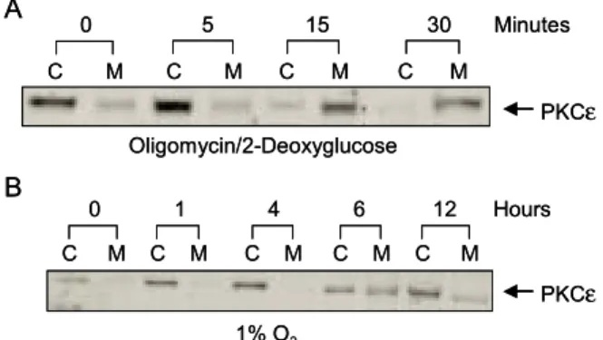
한 실험결과가 일치하는 것을 볼 수 있었다.100,104 Chemical hypoxia에 30분간 노출된 T84 cells에서 F-actin의 content는 136±2% control로 증가를 보였고, rhodamine-phalloidin 염색 후 면역형광현미경검사에서 는 actin cytoskeleton의 remodeling을 보이며, 특히 세포 의 기저외측(basolateral)에서 볼 수 있다. 또한 공초점 레이저현미경검사(confocal microscopy)에서 actin fila- ment의 cytoplasmic clumping (세포질 내 응결현상)과 함께 정상세포에서 보이는 stress fiber의 소실도 볼 수 있다. Chemical hypoxia (1μM oligomycinA plus 10 mM 2-deoxyglucose in glucose-free buffer)를 받은 T84 cell에 서 PKCε은 10분 이내에 세포질 분획에서 세포막 분 획으로 전위를 보였으며, 30∼60분 이내에 가장 높은 PKC 활성화(전위)를 보였다(Fig. 3A). 한편 PKCε의 허혈 후 활성화가 ATP의 고갈이 영향을 미치는 가를 알아보기 위해 hypoxic condition (1% O2)에 1∼12시간 동안 배양하여 세포하 분획 후 Western blot을 하여 PKCε이 세포질 분획에서 세포막 분획으로 전위(활성 화)됨을 확인하였다(Fig. 3B). 그러나 T84 cell에서 발 현된 PKC 동종효소 중 PKCα, -δ, -ζ는 chemical hypoxia와 저산소증(1% O2)의 조건에서 세포질 분획에 서 세포막 분획으로의 전위를 볼 수 없었다. 따라서 T84
human intestinal cell line에서 metabolic stress (즉, 허혈) 에 의해 PKCε이 활성화되는 것을 알 수 있었다.107 (2) PKC signaling in intestinal ischemic precondition- ing108: Sprague-Dawley rats 소장점막의 Western blot에 서 conventional(c) PKC 동종효소들 중에서 PKCα, -βI, -βII가 발현되었고 PKCγ는 발현되지 않았다.
Novel(n) PKC 동종효소들 중 PKCδ, -ε이 발현되었 고, PKCη, -θ는 발현되지 않았으며, atypical 동종효 소들 중 PKCι, -ζ가 발현되었다(PKCλ, -μ는 실 험에 포함되지 않았다). Rats의 상장간막동맥의 기시 부를 혈관감자로 10분간 허혈한 후 120분간 재관류하 여 IPC를 유발하였으며, 대조군은 모의 수술(sham- IPC)하였다.109 IPC 후 20분간 상장간막동맥을 재결찰 하였다. 허혈에 따른 소장(jejunum)의 조직학적 손상은 Parks' injury score110,111에 따라 점수화하였다. 10분간 허혈 손상을 받은 소장점막은 120분간의 재관류 후 조 직학적 소견에서 가역적으로 회복되었으며, 10분간 상 장간막동맥 결찰 후 소장점막의 PKCα, -δ, -ε의 동종효소들의 활성도(immunokinase assay)는 정상점막 에 비해 의미 있게 증가하였다. 그러나 그 외의 다른 PKC 동종효소인 PKCIβI, -βII, -ι, -ζ은 증가하 지 않았다. IPC protocol에 의한 전처치 후 후속의 20분 간 허혈 손상에 따른 조직학적 소견은 대조군은 소장 점막(villi)의 상부에서 하부까지 점막이 소실되는 점 막손상을 보였으나, 실험군(IPC)의 소장점막은 잘 보 존되어 있었으며, 소장점막 허혈 손상을 점수화하였을 때 대조군에 비해 실험군에서 의미 있는 감소를 보였 다(Fig. 4A, B).108 Gö6850 (conventional/ novel PKC 억 제제)로 전처치한 경우 IPC 후 2차 허혈에서 보존되었 던 소장점막 보호의 IPC 현상이 소실된 반면 Gö6976 (선택적 conventional PKC 억제제), rottlerin (PKCδ -specific 억제제), 생리식염수 등으로 전처치한 소장점 막은 잘 유지되어 있었으며, 조직손상을 점수화 하였 을 때에도 의미 있는 차이를 보였다(Fig. 4C∼F).108 본 실험은 심근조직에서처럼 선행의 일시적 장간막 허혈 이 후속의 이차적 허혈에 대해 소장(jejunum)의 점막과 조직을 보호하는 classic IPC 동물모델로 10분간의 일 차 허혈 후 재관류가 각각 30분, 60분이 경과하여 이차 허혈을 받은 소장점막은 IPC 효과가 없었다. 자세한 이유를 알 수 없지만 소장점막이 선행의 허혈로부터 조직학적 및 구조적 회복(restitutional repair)을 위해서 는 적어도 120분 이상의 재관류가 필요하였으며, pre- conditioning에 요구되는 단백질이나 cytokine 등의 활 성에 필요한 조직 반응 시간으로 생각된다.
Fig. 3. Time-dependent translocation of PKCε in T84 cell during metabolic stress. (A) When PKC isoforms are activated by phosphorylation, they translocate from cytosol(C) to membrane(M), and this has been the basis of the indirect measurement of their activation. When ischemic insult was applied to the cells in the form of oligomycin and 2-deoxyglucose, 30 minutes of oligomycin/2-deoxyglucose treatment induced significant translocation of the novel epsilon isoform to the membrane. (B) 12 hours of hypoxia (1% O2) also activates only PKC epsilon isoform. This PKC epsilon activation by hypoxia is also time-dependent, but occurs much more slowly compared to the ischemia model, starting at around 6 hours.
C M C M C M C M
0 5 15 30 Minutes
Oligomycin/2-Deoxyglucose
C M C M C M C M C M 0 1 4 6 12 Hours
1% O2
A
PKCε
PKCε B
C M C M C M C M
0 5 15 30 Minutes
Oligomycin/2-Deoxyglucose
C M C M C M C M C M 0 1 4 6 12 Hours
1% O2
A
PKCε
PKCε B
Mouse의 소장 점막에서 PKCα, -δ, -ζ, -η의 존재를 보고하였으며,112 rat intestinal epithelial cell culture model에서 면역형광법과 Western blot을 이용하 여 PKCα, -βII, -δ, -ε, -ζ의 발현을 보고하였 고,113 본 실험의 rat의 공장 점막에서는 PKCα, -βI, -βII, -δ, -ε, -ι, -ζ가 발현되었으며 PKCγ, -θ, -η는 발현되지 않았다(PKCλ, -μ는 실험에 포함되지 않았다). 비록 PKC 동종효소에 대한 각각에 대한 특이 억제제가 유용하지 않지만 subclass-특이 억 제제와 동종효소 특이 억제제들을 이용하여 각각의 동종효소에 대한 관계 여부를 확인할 수 있다. 가령, 예를 들면 Gö6850는 conventional과 novel PKC 동종효 소들을 모두를 억제하는 반면 Gö6976는 conventional PKC 동종효소만을 억제한다. Gö685049는 bisindolyla- leimide PKC 억제제의 하나로 PKCα, -βI, -δ, -ε,
-ζ를 억제하는 것으로 알려져 있다. Gö697649는 indolocarbazole PKC 억제제의 하나로 cPKCs와 nPKCs 구분하기에 매우 유용하다. 한편 Rottlerin (malloto- xin)48은 Mallotus philippinesis에서 추출된 자연산물로 저농도에서 PKC만을 억제한다. 그러나 in vivo 실험에 서는 약물의 실제 농도와 약물역동학적(pharmacokine- tics) 변화가 있을 수 있어 PKC 억제제의 실질적 효과 를 가늠하기 어렵지만 본 실험 팀은 T84 cell의 epithe- lial transport와 barrier function에서 각각의 PKC 억제제 농도를 이용하여 PKC 동종효소 들의 억제를 규명하여 입증한 바 있으며,103 Davis JM 등22은 PKC 억제제를 복강 내에 주입하여 전처치한 in vivo 실험에서 PKC 억제제의 효과를 입증한 바 있다. IPC를 받지 않고 이 차 허혈 손상을 받은 소장점막은 조직학적으로 점막 이 소실되는 등의 허혈 손상이 있었지만 IPC를 받은 Fig. 4. Microscopic findings of the rat small intestine. 1. Effect of prior IPC followed by subsequent ischemia A: 20-min of SMA occlusion without prior IPC (Sham-IPC), B: additional 20-min of SMAO in prior IPC animal. 2. Pretreated with various PKC inhibitors prior IPC and followed by subsequent 20-min of SMA occlusion. Effect of pretreated with PKC inhibitor on IPC. C; Saline (DMSO), D; Gö6976, E; Gö6850, F; Rottlerin. H&E stain.
IPC+Subsequent Ischemia Sham IPC+Subsequent Ischemia
Saline Go6976
Go6850 Rottlerin
A B
C D
E F
¨
¨
소장의 점막은 이차적 허혈 손상에 대해 저항을 보이 는 IPC 효과를 보였다 그러나 conventional과 novel PKC 동종효소들을 억제하는 Gö6850를 전처치한 경우 IPC 효과가 소실되었고, Gö6976 (conventional PKC 억 제제), rottlerin (specific nPKC 억제제), 생리식염수 등 으로 전처치한 경우 IPC 효과의 변화는 없었다. 일시 적 허혈(brief ischemia)로 유발된 소장점막의 허혈 손 상은 가역적이었으며, 일시적 허혈로 소장 점막의 PKC 동종효소들은 활성화되었다. 후속의 허혈 손상으 로 야기되는 소장점막의 손상은 선행의 IPC로 감소시 킬 수 있었으며, 이러한 IPC 현상은 selective cPKC 억 제제가 아닌 non-selective PKC 억제제로 소실시킬 수 있었다. 따라서 rat의 소장에서 ischemic preconditioning 은 존재하며 이는 nPKC 동종효소(특히 epsilon)의 활 성화와 관련이 깊으며, 이는 심근조직에서 보였던 preconditioning과 동일한 기전이라고 생각된다.108
결 론
1986년 심장 조직에서 처음 보고된 이래 ischemic preconditioning의 기전에서 일부 신호전달과정은 밝혀 진 바 있다. 다양한 종(species)간의 각기 다른 유발인 자가 있을 수 있지만, adenosine, bradykinin, opioids, free radical과 같은 주요 내인성 유발인자가 관여한다 는 것은 잘 알려진 사실이다. 그리고 또한 종 또는 실 험 모델 간의 차이가 있을 수 있지만 PKC, Tyrosine kinase 등과 같은 protein kinases가 관여한다는 사실은 널리 인정되고 있다. 하지만 preconditioning의 정확한 기전, upstream activator 혹은 downstream effector의 신 호전달체계에 대해서는 앞으로 더 많은 연구가 필요 하다. 또한 신체의 어느 부분에서도 관찰되는 PKC는 preconditioning 이외에도 다양한 생물학적 기전을 갖 는 protein kinase로 많은 종류의 동종효소가 있고 그 동종효소 각각의 기능과 역할도 조직에 따라 각각 다 르다고 알려져 있어 PKC signaling pathway의 연구가 요구되고 있고, 앞으로도 새로운 기능과 역할을 갖는 동종효소가 밝혀질 것으로 생각된다.
감사의 글
논문에 도움을 주신 Jeffrey B. Matthews, MD, Edward C. Mun, MD. J. Cecilia Song, Ph.D. (Department of Surgery, Beth Israel Deaconess Medical Center, Harvard Medical School, Boston, MA, USA)에게 감사
드립니다.
REFERENCES
1. Murry CE, Jennings RB, Reimer KA. Preconditioning with ischemia: a delay of lethal cell injury in ischemic myocardium. Circulation 1986;74:1124-36.
2. Ricciardi R, Meyers WC, Schaffer BK, Kim RD, Shah SA, Wheeler SM, et al. Protein kinase C inhibition abrogates hepatic ischemic preconditioning responses. J Surg Res 2001;97:144-9.
3. Lee HT, Emala CW. Protective effects of renal ischemic preconditioning and adenosine pretreatment: role of A1 and A3 receptors. Am J Physiol Renal Physiol 2000;
278:F380-7.
4. Lee HT, Emala CW. Protein kinase C and Gi/o proteins are involved in adenosine-and ischemic precondition- ing-mediated renal protection. J Am Soc Nephrol 2001;
12:233-40.
5. Lee HT, Emala CW. Preconditioning and adenosine pro- tect human proximal tubule cells in an in vitro model of ischemic injury. J Am Soc Nephrol 2002;13:2753-61.
6. Akimitsu T, Gute DC, Korthuis RJ. Ischemic precon- ditioning attenuates postischemic leukocyte adhesion and emigration. Am J Physiol 1996;271:H2052-9.
7. Hopper RA, Forrest CR, Xu H, Zhong A, He W, Rutka J, et al. Role and mechanism of PKC in ischemic pre- conditioning of pig skeletal muscle against infarction.
Am J Physiol Regul Integr Comp Physiol 2000;279:
R666-76.
8. Miner TJ, Tavaf-Motamen H, Stojadinovic A, Shea- Donohue T. Ischemia-reperfusion protects the rat small intestine against subsequent injury. J Surg Res 1999;
82:1-10.
9. Ytrehus K, Liu Y, Downey JM. Preconditioning protects ischemic rabbit heart by protein kinase C activation. Am J Physiol 1994;266:H1145-52.
10. Gray MO, Karliner JS, Mochly-Rosen D. A selective epsilon-protein kinase C antagonist inhibits protection of cardiac myocytes from hypoxia-induced cell death. J Biol Chem 1997;272:30945-51.
11. Qiu Y, Ping P, Tang XL, Manchikalapudi S, Rizvi A, Zhang J, et al. Direct evidence that protein kinase C plays an essential role in the development of late pre- conditioning against myocardial stunning in conscious rabbits and that epsilon is the isoform involved. J Clin Invest 1998;101:2182-98.
12. Ping P, Zhang J, Cao X, Li RC, Kong D, Tang XL, et al. PKC-dependent activation of p44/p42 MAPKs during myocardial ischemia-reperfusion in conscious rabbits.
Am J Physiol 1999;276:H1468-81.
13. Ping P, Zhang J, Huang S, Cao X, Tang XL, Li RC, et al. PKC-dependent activation of p46/p54 JNKs during ischemic preconditioning in conscious rabbits. Am J Physiol 1999;277:H1771-85.
14. Cohen MV, Baines CP, Downey JM. Ischemic precon- ditioning: from adenosine receptor of KATP channel.
Annu Rev Physiol 2000;62:79-109.
15. Korthuis RJ, Gute DCC, Cepinska G, Kvietys PR.
Cellular mechanisms of acute versus delayed precon- ditioning. Pathophysiology 1998;5:35-48.
16. Reimer KA, Murry CE, Yamasawa I, Hill ML, Jennings RB. Four brief periods of myocardial ischemia cause no cumulative ATP loss or necrosis. Am J Physiol 1986;
251:H1306-15.
17. Liu GS, Thornton J, Van Winkle DM, Stanley AW, Olsson RA, Downey JM. Protection against infarction afforded by preconditioning is mediated by A1 adenosine receptors in rabbit heart. Circulation 1991;84:350-6.
18. Sakamoto J, Miura T, Goto M, Iimura O. Limitation of myocardial infarct size by adenosine A1 receptor activation is abolished by protein kinase C inhibitors in the rabbit. Cardiovasc Res 1995;29:682-8.
19. Miki T, Cohen MV, Downey JM. Opioid receptor con- tributes to ischemic preconditioning through protein ki- nase C activa tion in rabbits. Mol Cell Biochem 1998;
186:3-12.
20. Baines CP, Goto M, Downey JM. Oxygen radicals rel- eased during ischemic preconditioning contribute to cardioprotection in the rabbit myocardium. J Mol Cell Cardiol 1997;29:207-16.
21. Goto M, Liu Y, Yang XM, Ardell JL, Cohen MV, Downey JM. Role of bradykinin in protection of is- chemic preconditioning in rabbit hearts. Circ Res 1995;
77:611-21.
22. Davis JM, Gute DC, Jones S, Krsmanovic A, Korthuis RJ. Ischemic preconditioning prevents postischemic P- selectin expression in the rat small intestine. Am J Physiol 1999;277:H2476-81.
23. Stojadinovic A, Kiang J, Smallridge R, Galloway R, Shea-Donohue T. Induction of heat-shock protein 72 protects against ischemia/reperfusion in rat small intes- tine. Gastroenterology 1995;109:505-15.
24. Sugden PH, Bogoyevitch MA. Intracellular signalling through protein kinases in the heart. Cardiovasc Res 1995;30:478-92.
25. Newton AC. Protein kinase C: structure, function, and regulation. J Biol Chem 1995;270:28495-8.
26. Simkhovich BZ, Przyklenk K, Kloner RA. Role of pro- tein kinase C as a cellular mediator of ischemic pre- conditioning: a critical review. Cardiovasc Res 1998;
40:9-22.
27. Jaken S. Protein kinase C isozymes and substrate. Curr
Opin Cell Biol 1996;8:168-73.
28. Mochly-Rosen D, Gorden AS. Anchoring proteins for protein kinase C: a means for isozyme selectivity. Faseb J 1998;12:35-42.
29. Nishizuka Y, The heterogeneity and differential expres- sion of multiple species of the protein kinase C family.
Biofactors 1988;1:17-20.
30. Dekker LV, Parker PJ. Protein kinase C-a question of specificity. Trends Biochem Sci 1994;19:73-7.
31. Hug H, Sarre TF. Protein kinase C isoenzymes: di- vergence in signal transduction? Biochem J 1993;291:
329-43.
32. Nishizuka Y. Protein kinase C and lipid signaling for sustained cellular responses. Faseb J 1995;9:484-96.
33. Nishizuka Y. The molecular heterogeneity of protein kinase C and its implication for cellular regulation.
Nature 1988;334:661-5.
34. Wetsel WC, Khan WA, Merchenthaler I, Rivera H, Hal- pern AE, Phung HM, et al. Tissue and cellular distri- bution of the extended family of protein kinase C iso- enzymes. J Cell Biol 1992;117:121-33.
35. Nishizuka Y. Intracellular signaling by hydrolysis of pho- spholipids and activation of protein kinase C. Science 1992;258:607-14.
36. Bacher N, Zisman Y, Berent E, Livneh E. Isolation and characterization of PKC-L, a new member of the protein kinase C-related gene family specifically expressed in lung, skin, and heart. Mol Cell Biol 1991;11:126-33.
37. Osada S, Mizuno K, Saido TC, Suzuki K, Kuroki T, Ohno S. A new member of the protein kinase C family, nPKC theta, predominantly expressed in skeletal muscle.
Mol Cell Biol 1992;12:3930-8.
38. Rennecke J, Johannes FJ, Richter KH, Kittstein W, Marks F, Gschwendt M. Immunological demonstration of protein kinase C mu in murine tissues and various cell lines. Differential recognition of phosphorylated forms and lack of down-regulation upon 12-O-tetradecanoy- lphorphol-13-acetate treatment of cells. Eur J Biochem 1996;242:428-32.
39. Blumberg PM, Leach KL, Konig B, Jeng AY, Sharkey NA. Receptors for the phorbol ester tumour promoters.
Ciba Found Symp 1985;116:205-23
40. Nishizuka Y. Studies and perspectives of protein kinase C. Science 1986;233:305-12.
41. Nishizuka Y. The Albert Lasker Medical Awards. The family of protein kinase C for signal transduction. Jama 1989;262:1826-33.
42. Kanashiro CA, Khalil RA. Signal transduction by protein kinase C in mammalian cells. Clin Exp Phamacol Phy- siol 1998;25:974-85.
43. Kraft AS, Anderson WB. Phorbol esters increase the amount of Ca2+, phospholipid-dependent protein kinase
associated with plasma membrane. Nature 1983;301:
621-3.
44. Mochly-Rosen D, Henrich CJ, Cheever L, Khaner H, Simpson PC. A protein kinase C isozyme is translocated to cytoskeletal elements on activation. Cell Regul 1990;
1:693-70.
45. Dorn GW 2nd, Mochly-Rosen D. Intracellular transport mechanisms of signal transducers. Annu Rev Physiol 2002;64:407-29.
46. Mochly-Rosen D. Localization of protein kinases by anchoring proteins: a theme in signal transduction.
Science 1995;268:247-51.
47. Nixon JS. The biology of protein kinase C inhibitors. In:
Parker PJ and Dekker LV, Editors, Protein Kinase C, R.G. Landes Company 1997. p. 205-36.
48. Gschwendt M, Muller HJ, Kielbassa K, Zang R, Kittstein W, Rincke G, et al. Rottlerin, a novel protein kinase inhibitor. Biochem Biophys Res Commun 1994;199:
93-8.
49. Martiny-Baron G, Kazanietz MG, Mischak H, Blumberg PM, Kochs G, Hug H, et al. Selective inhibition of protein kinase C isozymes by the indolocarbazole Gö6976. J Biol Chem 1993;268:9194-7.
50. Way KJ, Chou E, King GL. Identification of PKC- isoform-specific biological actions using pharmacological approaches. Trends Pharmacol Sci 2000;21:181-7.
51. Csukai M, Mochly-Rosen D. Pharmacologic modulation of protein kinase C isozymes: the role of RACKs and subcellular localisation. Pharmacol Res 1999;39:253-9.
52. Johnson JA, Gray MO, Chen CH, Mochly-Rosen D. A protein kinase C translocation inhibitor as an isozyme- selective antagonist of cardiac function. J Biol Chem 1996;271:24962-6.
53. Liu GS, Cohen MV, Mochly-Rosen D, Downey JM.
Protein kinase C-epsilon is responsible for the protection of preconditioning in rabbit cardiomyocytes. J Mol Cell Cardiol 1999;31:1937-48.
54. Dorn GW 2nd, Souroujon MC, Liron T, Chen CH, Gray MO, Zhou HZ, et al. Sustained in vivo cardiac protection by a rationally designed peptide that causes epsilon pro- tein kinase C translocation. Proc Natl Acad Sci USA 1999;96:12798-803.
55. Ron D, Mochly-Rosen D. Agonists and antagonists of protein kinase C function, derived from its binding proteins. J Biol Chem 1994;269:21395-8.
56. Ron D, Mochly-Rosen D. An autoregulatory region in protein kinase C: the pseudoanchoring site. Proc Natl Acad Sci USA 1995;92:492-6.
57. Lochner A, Marais E, Genade S, Moolman JA. Nitric oxide: a trigger for classic preconditioning? Am J Phy- siol Heart Circ Physiol 2000;279:H2752-65.
58. Schulz R, Cohen MV, Behrends M, Downey JM, Heusch
G. Signal transduction of ischemic preconditioning. Car- diovasc Res 2001;52:181-98.
59. Disatnik MH, Buraggi G, Mochly-Rosen D. Localization of protein kinase C isozymes in cardiac myocytes. Exp Cell Res 1994;210:287-97.
60. Kume, M, Yamamoto Y, Saad S, Gomi T, Kimoto S, Shimabukuro T, et al. Ischemic preconditioning of the liver in rats: implications of heat shock protein induction to increase tolerance of ischemia-reperfusion injury. J Lab Clin Med 1996;128:251-258.
61. Lloris-Carsi, JM, Cejalvo D, Toledo-Pereyra LH, Calvo MA, Suzuki S. Preconditioning: effect upon lesion mo- dulation in warm liver ischemia. Transplant Proc 1993;
25:3303-4.
62. Yin, DP, Sankary HN, Chong AS, Ma LL, Shen J, Foster P, et al. Protective effect of ischemic preconditioning on liver preservation-reperfusion injury in rats. Transplan- tation 1998;66:152-7.
63. Clavien, PA, Yadav S, Sindram D, Bentley RC. Pro- tective effects of ischemic preconditioning for liver resection performed under inflow occlusion in humans.
Ann Surg 2000;232:155-62.
64. Peralta, C, Hotter G, Closa D, Gelpi E, Bulbena O, Rosello-Catafau J. Protective effect of preconditioning on the injury associated to hepatic ischemia-reperfusion in the rat: role of nitric oxide and adenosine. Hepatology 1997;25:934-7.
65. Peralta C, Hotter G, Closa D, Prats N, Xaus C, Gelpi E, et al. The protective role of adenosine in inducing nitric oxide synthesis in rat liver ischemia precondi- tioning is mediated by activation of adenosine A2 re- ceptors. Hepatology 1999;29:126-32.
66. Arai, M, Thurman RG, Lemasters JJ. Contribution of adenosine A2 receptors and cyclic adenosine monopho- sphate to protective ischemic preconditioning of sinu- soidal endothelial cells against storage/reperfusion injury in rat livers. Hepatology 2000;32:297-302.
67. Nakayama, H, Yamamoto Y, Kume M, Yamagami K, Yamamoto H, Kimoto S, et al. Pharmacologic stimula- tion of adenosine A2 receptor supplants ischemic pre- conditioning in providing ischemic tolerance in rat livers.
Surgery 1999:126:945-54.
68. Carini, R, De Cesaris MG, Splendore R, Vay D, Dome- nicotti C, Nitti MP, et al. Signal pathway involved in the development of hypoxic preconditioning in rat hepato- cytes. Hepatology 2001;33:131-9.
69. Carini, R, Grazia De Cesaris M, Splendore R, Albano E. Stimulation of p38 MAP kinase reduces acidosis and Na+ overload in preconditioned hepatocytes. Febs Lett 2000;491:180-3.
70. Peralta, C, Bartrons R, Serafin A, Blazquez C, Guzman M, Prats N, et al. Adenosine monophosphate-activated