http://dx.doi.org/10.12671/jkfs.2016.29.2.114
114 Received July 6, 2015
Revised (1st) August 15, 2015, (2nd) November 8, 2015, (3rd) December 11, 2015
Accepted Ja nua ry 10, 2016
Address reprint requests to: Byong-Guk Kim, M.D., Ph.D.
Department of Orthopaedic Surgery, CHA Gumi Medical Center, 12 Sinsi-ro 10-gil, Gumi 39295, Korea
Tel: 82-31-780-5289ㆍFax: 82-31-708-3578 E-mail: [email protected]
Financial support: None. Conflict of interest: None.
Copyright ⓒ 2016 The Korean Fracture Society. All rights reserved.
This is an Open Access article distributed under the terms of the Creative Commons Attribution Non-Commercial License (http://creativecommons.org/licenses/
by-nc/4.0) which permits unrestricted non-commercial use, distribution, and reproduction in any medium, provided the original work is properly cited.
나선상 경골 간부 골절에 동반된 원위 경골 관절내 골절에서 컴퓨터 단층촬영의 유용성
변성은⋅이상준⋅김 욱⋅최영락⋅한수홍⋅김병국*
CHA 의과학대학교 분당차병원 정형외과, CHA 의과학대학교 구미차병원 정형외과*
Usefulness of Computed Tomography on Distal Tibia Intra-Articular Fracture Associated with Spiral Tibia Shaft Fracture
Seong-Eun Byun, M.D., Sang-June Lee, M.D., Uk Kim, M.D., Young Rak Choi, M.D., Soo-Hong Han, M.D., Ph.D., Byong-Guk Kim, M.D., Ph.D.*
Department of Orthopaedic Surgery, CHA Bundang Medical Center, CHA University, Seongnam, Department of Orthopaedic Surgery, CHA Gumi Medical Center, CHA University*, Gumi, Korea
Purpose: The purpose of this study is to evaluate the usefulness of computed tomography (CT) for spiral tibia shaft fracture by analyzing associated distal tibia intra-articular fractures diagnosed by CT only which met the indication of surgical fixation and were fixed.
Materials and Methods: Ninety-five spiral tibia shaft fractures with preoperative ankle plain radiographs and CT were analyzed retrospectively. The incidence and type of associated distal tibia articular fractures were evaluated by reviewing ankle plain radiography and CT. The number of fractures diagnosed by CT that correspond with the indication of fixation and that were actually fixed were analyzed.
Results: Among 95 spiral tibia shaft fractures, 62 cases (65.3%) were associated with distal tibia intra-articular fracture. There were 37 cases of posterior malleolar fracture, 5 cases of avulsion fracture of the distal anterior tibiofibular ligament, 5 cases of medial malleolar fracture, and 15 cases of complex fracture. Among 52 posterior malleolar fractures including complex fracture, 20 cases were diagnosed by ankle plain radiograph. Of these 20 cases, 16 posterior malleolar fractures (80.0%) met the indication of surgical fixation, and 14 cases were actually fixed with a screw. Among 32 posterior malleolar fractures diagnosed by CT only, 26 cases (81.3%) met the indication of surgical fixation and 18 cases (56.3%) were fixed by screw.
Conclusion: Approximately 50% of associated fractures were diagnosed by CT only and more than 80% of associated posterior malleolar fractures met the indication of surgical fixation and among these fractures, 18 cases (56.3%) were actually fixed by screw. This result suggests that CT is useful in diagnosis and treatment of distal tibia intra-articular fracture associated with spiral tibia shaft fracture.
Key Words: Tibia fracture, Intra-Articular fracture, Posterior malleolus fracture, Computed tomography
서 론
나선상 경골 간부 골절은 경골 간부 골절의 약 17%에 해당하며,1) 원위 경골 관절내 골절이 동반되는 경우가 많 은 것으로 알려져 있다.1-6) 특히 원위 경골의 나선상 골절
Fig. 1. Measurement of relative length of posterior malleolar fragment. A: anteroposterior length of articular surface, B:
anteroposterior length of posterior malleolar fragment.
Table 1. Types of Associated Distal Tibia Intra-Articular Frac- ture in Tibia Shaft Fracture
Variable PM
fracture ATFL avulsion
fracture MM fracture
Combined fracture Total
Plain radiograph 15* 5* 5 5 30
CT 23 5 0 4 32
Total 37* 5* 5 15* 62
Values are presented as number only. *One posterior malleolar fracture and 5 anterior tibio-fibular ligament avulsion fractures dia- gnosed by plain radiograph were revealed as combined injuries after reviewing computed tomography and classified as com- bined fracture in ‘total’ line. PM: Posterior malleolus, ATFL:
Anterior tibio-fibular ligament, MM: Medial malleolus, CT: Com- puted tomography.
에서 원위 경골 관절내 골절이 주로 동반되며 그 빈도는 50%-60%에 이르는 것으로 보고되었다.4,5) 동반된 원위 경 골 관절내 골절을 수술 전 확인하지 못할 경우 좋지 않은 결과를 초래할 수 있어 유의하여야 한다. Boraiah 등2)은 단순방사선 검사에서 확인되지 않은 비전위 후과 골절이 수술 후 전위된 경우를 보고하였으며, Georgiadis 등7)은 방사선 검사에서 확인되지 않은 4예의 비전위 후과 골절이 골수내정 고정 수술 중 전위되었음을 보고하였다. 또한 관 절면의 손상으로 인한 이차성 관절염과의 연관성 또한 보 고되어 있다.8) 따라서 수술 전 경골 간부 골절과 동반된 원위 경골 관절내 골절의 진단은 중요하나 단순방사선 검 사로는 진단이 어려운 경우가 많아 동반 골절의 빈도가 높 은 나선상 골절의 경우 수술 전 컴퓨터 단층촬영 (computed tomography, CT)이 권고되고 있다.1,2,4,9)
이에 저자들은 경골 간부 나선형 골절 환자 중 발목관절 의 단순방사선 사진과 CT 촬영을 모두 시행한 환자를 대 상으로 CT에서만 확인 가능하였던 동반 원위 경골 관절내 골절의 양상과 이 중 수술적 고정의 적응에 해당하는 경우 및 수술적 고정이 시행된 증례를 후향적으로 분석하여 경 골 간부 나선형 골절에서 발목관절 CT 촬영의 필요성을 검증하고자 하였다.
대상 및 방법
2010년 10월부터 2015년 3월까지 분당차병원에서 수술 을 시행 받은 107예의 경골 간부 나선상 골절을 대상으로 후향적으로 연구를 시행하였다. 선정 기준은 경골 간부의 나선상 골절(AO/OTA 분류 42-A1, 42-B1, 42-C1 골절) 중
수술 전 족관절 단순방사선 검사 및 족관절의 CT 촬영이 시행된 경우로 하였다. AO/OTA 분류상 43 및 44 골절에 해당하는 환자들은 제외하였으며, 골성숙이 완료되지 않은 환자(4예) 역시 제외하였다. 총 95예가 대상이 되었으며, 환자들의 평균 연령은 49.7세(16-92세)였고 남자 50명, 여 자 45명이었다.
수술 전 족관절 단순방사선 검사(전후면상, 측면상, 사면 상)와 CT를 통하여 원위 경골의 관절내 골절을 확인하고 빈도 및 종류를 분석하였다. 원위 경골 관절내 골절을 후 과 골절, 내과 골절, 원위 경비 인대 견열 골절 및 두 가 지 이상이 동반된 복합골절로 분류하였다.
동반 후과 골절이 수술적 고정의 적응증에 해당되는지를 측정하기 위해 CT의 축상면 중 원위 경골 관절면이 가장 넓게 보이는 상을 분석하였다. CT의 축상면상에서 골편의 전위 정도를 측정하여 2 mm 이상의 전위를 보이는 경우 에 수술적 고정의 적응증에 해당한다고 판정하였다. 같은 상에서 경골 원위부 횡축에 수직인 가장 긴 거리를 이용하여 관절면에 대한 골절편의 상대적 비율을 측정하여(Fig. 1), 후과 골절이 관절면의 25% 이상을 침범하는 경우에 수술 적 고정의 적응증에 해당한다고 판단하였다. 후과 골절의 분석에는 복합골절에 동반된 후과 골절까지 포함하여 시행 하였다.
본 연구는 CHA 의과학대학교 분당차병원 Institutional Review Board의 승인을 받고 진행되었다(BD2015-111).
결 과
전체 95예 중 62예(65.3%)에서 원위 경골의 관절내 골절 이 확인되었다. 이 중 후과 골절이 37예, 경비 인대 견열
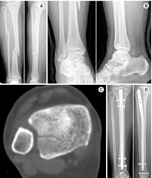
Fig. 2. (A) Preoperative tibia anteroposterior (AP) and lateral image of a 61-year-old male with an AO/OTA 42-A1 type fracture. (B) Preoperative ankle AP and lateral image showing a lateral malleolar fracture and no associated distal tibia intra- articular fracture. (C) Axial im- age of computed tomography showing a minimally displaced posterior malleolar fracture ex- ceeding 25% of the articular surface and an avulsion frac- ture of the anterior tibio- fibular ligament. (D) AP and lateral image after plating with screw fixation of the posterior malleolar fragment and Kirschner wire fixation of the avulsion fracture of the anterior tibio- fibular ligament.
Table 2. Component of Associated Combined Fractures PM & ATFL
avulsion fracture
PM &
MM fracture
MM & ATFL avulsion fracture
No. of combined fracture 12 3 0
PM: Posterior malleolus, ATFL: Anterior tibio-fibular ligament, MM: Medial malleolus.
골절 5예, 내과 골절 5예, 복합골절이 15예로 확인되었다 (Table 1). 복합골절 15예 모두에서 후과 골절이 동반되었 으며, 12예가 후과 골절과 원위 경비 인대 견열 골절이 동
반된 예였다(Table 2).
단순방사선 검사에서 동반 원위 경골 관절내 골절이 확 인된 환자는 총 30예(31.6%)로, 후과 골절 15예, 경비 인 대 견열 골절 5예, 내과 골절 5예, 복합 골절이 5예로 확 인되었으며, 복합골절 중 5예에서 후과 골절이 동반되었다 (Table 1). 이 중 후과 골절 1예 및 경비 인대 견열 골절 5예는 단순방사선 사진상에서는 단독 골절이었으나 CT 결 과 다른 원위 경골 관절내 골절이 동반된 복합골절로 확인 되었으며, 이 중 원위 경비 인대 견열 골절에 동반된 골절 은 모두 후과 골절이었다.
단순방사선 검사에서 동반 원위 경골 골절이 확인되지
Fig. 3. (A) Preoperative tibia anteroposterior (AP) and lateral image of a 51-year-old female with an AO/OTA 42-A1 type fracture. (B) Preoperative ankle AP and lateral image showing no associated distal tibia intra- articular fracture. (C) Axial im- age of computed tomography showing a minimally displaced posterior malleolar fracture ex- ceeding 25% of the articular surface. (D) AP and lateral im- age after intramedullary nailing with fixation of the posterior malleolar fragment.
Table 3. Comparison between PM Diagnosed by Plain Radio- graph and CT
Variable
Diagnosed by radiograph
(n=20)
Diagnosed by CT (n=32) PM mets indication of surgical fixation 16 26
PM fragment size >25% 16 26
PM >2 mm of gap 2 0
PM fixed 14 18
Values are presented in number only. PM: Posterior malleolar fracture, CT: Computed tomography.
않고, CT를 통하여 확인된 환자는 총 32예(33.7%)로 후과 골절이 23예, 원위 경비 인대 견열 골절 5예, 복합골절 4
예였으며, 복합골절 중 4예에서 후과 골절이 동반되었다 (Table 1).
95예의 나선상 경골 간부 골절 중 73예에서 금속정 고 정술을 시행하였으며, 22예에서 금속판 고정술을 시행하였 다. 동반 골절 중 내과 골절의 경우는 8예 모두에서 나사 못 고정을 시행하였다. 원위 경비 인대 견열 골절의 경우, 복합골절까지 포함한 17예 중 나사못 고정이 가능할 정도 의 골편 크기를 보인 5예에서 고정을 시행하였으며, 1예에 서는 Kirschner 강선을 이용하여 고정하였다(Fig. 2). 단순 방사선 검사로는 확인할 수 없고, CT에서만 확인이 된 원 위 경비 인대 견열 골절 7예 중 고정이 가능할 정도의 골 편 크기를 보인 2예에 대해 고정을 시행하였다(Fig. 2).
복합골절에 동반된 후과 골절까지 포함한 총 52예의 동 반 후과 골절 중 20예(38.5%)는 단순방사선 검사에서 확인
이 되었으나 32예(61.5%)는 CT에서만 확인이 가능하였다.
단순방사선 검사를 통해 진단된 동반 후과 골절 20예 중 16예(75%)가 수술적 고정의 적응에 해당하는 골절로 확인 되었으며 이 중 14예에서 실제 수술적 고정이 시행되었다.
CT를 통해 진단된 골절 32예 중에서는 26예(81.3%)가 수 술적 고정의 적응에 해당하는 골절로 확인되었으며 이 중 18예에서 수술적 고정이 시행되었다(Table 3, Fig. 2, 3).
고 찰
경골 간부 골절에 동반된 원위 경골 관절내 골절의 빈도 는 9.7%-49.2%로 다양하게 보고되고 있다.1,2,4-6) 나선상 골 절의 경우에는 이 동반 골절의 빈도가 더욱 높게 보고되고 있다.4,5) Purnell 등4)은 원위 경골 간부 골절 중 43%의 증 례에서 원위 경골골절이 동반되었으나 나선상 골절만을 대 상으로 하였을 때는 56%의 증례에서 동반골절이 있다고 하였다. Schottel 등5)도 71예의 경골간부 골절을 분석한 후 향적 연구에서 경골 간부 골절에서 49.2%의 원위 경골 골 절이 동반되었으며, 이 중 원위 나선상 골절의 경우 60%의 증례에서 원위 경골의 골절이 동반되었다고 보고하였다.
나선상 경골 간부 골절을 대상으로 한 본 연구에서는 65%
의 증례에서 원위 경골 관절내 골절이 동반되어 기존 연구 와 비슷한 결과를 보였으며, 동반 원위 골절 중 후과 골절 의 비중이 가장 높아 기존 연구와 같은 결과를 보였다.4,5,9) 경골 간부 골절에 동반된 후과 골절 등 원위 경골 관절 내 골절을 진단하지 못하는 경우 술 후 불량한 예후를 초 래할 수 있다. Boraiah 등2)은 수술 후 재활과정에서 수술 전 확인되지 않은 비전위 후과 골절이 전위된 경우를 보고 하였으며, Georgiadis 등7)은 수술 전 단순방사선 검사에서 발견되지 않았던 4예의 비전위 후과 골절이 경골 간부 골 절에 대한 골수내 정 고정 수술 중 전위되었음을 보고하였 으며, 과도한 확공이나 지나치게 굵거나 긴 골수정의 사용 시 전위의 위험이 높다고 하였다. 수술 전 인지하지 못한 동반 골절이 수술 중 전위가 될 경우 수술이 더 어려워지 며, 많은 시간이 소요된다.7) 또한 전위가 되지 않더라도 후과 골절이 있는 경우 발목관절의 안정성 및 정상적인 역 동성을 잃게 되며 관절면의 손상으로 인한 이차성 관절염 과의 관련성이 보고된 바 있어 주의를 요한다.8,10)
따라서 나선상 경골 간부 골절에서 수술 전 동반된 원위 경골 관절내 골절을 진단하는 것이 중요하나 단순방사선 검사로 확인되지 않는 경우가 많다. Schottel 등5)은 CT를 통해 진단한 동반 골절 35예 중 8예만을 단순방사선 검사 에서 확인할 수 있다고 하였으며, Purnell 등4)은 단순방사 선 검사에서 방사선과 전문의가 고해상도 화면을 이용한 경우에도 CT에서 발견된 후과 골절의 30%를 발견하지 못
하였다고 보고하였다. Hou 등1)은 전향적 연구에서 34예의 후과 골절 중 단 3예만이 술 전 단순방사선 검사를 통해 진단되었다고 하였으며, Kim 등9)도 20예 중 6예의 족관절 골절은 CT에서만 진단할 수 있었다고 하였다. 본 연구에서 도 나선상 경골 간부 골절에 동반된 원위 경골 관절내 골 절 중 62예 중 32예가 족관절 단순방사선 검사에서 확인되 지 않았으며, 이 중 단순방사선 검사에서는 단독 동반 골 절이었으나 CT에서 복합 골절로 확인된 경우까지 포함한다 면 이 비율은 더 늘어나게 되어 후과 골절의 경우 60% 이 상이 단순방사선 검사에서 진단되지 않고, CT를 통하여 확 인되었다.
CT를 통한 동반 원위 경골 골절의 진단만큼이나 중요한 것이 확인된 동반 골절의 수술적 고정 여부이다. 동반 골 절 중 가장 흔한 후과 골절의 경우 일반적으로 25% 이상 의 관절침범, 2 mm 이상의 전위, 거골의 아탈구 등이 수 술 적응증으로 권고되고 있으며,11) 경골 간부에 동반된 후 과 골절에서도 이러한 적응증을 사용하여 후과 골절을 고 정하였다고 보고된 바 있다.4,9)
본 연구 결과 2 mm 이상의 전위를 보이는 동반 후과 골절은 단순방사선 검사에서 확인된 골절 2예밖에 없었다.
Kim 등9) 역시 경골 간부 골절에 동반된 11예의 후과 골절 에서 2 mm 이상의 전위를 보이는 경우가 한 예도 없었다 고 하였다. 이는 나선상 골절의 손상 기전이 저 에너지의 회전력에 의한 것이므로 원위 경골 관절내 골절이 발생하 여도 전위가 많이 발생하지 않았던 것으로 보인다.3,12)
골편의 크기를 나타내는 골편의 관절면에 대한 상대적 비율이 수술적 고정의 적응이 되는 기준인 25%를 넘는 후 과 골절의 비율은 단순방사선 검사로 진단된 경우 80%, CT로 진단된 골절의 경우 81.3%로 고정이 필요한 경우가 대부분이었다.
동반된 후과 골절의 60% 이상이 단순방사선 검사에서 확인되지 않은 것을 고려하면, 수술적 고정의 적응이 되는 동반 후과 골절의 수가 단순방사선 검사에서 진단이 된 경 우보다 그렇지 않은 경우에 더 많았다. 적응증에 해당하는 것으로 확인된 증례 중 모든 증례에서 실제로 수술적 고정 이 시행되지는 않았으나 단순방사선 검사에서는 확인되지 않고 CT를 통해 확인된 후과 골절 18예에서 수술적 고정 이 시행되었다. 또한 원위 경비 인대 견열 골절의 경우에 도 단순방사선 검사에서 확인되지 않았던 2예에서 수술적 고정이 시행되었다. 이들 증례는 CT를 통하여 수술적 고정 이 필요한 동반 골절을 확인하고 수술까지 시행함으로써 불량한 예후를 초래할 수 있는 합병증을 예방한 경우라 할 수 있다.
본 연구의 제한점으로는 첫째, 후향적 연구로 동반골절 의 고정에 동일한 적응증이 적용되지 않은 증례들을 분석
한 점이다. 둘째, 예후와의 관련성을 분석하지 못한 점으로 추후 장기적인 수술 후 추시 관찰을 시행한 추가 연구가 필요할 것으로 생각된다.
결 론
본 연구 결과 나선상 경골 간부 골절에 동반된 원위 경 골 관절내 골절 중 약 1/2이 CT를 통하여 진단되었다. 동 반 후과 골절의 경우 단순방사선 검사에서 진단되지 않은 비전위 후과 골절 18예(56.3%)에서 수술적 고정이 시행되 었다. 따라서 나선상 경골 간부 골절의 경우 동반 원위 경 골 관절내 골절에 대한 적절한 치료를 위해 술 전 발목 관 절 CT 촬영이 필요할 것으로 생각된다.
References
1) Hou Z, Zhang Q, Zhang Y, Li S, Pan J, Wu H: A oc- cult and regular combination injury: the posterior malleo- lar fracture associated with spiral tibial shaft fracture. J Trauma, 66: 1385-1390, 2009.
2) Boraiah S, Gardner MJ, Helfet DL, Lorich DG: High association of posterior malleolus fractures with spiral dis- tal tibial fractures. Clin Orthop Relat Res, 466:
1692-1698, 2008.
3) Kukkonen J, Heikkilä JT, Kyyrönen T, Mattila K, Gullichsen E: Posterior malleolar fracture is often asso- ciated with spiral tibial diaphyseal fracture: a retrospective study. J Trauma, 60: 1058-1060, 2006.
4) Purnell GJ, Glass ER, Altman DT, Sciulli RL, Muffly MT, Altman GT: Results of a computed tomography
protocol evaluating distal third tibial shaft fractures to as- sess noncontiguous malleolar fractures. J Trauma, 71:
163-168, 2011.
5) Schottel PC, Berkes MB, Little MT, et al: Predictive ra- diographic markers for concomitant ipsilateral ankle in- juries in tibial shaft fractures. J Orthop Trauma, 28:
103-107, 2014.
6) van der Werken C, Zeegers EV: Fracture of the lower leg with involvement of the posterior malleolus; a ne- glected combination? Injury, 19: 241-243, 1988.
7) Georgiadis GM, Ebraheim NA, Hoeflinger MJ:
Displacement of the posterior malleolus during intra- medullary tibial nailing. J Trauma, 41: 1056-1058, 1996.
8) Stuermer EK, Stuermer KM: Tibial shaft fracture and ankle joint injury. J Orthop Trauma, 22: 107-112, 2008.
9) Kim JW, Choi HJ, Lee DH, Kim YC: Ankle fracture associated with tibia shaft fractures. J Korean Fract Soc, 27: 136-143, 2014.
10) Jaskulka RA, Ittner G, Schedl R: Fractures of the pos- terior tibial margin: their role in the prognosis of malleo- lar fractures. J Trauma, 29: 1565-1570, 1989.
11) Macko VW, Matthews LS, Zwirkoski P, Goldstein SA:
The joint-contact area of the ankle. The contribution of the posterior malleolus. J Bone Joint Surg Am, 73:
347-351, 1991.
12) Robinson CM, McLauchlan GJ, McLean IP, Court-Brown CM: Distal metaphyseal fractures of the ti- bia with minimal involvement of the ankle. Classification and treatment by locked intramedullary nailing. J Bone Joint Surg Br, 77: 781-787, 1995.
Copyright ⓒ 2016 The Korean Fracture Society. All rights reserved.
This is an Open Access article distributed under the terms of the Creative Commons Attribution Non-Commercial License (http://creativecommons.org/licenses/
by-nc/4.0) which permits unrestricted non-commercial use, distribution, and reproduction in any medium, provided the original work is properly cited.
http://dx.doi.org/10.12671/jkfs.2016.29.2.114
나선상 경골 간부 골절에 동반된 원위 경골 관절내 골절에서 컴퓨터 단층촬영의 유용성
변성은⋅이상준⋅김 욱⋅최영락⋅한수홍⋅김병국*
CHA 의과학대학교 분당차병원 정형외과, CHA 의과학대학교 구미차병원 정형외과*
목 적: 경골 간부 골절 중 나선상 골절은 경골 원위 관절내 골절이 동반되는 빈도가 높으나 단순방사선 검사만으로 발견되지 않아 적절히 치료하지 못하는 경우가 보고되고 있다. 본 연구에서는 computed tomography (CT)에서만 확인 가능하였던 원위 경골 관절내 동반 골절의 양상 및 수술적 고정의 적응에 해당하였거나 시행했던 경우를 분석하여, 경골 간부 나선형 골절에 서 발목관절 CT 촬영의 필요성을 검증하고자 하였다.
대상 및 방법: 수술 전 족관절 단순방사선 검사 및 CT를 시행한 95예의 나선상 경골 간부 골절을 대상으로 CT에서만 진단된 동반 원위 경골 관절내 골절의 양상을 파악하고, 이 중 수술적 고정의 적응에 해당하였거나 수술적 고정을 시행한 증례를 후향적 으로 분석하였다.
결 과: 원위 경골의 관절내 골절이 62예에서 확인되었다. 복합골절을 포함한 52예의 후과 골절 중 20예는 단순방사선 검사로 진단되었고, 이 중 16예가 수술적 고정의 적응증이 되었으며, 14예에서 시행되었다. CT로 진단된 32예의 후과 골절 중 26예에 서 수술적 고정의 적응증이 되었고, 이 중 18예에서 시행되었다.
결 론: CT는 나선상 경골 간부 골절에서 동반골절의 진단율을 높이며, 진단된 후과 골절의 경우 수술적 고정이 시행된 빈도가 높아 나선상 경골 간부 골절에서 CT의 시행이 필요할 것으로 보인다.
색인 단어: 경골 골절, 관절내 골절, 후과 골절, 컴퓨터 단층촬영
접수일 2015. 7. 6 수정일 1차 2015. 8. 15, 2차 2015. 11. 8, 3차 2015. 12. 11 게재확정 2016. 1. 10 교신저자 김병국
39295, 구미시 신시로10길 12, CHA 의과학대학교 구미차병원 정형외과 Tel 031-780-5289, Fax 031-708-3578, E-mail [email protected]
120