pISSN 2288-9272 eISSN 2383-8493 J Oral Med Pain 2017;42(2):29-34 https://doi.org/10.14476/jomp.2017.42.2.29
Relationships between Intermittent Locking History and Self-Reported Bruxism in Temporomandibular Joint
Myeong-Ok Lee, Yeon-Hee Lee, Soo-Kyung Kang, Yang-Hyun Chun, Jung-Pyo Hong, Q-Schick Auh
Department of Oral Medicine, School of Dentistry, Kyung Hee University, Seoul, Korea
Received May 29, 2017 Revised June 26, 2017 Accepted June 26, 2017
Purpose: To evaluate aggravating factors of intermittent locking among temporomandibular joint using magnetic resonance imaging (MRI) and diagnostic criteria for temporomandibular disorder (DC/TMD) diagnosis.
Methods: A retrospective analysis was conducted of 35 patients with intermittent locking his- tory but normal intra-articular findings between September 2012 and June 2015 in Kyung Hee University Dental Hospital. A standardized DC/TMD assessment was performed on subjects with MRI findings. Clinical findings were assessed on the basis of maximum mouth opening (active & passive), self-reported habits, patients’ age, gender, systemic diseases at the initial visit. First, chi square test was used to examine differences with variables and then risk factors for intermittent locking were assessed using multivariate logistic regression.
Results: Self-reported bruxism was strongly associated with intermittent locking history.
Conclusions: The new DC/TMD protocol is intended for use within any clinical setting and sup- ports the full range of diagnostic activities from screening to definitive evaluation and diag- nosis. Self-reported sleep bruxism has been associated with a higher likelihood of intermittent locking. Comorbidity is therefore a factor that must be assessed. It is necessary to consider the amount of contact of the teeth and the duration.
Key Words: Bruxism; Diagnostic criteria for temporomandibular disorder; Intermittent locking;
Magnetic resonance imaging; Temporomandibular joint disorders
Correspondence to:
Q-Schick Auh
Department of Oral Medicine, School of Dentistry, Kyung Hee University, 26 Kyungheedae-ro, Dongdaemun-gu, Seoul 02447, Korea
Tel: +82-2-958-9355 Fax: +82-2- 961-1457 E-mail: [email protected]
JOMP
Journal of Oral Medicine and PainCopyright Ⓒ 2017 Korean Academy of Orofacial Pain and Oral Medicine. All rights reserved.
CC This is an open-access article distributed under the terms of the Creative Commons Attribution Non-Commercial License (http://creativecommons.org/licenses/by-nc/4.0/),
INTRODUCTION
Temporomandibular disorders (TMDs) are a comprehen- sive term of diseases that include many clinical problems in the masticatory muscles and temporomandibular joint (TMJ) region, which show morbidity rates of approximately 5% to12% of the population. TMDs are also a major cause of non-odontogenic pain in the orofacial region1) and may cause jaw pain, headache, ear ache, facial pain, mandibu- lar movement restriction, asymmetric mandibular move- ment form and joint sound, clicking, and crepitus as well as oral habits, such as teeth grinding, non-painful masticatory muscular hypertrophy, and abnormal tooth wear.2-4)
The research diagnostic criteria for TMD (RDC/TMD)
introduced by Dworkin and LeResche5) in 1992 show scien- tifically proved reliability in the evaluation of TMDs’ clini- cal symptoms and signs and includes the evaluation of be- havioral, psychological, and psychosocial elements.6) In ad- dition, it presents clinical tests methods that show reliability in the evaluation of TMDs’ symptoms.7) Recently, in 2014, a revised version, diagnostic criteria for TMD (DC/TMD) was introduced, in which a new diagnostic criterion, anterior disc displacement with reduction with intermittent locking is presented.6)
Anterior disc displacement with reduction is a joint dis- placement condition that occurs the most frequently in the TMJ. It is a phenomenon in which the articular disc is located anterior to the condylar head when the mouth is
closed but is reduced to the region above the condylar head when the mouth is opened. This condition does not cause any pain in most cases but does cause joint sounds when the mouth is opened, closed, or opened/closed.8,9)
However, this condition becomes progressive sometimes leading to trismus accompanied by intermittent pain, which is called intermittent locking. Intermittent locking is a pre- liminary stage of complete locking, which may lead to chronic trismus if not preemptively treated.10)
Thus far, micro-trauma, bruxism, excessive movements, and degenerative changes have been suspected as causes of anterior articular disc displacement with reduction.11) However, studies of the causes of or risk factor for intermit- tent locking are still rare. In the recent experiment, it was reported that excessive masticatory movements cause tem- porary intermittent locking.12) This suggests that oral habits may cause intermittent locking. Therefore, the purpose of the present study was to evaluate factors that may cause in- termittent locking in TMJs using the DC/TMD and magnetic resonance imaging (MRI) which is the diagnostic reference criterion of the DC/TMD.
MATERIALS AND METHODS
1. Subjects
This retrospective cross-sectional study protocol was ap- proved by the Institutional Review Board of Kyung Hee University Dental Hospital (KHD IRB 1612-4) and is in com- pliance with the Hesinki Declaration. The present study was conducted with 35 patients (male 12, female 23) with inter- mittent locking history, who showed normal joint-disc re- lationships in clinical tests, radiography, and MRI scanning
among those patients that visited the Department of Oral Medicine, Kyung Hee University Dental Hospital between September 2012 and June 2015 retrospectively (Fig. 1). The ages of the subjects ranged from 12 to 81 years.
2. Clinical Examination
As test items, experiences in injuries and whether the subjects had any oral habit or not were described by the subjects and oral habits, such as clenching, lip biting, nail biting, and tongue biting were examined. In addition, max- imum opening (passive, active), ages, and genders were re- corded (Table 1).
Normal joint-disc relationships were clinically identified
Fig. 1. A flow sheet for the reliability assessment of the magnetic resonance imaging (MRI) and diagnostic criteria for temporo mandibular disorder (DC/TMD) based diagnosis of the temporomandibular joint disc displacement.
Reconfirmation through MRI
The confirmation of oral habits, maximum opening, age, gender by an examiner
Multiple regression analysis
Compared the relationships between intermittent locking and variables
35 Subjects (male 12, female 23) with intermittent locking history but, normal joint disc displacement diagnosed by DC/TMD
Table 1. The gender distribution of patients with bruxism, clenching, morning stiffness, morning lock among normal joint disc displacement groups (n=35)
Group Gender Total Bruxism Clenching Morning stiff Morning lock
Yes Yes Yes Yes
Normal Male 5 (14.3) 0 (0) 0 (0) 0 (0) 0 (0)
Female 18 (51.4) 0 (0) 3 (8.6) 0 (0) 1 (2.9)
Total 23 (65.7) 0 (0) 3 (8.6) 0 (0) 1 (2.9)
Intermittent locking (history) Male 7 (20.0) 2 (5.7) 1 (2.9) 2 (5.7) 0 (0)
Female 5 (14.3) 0 (0) 0 (0) 2 (5.7) 2 (5.7)
Total 12 (34.3) 2 (5.7) 1 (2.9) 4 (11.4) 2 (5.7)
Values are presented as number (%).
using the DC/TMD and were reconfirmed through radiog- raphy and MRI. In the maximum intercuspal position, the MRI findings of normal disc position in the sagittal plane were defined as the posterior band of the disc being at the 12 o’clock position relatively to the condyle and on full mouth opening, the intermediate zone of the disc is located between the condylar head and the articular eminence.
During the physical examination, the subjects were in- spected for the absence of clicking, popping, snapping noise during both opening and closing movements with palpa- tion. Intermittent locking was identified using subjects’
medical histories whether jaw locks with limited mouth opening, even for a moment, and then unlock in the last 30 days.
The time frame for assessing selected biomechanical in- tra-articular disorders is in ‘the last 30 days’ since the stated sensitivity and specificity of these criteria were established using this time frame. Although the specific time frame can be dependent on the context in which the noise or bio- mechanical complaints are being assessed, the validity of
this diagnosis based on different time frames has not been established.7)
3. Statistical Analyses
Multiple regression analyses were conducted for risk fac- tors for intermittent locking. The analyses included various variables included in clinical tests. In addition, t-tests and chi-squared post hoc tests of individual variables were con- ducted. The significance level was set to p=0.05. Statistical processing was conducted using the SPSS ver. 16.0 software (SPSS Inc., Chicago, IL, USA).
RESULTS
Cross analyses of individual clinical variables were con- ducted through t-tests and chi-squared tests. Significance level was set to p-value<0.05. According to the results of chi- squared tests, self-reported bruxism/clenching was strongly associated with intermittent locking history (Table 2).
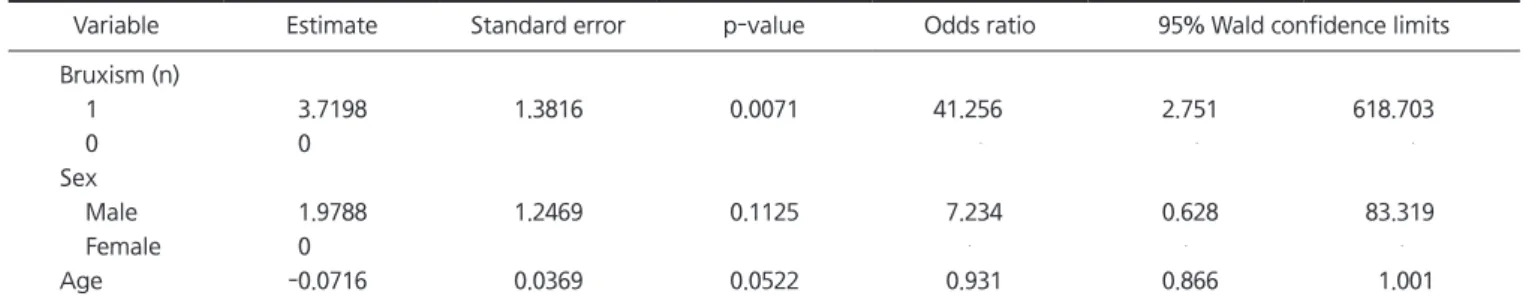
Logistic-regression analyses were conducted and ccord- ing to the results, it can be seen that those that have self- reported bruxism/clenching symptoms are much more like- ly to show intermittent locking history than those that have no bruxism/clenching history. Weak correlations between age and intermittent locking were observed. The correla- tions will be examined in the discussion section (Table 3).
DISCUSSION
A hypothesis in the present study is that among oral hab- its, bruxism may increase the possibility of intermittent locking. According to the results of the present study, self- reported bruxism/clenching was a risk factor that can make even the normal TMJ skip the stage of anterior articular
Table 2. t-test and chi-square test of risk factors for reported intermittent locking
Variable Normal Intermittent locking p-value
Age (y) 50.44±14.32 33.90±19.33 0.007a
Active range of motion (o)
41.26±8.16 35.08±12.53 0.087a
Passive range of motion (o)
45.39±9.14 40.50±9.99 0.155a
Sex
Female 18 (78.3) 5 (21.7) 0.059b
Male 5 (41.7) 7 (58.3)
Bruxism
No 19 (86.4) 3 (13.6) 0.002b
Yes 4 (30.8) 9 (69.2)
Values are presented as mean±standard deviation or number (%).
aBy t-test. bBy chi-square test.
Table 3. Multiple logistic regression test of risk factors for reported intermittent locking
Variable Estimate Standard error p-value Odds ratio 95% Wald confidence limits
Bruxism (n)
1 3.7198 1.3816 0.0071 41.256 2.751 618.703
0 0
Sex
Male 1.9788 1.2469 0.1125 7.234 0.628 83.319
Female 0
Age -0.0716 0.0369 0.0522 0.931 0.866 1.001
disc displacement with reduction and develop intermittent locking. This means that bruxism/clenching may tempo- rarily lead to anterior displacement that causes temporary locking. Temporary locking should be clinically carefully observed because it is a predicative factor for articular disc displacement without reduction.13)
The hypothesis that bruxism/clenching may be a cause of orofacial pain has been always held at clinics thus far.
A study indicated that whereas 55% of TMDs patients re- ported teeth grinding, only 15% of the control group did so.14) Intermittent mild teeth grinding may be associated with masticatory muscle pain in the morning.15) Therefore, even if bruxism/clenching is not a definite cause of TMDs, whether or not teeth bruxism/clenching and TMDs coexist should be evaluated without fail.
Joint sounds are a common finding in general people.
In most cases,16-18) joint sounds do not seem to be associ- ated with pain or reduced joint mobility. If all noisy joints progress to severe disorders, all joints accompanied by joint sounds should be treated without fail. However, the fact that joint sounds that do not change over time exist indi- cate that the TMJ may be adapted to less than best joint- disc relationships.
According to a very long-term study conducted by de Leeuw et al.,19) joint sounds remained in 54% of patients 30 years after nonsurgical treatment of disorders in the ar- ticular capsule. Although this finding indicates that joint sounds remained in many patients, none of those patients felt discomfort of dysfunction. As with other studies cited in the present paper, this study suggests that joint sounds have nothing to do with pain or TMJ dysfunction.
These studies lead to the thought that not all joint sounds are progressive or require treatment. According to sever- al studies,20-22) only 7% to 9% of disorders in the articular capsule identified based on joint sounds were progressive.
Another study indicated that disorders in the articular cap- sule would rarely progress into articular disc displacement without reduction. However, if any disorder in the articular disc brings about serious articular disc displacement with- out reduction, it may progress into intermittent locking.23)
Recently, several studies reported that intermittent lock- ing is highly likely to develop into anterior disc displace- ment without reduction.24,25) To make a deduction, some of
those patients that were asymptomatic in the clinical tests during the present experiment but reported histories of in- termittent locking might have already shown anterior disc displacement without reduction. Once the condition has progressed into anterior disc displacement without reduc- tion, the amount of opening becomes 25-30 mm and hard end feeling appears at the maximum opening position. That is, even if soft and continuous force is applied to the man- dibular incisor, the opening hardly increases.26)
A team consisting of international clinical research spe- cialists developed the RDC/TMD to relieve shortcomings in diverse existing diagnostic methods and for scientific evaluation of TMDs with common tools that can be ap- plied to research and diagnosis. The RDC/TMD uses clinical tests that include behavioral, psychological, and psychoso- cial elements and medical history collection and has been proved to be reliable. In addition, it is a method of which the reliability, sensitivity, validity, and specificity have been verified to the extent that it shows significant differences among muscular pain, articular pain, and headache due to TMDs.5)
However, problems in the RDC/TMD version have been raised based on diverse rationales and methodologies.
Currently, the DC/TMD DC have been presented through massive revision procedures to enable efficient approach to the substance of the disease. The DC/TMD was introduced in the Journal of Oral & Facial Pain and Headache, 2014 by Schiffman et al.7) and is now being diffused in diverse languages.
The largest change made in DC/TMD in relation to articu- lar disc disorders is the addition of intermittent locking. The diagnosis that was included in anterior disc displacement with reduction in RDC/TMD was subdivided. This implies the fact that intermittent locking is a preliminary stage of complete locking with pathophysiological or clinical impor- tance that is differentiated from anterior articular disc dis- placement with reduction without intermittent locking. A diagnostic reference criterion for articular disc displacement under the DC/TMD DC is MRI scanning.
In the present experiment, the relationships between the history of intermittent locking and diverse clinical variables were evaluated with patients that showed normal articular- disc relationships in clinical tests and MRI scanning. In the
case of patients with bruxism/clenching, unlike general pro- gression, the stage of anterior disc displacement with reduc- tion was skipped and the conditions of intermittent locking were immediately observed. Given the foregoing, bruxism/
clenching seems to combine orthopedic instability and joint loads to act as a causative agent.
These changes actually begin at the cell level and gradu- ally progress to appear clinically. That is, if abnormally large and continuous loads imposed on joint tissues ex- ceed the functional limit of joint tissues, the joint tissues will begin to collapse. When joint loads exceed the func- tional limit, the collagen fibrils are broken down into small pieces and the rigidity of collagen net structures decreases.
Consequently, proteoglycan-water gel swelling occurs and the effusion flows out to the glenoid cavity leading to the softening of the articular surface, which is called chondro- malacia.27) When the foregoing led to changes in the fric- tional characteristics of the articular surface making the articular surface viscose, mechanical changes occur in con- dylar head-disc movements. The continuous sticking seems to cause strain deformation to the disc ligament when joint motions occur eventually resulting in intermittent locking.
Another interesting result of the present experiment is that the possibility of intermittent locking is slightly high- er among younger patients. Although accurate causal re- lationships might be identified only by evaluating both masticatory and swallowing activities, intermittent locking seems to be associated with the reduction in masticatory force overtime in lifetime. Since it is assumed that 58.71 lb of force is applied to the teeth during a single mastication cycle for 115 ms on average,28) a force of 6.75 lb/s can be regarded to act during each mastication.29) If it is assumed that 1,800 times of mastication occur per day on average,30) the entire bite force/time activity per day can be estimated to be 12,150 lb/s. Swallowing force should be also consid- ered. Since each person swallows food approximately 146 times per day,31) and a force of 66.51 lb acts for 522 ms on the teeth per swallowing,29) a force of 5,068 Ib/s is used per day. Therefore, the total force/time activity due to mastica- tion and swallowing can be regarded to be 17,200 lb/s per day. Given that the foregoing varies greatly among indi- viduals, it can be understood that the foregoing may cause changes to individual tissues in the masticatory system.
Despite its limitations that rely on self-report for brux- ism/clenching, this is one of the few studies of onset of in- termittent locking in TMJ. One of the unique advantages of this study was the use of DC/TMD to determine if the intra- articular problem met specified diagnostic criteria. Other studies examining risk factors for intermittent locking on- set have lacked such diagnostic assessment. We were able to confirm that bruxism/clenching for onset of intermittent locking in normal TMJ features. These findings suggest that individuals who have bruxism/clenching may have an un- derlying vulnerability to intermittent locking that involves limited mouth opening.
In cases where bruxism/clenching habits were suspected, the possibility for normal articular-disc relationships to skip the stage of anterior disc displacement with reduction and develop intermittent locking was statistically significantly higher. That is, bruxism/clenching was closely associated with intermittent locking. Therefore, the correlations be- tween the two conditions should be considered without fail during initial DC/TMD diagnosis.
In addition, weak negative correlations between intermit- tent locking and ages were observed. This is assumed to be associated with biting force for now. However, the evalua- tion of mastication and swallowing activity, that is, consid- eration of two elements; the amount of contact of the teeth and the duration, may be necessary later.
CONFLICT OF INTEREST
No potential conflict of interest relevant to this article was reported.
REFERENCES
1. Okeson JP. Bell’s orofacial pain: the clinical management of oro- facial pain. 6th ed. Chicago: Quintessence Pub.; 2005.
2. de Leeuw R. Orofacial pain: guidelines for assessment, diagnosis, and management. 4th ed. Chicago: Quintessence Pub.; 2008. pp.
129-204.
3. Magnusson T, Egermark I, Carlsson GE. A longitudinal epidemio- logic study of signs and symptoms of temporomandibular disor- ders from 15 to 35 years of age. J Orofac Pain 2000;14:310-319.
4. Magnusson T, Egermarki I, Carlsson GE. A prospective investiga- tion over two decades on signs and symptoms of temporoman- dibular disorders and associated variables. A final summary. Acta
Odontol Scand 2005;63:99-109.
5. Dworkin SF, LeResche L. Research diagnostic criteria for tem- poromandibular disorders: review, criteria, examinations and specifications, critique. J Craniomandib Disord 1992;6:301-355.
6. Oh JT, Kim W, Chung SC. A study of characteristics of TMD using RDC/TMD. J Oral Med Pain 2004;29:177-185.
7. Schiffman E, Ohrbach R, Truelove E, et al. Diagnostic Criteria for Temporomandibular Disorders (DC/TMD) for clinical and research applications: recommendations of the international RDC/TMD Consortium Network and Orofacial Pain Special Interest Group. J Oral Facial Pain Headache 2014;28:6-27.
8. Huddleston Slater JJ, Lobbezoo F, Onland-Moret NC, Naeije M.
Anterior disc displacement with reduction and symptomatic hy- permobility in the human temporomandibular joint: prevalence rates and risk factors in children and teenagers. J Orofac Pain 2007;21:55-62.
9. Farrar WB. Craniomandibular practice: the state of the art; defi- nition and diagnosis. J Craniomandibular Pract 1982;1:4-12.
10. Kurita K, Westesson PL, Yuasa H, Toyama M, Machida J, Ogi N. Natural course of untreated symptomatic temporoman- dibular joint disc displacement without reduction. J Dent Res 1998;77:361-365.
11. Stegenga B, de Bont LGM. TMJ disc derangements. In: Laskin DM, Greene CS, Hylander WL, eds. Temporomandibular disorders:
an evidence-based approach to diagnosis and treatment. Chicago:
Quintessence Pub.; 2006. pp. 125-136.
12. Kalaykova S, Lobbezoo F, Naeije M. Effect of chewing upon disc reduction in the temporomandibular joint. J Orofac Pain 2011;25:49-55.
13. Emshoff R, Rudisch A, Innerhofer K, Brandlmaier I, Moschen I, Bertram S. Magnetic resonance imaging findings of internal derangement in temporomandibular joints without a clini- cal diagnosis of temporomandibular disorder. J Oral Rehabil 2002;29:516-522.
14. Yachida W, Castrillon EE, Baad-Hansen L, et al. Craniofacial pain and jaw-muscle activity during sleep. J Dent Res 2012;91:562- 567.
15. Rompré PH, Daigle-Landry D, Guitard F, Montplaisir JY, Lavigne GJ. Identification of a sleep bruxism subgroup with a higher risk of pain. J Dent Res 2007;86:837-842.
16. Sato S, Goto S, Nasu F, Motegi K. Natural course of disc displace- ment with reduction of the temporomandibular joint: changes in clinical signs and symptoms. J Oral Maxillofac Surg 2003;61:32- 34.
17. Kamisaka M, Yatani H, Kuboki T, Matsuka Y, Minakuchi H. Four-
year longitudinal course of TMD symptoms in an adult popula- tion and the estimation of risk factors in relation to symptoms. J Orofac Pain 2000;14:224-232.
18. Egermark I, Carlsson GE, Magnusson T. A 20-year longitudinal study of subjective symptoms of temporomandibular disorders from childhood to adulthood. Acta Odontol Scand 2001;59:40- 48.
19. de Leeuw R, Boering G, Stegenga B, de Bont LG. Clinical signs of TMJ osteoarthrosis and internal derangement 30 years after non- surgical treatment. J Orofac Pain 1994;8:18-24.
20. Randolph CS, Greene CS, Moretti R, Forbes D, Perry HT. Con- servative management of temporomandibular disorders: a post- treatment comparison between patients from a university clinic and from private practice. Am J Orthod Dentofacial Orthop 1990;98:77-82.
21. Greene CS, Laskin DM. Long-term evaluation of treatment for myofascial pain-dysfunction syndrome: a comparative analysis. J Am Dent Assoc 1983;107:235-238.
22. Greene CS, Laskin DM. Long-term status of TMJ clicking in pa- tients with myofascial pain and dysfunction. J Am Dent Assoc 1988;117:461-465.
23. Brooke RI, Grainger RM. Long-term prognosis for the clicking jaw. Oral Surg Oral Med Oral Pathol 1988;65:668-670. Erratum in: Oral Surg Oral Med Oral Pathol 1989;67:131.
24. Westesson PL, Lundh H. Arthrographic and clinical character- istics patients with disk displacement who progressed to closed lock during a 6-month period. Oral Surg Oral Med Oral Pathol 1989;67:654-657.
25. Kalaykova S, Lobbezoo F, Naeije M. Two-year natural course of anterior disc displacement with reduction. J Orofac Pain 2010;24:373-378.
26. Okeson JP. Management of temporomandibular disorders and oc- clusion. 7th ed. St. Louis: Elsevier; 2014. pp. 271-278.
27. Stegenga B, de Bont LG, Boering G, van Willigen JD. Tissue re- sponses to degenerative changes in the temporomandibular joint:
a review. J Oral Maxillofac Surg 1991;49:1079-1088.
28. Gibbs CH, Mahan PE, Lundeen HC, et al. Occlusal forces during chewing--influences of biting strength and food consistency. J Prosthet Dent 1981;46:561-567.
29. Lundeen HC, Gibbs CH. Advances in occlusion. Boston: John Wright; 1982. pp. 211.
30. Graf H. Bruxism. Dent Clin North Am 1969;13:659-665.
31. Flanagan JB. The 24-hour pattern of swallowing in man. J Dent Res 1962;42:1072.